Abstract
Interactions between two cells or between cell and extracellular matrix mediated by protein–carbohydrate interactions play pivotal roles in modulating various biological processes such as growth regulation, immune function, cancer metastasis, and apoptosis. Galectin-3, a member of the β-galactoside-binding lectin family, is involved in fibrosis as well as cancer progression and metastasis, but the detailed mechanisms of its functions remain elusive. This review discusses its structure, carbohydrate-binding properties, and involvement in various aspects of tumorigenesis and some potential carbohydrate ligands that are currently investigated to block galectin-3 activity.
Keywords: galectin-3, angiogenesis, apoptosis, tumorigenesis, TF disaccharide
Introduction
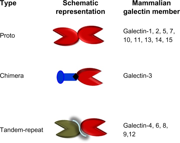
In recent years, protein–carbohydrate interactions have been considered as very important for the modulation of cell–cell and cell–extracellular matrix (ECM) interactions, which, in turn, mediate various biological processes such as cell activation, growth regulation, cancer metastasis, and apoptosis. Thus, the identification and expression of carbohydrate-binding proteins (lectins) and their partners (carbohydrate ligands) and the detailed understanding of the molecular mechanisms and downstream effects of these protein–carbohydrate interactions are subjects of current intense research. Galectins, a family of at least 15 β-galactoside-binding proteins, are involved in growth development as well as cancer progression and metastasis.1–5 However, the detailed mechanisms of these functions remain elusive. Based on their subunit structures, galectins are classified into three types: proto, chimera, and tandem repeat (Fig. 1).5 Prototype galectins contain one carbohydrate-recognition domain (CRD) per subunit. Galectins-1, -2, -5, -7, -10, -11, -13, -14, and -15 are examples of prototype galectins, of which galectins-1, -2, and -7 are dimers. Tandem repeat-type galectins (eg, galectins-4, -6, -8, -9, and -12) contain two CRDs joined by a linker peptide. Galectin-3 is the only representative of the chimera-type galectin, which has one CRD at the C-terminal end. Galectin-3 is one of the most studied member of the galectin family.2,6–9 As a multi functional protein with increased or decreased expression in many types of human cancers, CRD-dependent or CRD-independent functions, and also its cellular locations (cell surface, nucleus, cytoplasm, mitochondria, and endosomal compartment),10 galectin-3 has generated significant interest in cancer research over the past decades. This review describes its structure, carbohydrate-binding properties, transcriptional regulation, and its involvement in various aspects of tumorigenesis. Some potential carbohydrate ligands that are currently investigated to block galectin-3 function are also discussed.
Figure 1.

Classification of galectins. Schematic representation of proto-, chimera-, and tandem repeat-type galectins. They are numbered according to the order of their discovery.
Structure of Galectin-3
Primary structure
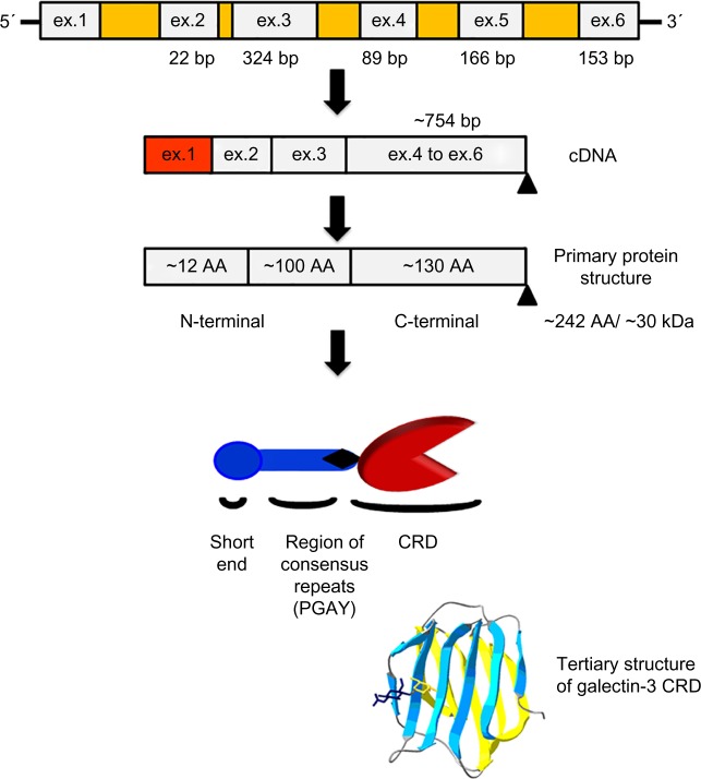
Galectin-3 (previously known as Mac-2, L-29, L-31, L-34, immunoglobulin E-binding protein, CBP35, and CBP30) contains three structurally distinct domains: a highly conserved 12-amino acid short N-terminal domain (ND),7 proline- and glycine-rich long ND, and a C-terminal CRD (Fig. 2).11 Galectin-3 is a monomer but can form multimer at certain circumstances such as at high concentration.12 The short ND may have roles in secretion and apoptosis. Deletion of short ND blocks secretion of galectin-3,8 while mutation of the conserved Ser6 affects galectin-3 antiapoptotic signaling activity.13 The long ND is responsible for multimerization of galectin-3 and shows positive cooperativity in carbohydrate binding.12 This has been asserted from the observation that matrix metalloproteinases, MMP-2 and MMP-9, cleave galectin-3 at the position Ala62–Tyr63, resulting in 22 kDa fragment, which fails to self-associate.14 The C-terminal domain of galectin-3 is composed of about 130 amino acids. It forms a globular structure like other galectins7 and accommodates whole carbohydrate-binding site responsible for lectin activity.15,16 Within the CRD, particularly interesting amino acid sequence is NWGR. This motif is highly conserved within the BH1 domain of the Bcl-2 family proteins and is responsible for the antiapoptotic activity of both Bcl-2 and galectin−3.17 The NWGR motif is also involved in the self-association of galectin-3 molecules through the CRDs in the absence of saccharide ligands.18
Figure 2.
Structure of galectin-3. Schematic representation of nucleotide (genomic and cDNA) and protein (primary and tertiary) structures.
Genomic and complementary DNA structures
The human galectin-3 gene (LGALS3) is located on locus q21–q22 of chromosome 1419 and is about 17 kb long containing six exons and five introns (Fig. 2).20 Exon 1 contains the major part of the 5′ untranslated sequence of messenger RNA (mRNA), while exon 2 houses the remaining part of the 5′ untranslated region, the translation initiation site and codon sequence for the first six amino acids, including the initial methionine. Exon 3 comprises long ND, while exons 4–6 house the CRD. Size of human galectin-3 mRNA (transcript variant 1) is 1017 bp – of which the open reading frame consists of 753 bp (NM_002306.3). Production of alternative transcripts arising from an internal promoter in the intron II is known in peripheral blood leukocytes.21,22 These transcripts arise from an internal gene embedded within LGALS3, named galig (galectin-3 internal gene).22 Galig’s CRD is incapable of binding carbohydrates as it contains two overlapping open reading frames out of frame within the lectin coding sequence. However, the galig protein promotes cytochrome c release upon direct interaction with the mitochondria.23
Three-dimensional structure
Galectin-3 CRD was crystallized in complex with Thomsen–Friedenreich (TF) antigen (Galβ1-3GalNAcα1-O-Ser/Thr), lactose, TF p- nitrophenyl (TFN), or GM1.24–26 The general folds of galec-tin-3 CRD in these complexes show a high similarity to the previously reported structures.24 The CRD adopts a typical galectin fold in which six-stranded (S1–S6) and five-stranded (F1–F5) antiparallel β-sheets jointly formed a β-sandwich structure. The S1–S6 β-strands constitute a concave surface on which TF antigen and other glycans are bound. All these structural features are like those previously reported.24–26 In the present structures, the residues involved in TF binding are located on S4–S6 β-strands and the loop connecting S4 and S5. Electron density maps show that TF antigen, TFN, and GM1 in the complexes are all well-ordered, and carbohydrate rings of TFs are in the chair conformations.
Carbohydrate-Binding Properties of Galectin-3 and its Ligands
Although all galectins bind β-galactoside, their ability to discriminate among carbohydrate structures is striking. For most galectins, N-acetyllactosamine (Galβ1, 4GlcNAc) is 5–10 times more active than lactose,27–31 and so N-glycans are good ligands. Interestingly, a striking difference was observed between interactions of galectin-1 and -3 toward the TF disaccharide (TFD, Galβ1, 3GalNAc) found in O-glycans.26–29 On isothermal titration calorimetry assays, galectin-3 was found to interact with TF antigen with 100-fold higher affinity compared to galectin-1.26 The basis for the variable binding profiles of these galectins has been explained by their three-dimensional (3D) structures.26,32,33 Although galectins lack a typical secretory signal peptide,34 they are present not only in the cytosol but also in the ECM.35,36 In the extracellular space, galectins bind to β-galactoside-containing glycoproteins of ECM and cell surface. Extracellular galectin-3 binds laminin,37,38 fibronectin,39 CD29,40 CD66,41 α1β1 integrin,39 and Mac-2-binding protein.42 Intracellularly, galectin-3 binds gemin 4,17 Bcl-2,43 nucling,44 synexin,45 and β-catenin46,47 via protein–carbohydrate or protein–protein interactions.
Transcriptional Regulation
Although a large body of data about galectin-3 expression are available in the literature, the mechanisms of regulation of galectin-3 expression are not well understood. However, the expression of galectin-3 depends on cell type, external stimuli, and environmental conditions and involves numerous transcription factors and signaling pathways.7 Galectin-3 expression may serve as differentiation marker for certain cell types. For example, the differentiation of the human monocytes or promyelocytic cell line HL-60 to macrophage-like cells induced by phorbol ester is accompanied by increased expression of galectin-3.48 Galectin-3 expression is upregulated in phagocytic macrophages and thus considered as a “macrophage activation marker.”49 Galectin-3 expression is also elevated in microglia and macrophages activated by phagocytosis of myelin or when exposed to granulocyte-macrophage colony-stimulating factor.50 In contrast, the activation of human monocytes by lipopolysaccharide and interferon-γ is accompanied by decrease of galectin-3 expression.51 The reduced expression of galectin-3 was also observed in monocytic THP-1 cells treated with nonsteroidal52 or corticosteroidal anti-inflammatory drugs.53 Interestingly, galectin-3 expression is absent or barely detected in the resting lymphocytes,54,55 but the activated B- and T-cells induce galectin-3 expression.55 Galectin-3 could also be considered as a transformation marker since the galectin-3 expression is increased in fully ras-transformed fibroblasts, when cells have lost their anchorage-dependent growth.56
In the promoter region of the galectin-3 gene, several regulatory elements such as five putative Sp1-binding sites (GC boxes), five cAMP-dependent response element (CRE) motifs, four Adaptor Protein-1 (AP-1)- and one AP-4-like sites, two nuclear factor-kappa B (NF-κB)-like sites, one sis-inducible element (SIE), and a consensus basic helix–loop– helix core sequence are found.19 The presence of multiple GC box motifs for binding ubiquitously expressed Sp1 transcription factor is a characteristic of constitutively expressed “housekeeping” genes. The activation of the Sp1-binding transcription factor is responsible for galectin-3 induction by Tat protein of HIV.57 The SIE that binds sis-inducible factors was suggested to be a possible candidate for the growth-induced activation of galectin-3 gene expression, caused by the addition of serum. The presence of CRE and NF-κB-like site in the galectin-3 promoter suggests that the activation of galectin-3 expression could be regulated through the signaling pathways involving the CRE-binding protein (CREB) or the NF-κB transcription factor. The CREB/Activating transcription factor (ATF) and the NF-κB/Rel transcription factor pathways may be involved in the regulation of galectin-3 expression by the Tax protein during Human T-lymphotropic virus-1 (HTLV-1) infection of T-cells.58 The involvement of the NF-κB transcription factor in the regulation of galetin-3 expression, as well as the Jun protein, a component of AP-1 transcription factor has recently been confirmed.59 The regulation of galectin-3 expression through the NF-κB transcription factor was shown to be mediated by nucling, a novel apoptosis-associated protein, which interferes with NF-κB via the nuclear translocation process of NF-κB/p65, thus inhibiting galectin-3 expression on both protein and mRNA level.60,61 In skeletal tissues, the regulation of galectin-3 expression is mediated by the transcription factor Runx2.6 Very recently, galectin-3 expression is found to be regulated in pituitary and prostate tumors by methylation of CpG islands in promoter region.62–65 Galectin-3 was shown to be highly expressed in androgen-independent PC-3 and DU-145 human prostate cancer cell lines but weakly expressed in androgen-dependent Androgen-sensitive human prostate adenocarcinoma (LNCaP) prostate cancer cells.64 Treatment of LNCaP cells with azacytidine (DNA methyltransferase inhibitor) showed restored expression of galectin-3, indicating that the promoter methylation is responsible for galectin-3 gene silencing.64 We have also demonstrated DNA methylation on the galectin-3 promoter in LNCaP cells following polymerase chain reaction (PCR) amplification of the bisulfate-treated DNA and cloning and sequencing of the PCR product.62
Role of Galectin-3 in Cell–Cell and Cell–ECM Interactions
Numerous studies indicate that galectin-3 has important roles in normal development and tumorigenesis through regulating cell proliferation, apoptosis, cell adhesion, invasion, angiogenesis, and metastasis by binding to the cell surface β-galactose-containing glycoconjugates or glycolipids.3,4,66 However, galectin-3 is also known to have CRD-independent functions intracellularly at the mitochondria.16,67–69
Galectin-3 expression in normal tissues: role of galectin-3 in growth development
Galectin-3 is developmentally regulated and expressed in many tissues of adults.70,71 During mouse embryogenesis, galectin-3 first appears at the fourth day of gestation in the trophectoderm of blastocyst, followed by its expression in the notochord cells between 8.5 and 11.5 days of gestation.70 In later stages of mouse development, galectin-3 is expressed in the cartilage, ribs, facial bones, suprabasal layer of epidermis, endodermal lining of the bladder, larynx, and esophagus.7 In adults, galectin-3 is mainly expressed in the epithelial cells such as small intestine,72 colon,73 cornea,74,75 kidney,76 lung,77 thymus,78 breast,79 and prostate.80 The expression of galectin-3 is also detected in the ductal cells of salivary glands,81 pancreas,82 kidney,83 eye,84 and intrahepatic bile ducts.85 Regarding cell type, galectin-3 expression is observed in fibroblasts,86 chondrocytes and osteoblasts,43 osteoclasts,87 keratinocytes,88 Schwann cells,89 gastric mucosa,90 endothelial cells,91 and also immune-related cells such as neutrophils,92 eosinophils,93 basophils and mast cells,94 Langerhans cells,88,95 dendritic cells,96 as well as mono-cytes51 and macrophages from different tissues.3,6,97,98
Galectin-3 promotes tumor progression and metastasis: changes in cellular localization of galectin-3
Galectin-3 is expressed in many tumors and possibly plays an important role in tumor progression and metastasis.6,7,9,43,79,80,98,99 However, the intensity of the galectin-3 expression in tumors depends on the type of tumor, its invasiveness, and metastatic potential.44,45 For example, increased expression of galectin-3 is observed in colon, head and neck, gastric, endometrial, thyroid, liver, bladder cancers, and breast carcinomas.46,47,79,99–101 Galectin-3-transfected human breast cancer cells BT549, which is galectin-3 null, after intrasplenic injection, formed metastatic colonies in the liver, while gal3 null BT549 cells did not.102 Change in cellular localization of galectin-3 is also observed during progression of various cancers. For example, downregulation of galectin-3 expression has been demonstrated in colorectal cancer, with increased cytoplasmic expression of galectin-3 at more advanced stages.44,45,103 In tongue cancer, nuclear galectin-3 is decreased, but cytoplasmic galectin-3 is increased during progression from normal to cancer.44,45 The decreased expression of galectin-3 was also observed in prostate,64,80,104 kidney,105 and pituitary cancers.63 In prostate cancer, although galectin-3 is downregulated, its nuclear exclusion and cytoplasmic localization are correlated with disease progression.64,80,106 Phosphorylation of galectin-3 at Ser6 regulates its nuclear export.107 Recent data by us and others indicated that decreased expression of galectin-3 in pituitary and prostate tumors is, in part, due to its galectin-3 promoter methylation.62–65 Galectin-3 expression in gastric, liver, lung, bladder, and head and neck cancers was significantly increased compared to the normal tissues and correlated with the progression of clinical stages and metastases.101–105
Cytoplasmic galectin-3 inhibits apoptosis
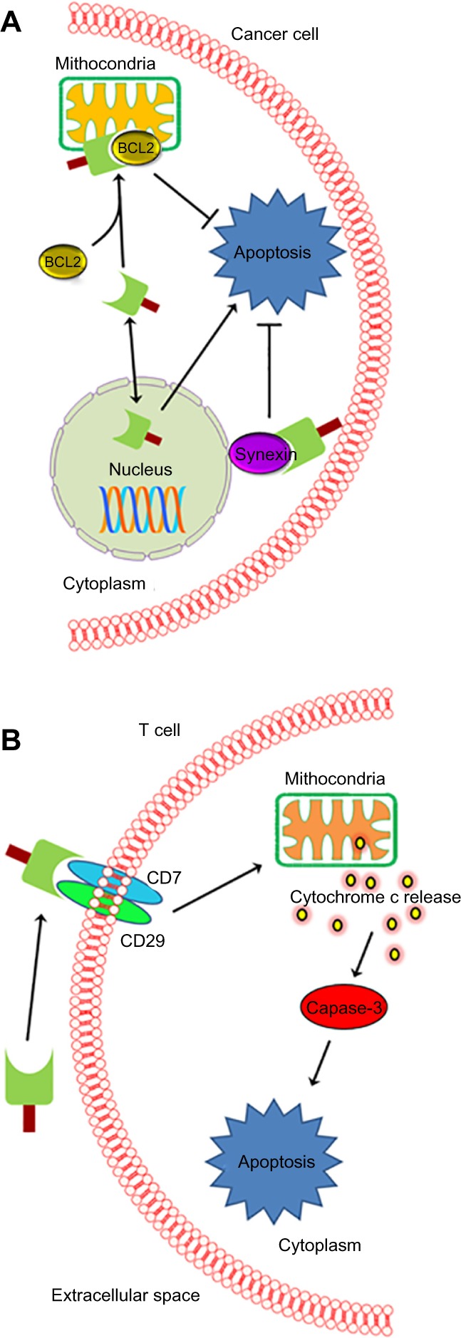
The anti-apoptotic functions of cytoplasmic galectin-3 has been consistently shown in many types of cancer cells, including breast, prostate, thyroid, bladder, colorectal, pancreatic, gastric, myeloid leukemia, neuroblastoma, and some B-cell lymphoma.68,108–114 However, galectin-3 seems to induce apoptosis in other B-cell lymphomas.115 For this antiapoptotic function of galectin-3, several mechanisms have been proposed (Fig. 3A).6
Figure 3.

Function of galectin-3 in apoptosis. Schematic representation of (A) intracellular and (B) extracellular function of galectin-3. (A) Nuclear galectin-3 is apoptotic, while cytoplasmic galectin-3 is shown antiapoptotic. (B) Galectin-3 mediated apoptosis of T-cells.
For example, galectin-3 acts as a specific binding partner for activated K-Ras, which promotes strong activation of phosphoinositide 3-kinase.105 Galectin-3 is the only member of its family that contains the NWGR antideath domain. In particular, the NWGR motif presented at the ND of galec-tin-3 shows a strong homology with the groove–BH1 motif interface of the Bcl-2 protein family, which appears to be essential for its antiapoptotic functions.67 Bcl-2 translocation to the mitochondrial membrane blocks apoptosis and cytochrome c release.106 Cytochrome c release and nitric oxide-induced apoptosis were blocked in galectin-3-transfected BT549 human breast carcinoma cells.107 Moreover, galectin-3 binds Bcl-2 protein in vitro and inhibits mitochondrial apoptotic response.16,67 Interestingly, synexin (annexin 7) is required for galectin-3 prevention of mitochondrial damage.45 Recently, targeting and/or co-targeting Bcl-2 and Bax was proposed as promising strategies for cancer therapy.116–118 In this context, a combination therapy with depletion of galectin-3 and Bcl-2 inhibitor and/or Bax activator might exhibit strong synergy against metastatic cancer.
Extracellular galectin-3 secreted from tumor cells induces apoptosis of cancer-infiltrating T-cells: possible role of galectin-3 in the immune escape mechanism during tumor progression
Recent studies revealed that galectin-3 can induce apoptosis of activated T-cells or is responsible for deficient T-cell functions (Fig. 3B).9,39,119 Interestingly, galectin-3-null T-cell lines, such as Jurkat, CEM, and MOLT-4 cells, were significantly more sensitive to exogenous galectin-3 than galectin-3-expressing lines SKW6.4 and H9. For example, galectin-3-transfected Jurkat cells were found more resistant to apoptosis induced by anti-Fas antibodies or staurosporine (protein kinase inhibitor) compared to the nontransfected control cells.17,120 These differences are probably due to a balance between the antiapoptotic activity of intracellular galec-tin-3 and proapoptotic activity of extracellular galectin-3. Extracellular galectin-3 can also induce apoptosis in human T-cells including human peripheral blood mononuclear cells and activated mouse T-cells.39 This would imply that tumor cells defend themselves against infiltrating T-cells by secreting galectin-3. Two major signaling pathways, one via death receptors Fas (apo-1/CD95) and the other using TRAIL (TNF-related apoptosis inducing ligand or Apo2-L), are known for extrinsic apoptotic signals.121,122
Cell surface glycoproteins, such as CD29, CD7, CD95, CD98, and T-cell receptor have been shown to associate with galectin-3, which may mediate induction of apoptosis by extracellular galectin-3.68,123 For example, extracellular galetin-3 binds to the CD29/CD7 complex, which triggers the activation of an intracellular apoptotic signaling cascade followed by mitochondrial cytochrome c release and activation of caspase-3.9
Galectin-3 mediates homotypic and heterotypic aggregation and promotes angiogenesis, tumor cells endothelial interactions, and tumor metastasis: role of TF antigen in cancer metastasis
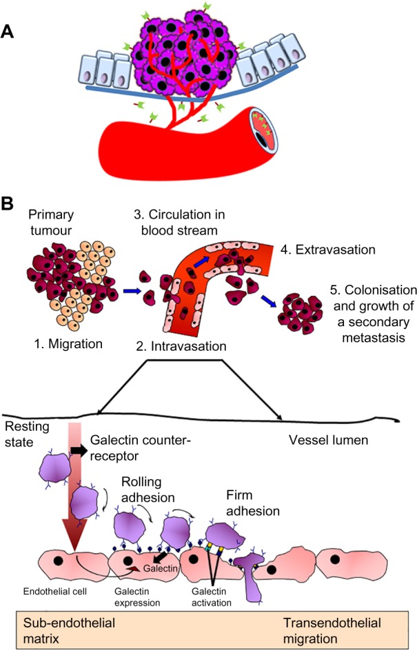
Several studies suggest that galectin-3 promotes tumor angiogenesis and metastasis in many cancers (Fig. 4A).124 Disruption of galectin-3 expression could impair tumoral angiogenesis by reducing Vascular endothelial growth factor (VEGF) secretion from TGFβ1-induced macrophages.125 Once the primary tumor is established, the formation of secondary tumors by circulating cancer cells requires embolization by aggregating with other tumor cells in microcapillaries followed by extravasation at secondary sites (Fig. 4B). In the first step of extravasation, cells bind to endothelial cells through protein–carbohydrate interactions and penetrate through the layers of endothelial cells and basement membrane. It was shown that cell surface galectin-3 mediates homotypic cell adhesion by binding to soluble complementary glycoconjugates.126 Interactions of metastatic cancer cells with vasculatory endothelium are critical during early stages of cancer metastasis.127 Galec-tin-3 mediates homotypic and heterotypic aggregation and promotes interactions between tumor cells and endothelial cells, angiogenesis, and tumor metastasis.2,4,6 It has been shown that galectin-3 expressed in activated endothelium participates in docking of cancer cells including breast and prostate cancers on capillary endothelium by specifically interacting with cancer cells-associated TFD (Galβ1, 3GalNAc).128–130 The TFD, present in the core I structure of mucin-type O-linked glycan, is generally masked by sialic acid in normal cells but is exposed or nonsialylated in malignant and premalignant epithelia.129,130 Circulating galectin-3 has been shown to increase cancer cell homotypic aggregation by interaction with TFD on the cancer-associated transmembrane mucin protein MUC1.131,132 Significance of galectin-3 in homotypic and heterotypic cell–cell interactions was also demonstrated by using 3D co-cultures of endothelial and epithelial cells.79
Figure 4.

Function of galectin-3 in tumor angiogenesis and metastasis. (A) Schematic representation of galectin-3-mediated tumor cell angiogenesis. (B) Schematic representation of galectin-3-mediated tumor–endothelial cell interactions and tumor cell extravasation (adapted from PhD dissertation of Dr. Hannah Jane Lomax-Browne – Breast Cancer Research Group, Department of Surgery, University College, London).
Many proinflammatory cytokines are overexpressed in cancer condition and are increasingly realized to play critical roles in promoting various steps in cancer progression and metastasis.133,134 Recent studies have indicated that over expression of galectin-3 in cancer induces secretion of several proinflammatory cytokines, hence, indirectly involved in promoting metastasis.135 Galectin-3 at pathological concentrations found in patients with metastatic colon cancer induces secretion of interleukin-6, Granulocyte-colony stimulating factor (G-CSF), soluble Intercellular Adhesion Molecule 1 (sICAM-1), and Granulocyte-macrophage colony-stimulating factor (GM-CSF) from blood vascular endothelial cells in vitro and in mice.135 These cytokines interact with the vascular endothelium to increase the expressions of a number of endothelial cell surface adhesion molecules, such as E-selectin, Intercellular Adhesion Molecule 1 (ICAM-1), and Vascular cell adhesion molecule 1 (VCAM-1), resulting in increased cancer cell–endothelial adhesion and increased endothelial cell migration and tubule formation. Downregulation of galectin-3 via RNA interference decreases production of proinflammatory cytokines in monocyte-derived dendritic cells,136 suggesting that depletion of galectin-3 could be a promising strategy for cancer therapy. Galectin-3 was also shown to stimulate capillary tube formation of human umbilical vein endothelial cells in vitro and angiogenesis in vivo, which was inhibited by specific sugars and antibodies. Overexpression of galectin-3 in galectin-3 nonexpressing prostate cancer cell line LNCaP induced in vivo tumor growth and angiogenesis.112
Galectin-3 Antagonists for Cancer Therapy
There have been a few attempts to use naturally occurring substances to control and prevent cancer metastasis. Modified citrus pectin (MCP), a pH-modified soluble β-galactosyl-containing polysaccharide obtained from the peel of citrus fruits, has been claimed to be an effective antimetastatic drug for many cancers.137 The MCP was shown to inhibit in vitro tumor cell adhesion to endothelium138 and homotypic aggregation as well as in vivo formation of metastatic deposits of human breast and prostate carcinoma cells in lungs and bones.139 Another polysaccharide derived from citrus pectin, GCS-100, has been shown to have great potential to treat multiple myeloma cells, including those resistant to dexamethasone, melphalan, or doxorubicin.140,141 It modulates myeloid leukemia cell differentiation protein-1 (MCL-1), Phorbol-12-myristate-13-acetate-induced protein 1 (NOXA), and cell cycle to induce myeloma cell death.141
Tumor cells released galectin-3 was found to bind glycosylated receptors at the surface of tumor-infiltrating lymphocytes (TIL), forming glycoprotein–galectin lattices that could reduce the motility and functionality of TILs.142 Disruption of glycoprotein–galectin-3 lattices using anti-galectin-3 antibodies, or galectin-3 antagonists such as N-acetyllactosamine, GCS-100 (MCP-derived polysaccharide), or GM-CT-01 (a galactomannan from guar gum) boosted cytokine secretion by TIL (corrected impaired TIL).142,143 As TFD is found exposed mostly on tumor cell surface (masked in normal cells) and also tumor–endothelial cell interactions required for metastasis are mediated by endothelium-associated galectin-3 and cancer cell-associated TFD, we reasoned that exogenous TFD would be more effective and specific to block galectin-3-mediated tumorigenesis. We purified TFD-containing glycopeptide from cod fish and showed that TFD compound could inhibit tumor–endothelial cell interactions and angiogenesis.27 Moreover, the purified TFD compound also blocked T-cell apoptosis mediated by either recombinant, tumor-associated, or cancer patient serum-associated galectin-3.27 It is important to point out that while these galectin-3 antagonists described above are able to limit activities of the extracellular galectin-3, they seem to be ineffective in targeting intracellular galectin-3. As intracellular galectin-3 can also contribute to the process of tumor progression through their CRD-dependent and CRD-independent intracellular ligands, alternative strategies to better develop galectin-3 antagonists or combination of galectin-3 antagonist and small interfering RNA are needed to target both intracellular and extracellular galectin-3.10
Concluding Remarks
Numerous studies have indicated that galectin-3 is involved in multiple stages of cancer progression and metastasis and may render anticancer activities in several ways. First, the intracellular (cytoplasmic) galectin-3 is antiapoptotic providing survival advantage to cancer cells. Second, galectin-3 promotes tumor neoangiogenesis. Third, the extracellular galectin-3 is involved in homotypic aggregation. Fourth, tumor–endothelial cell interactions required for metastasis are believed to be mediated by endothelium-associated galectin-3 and cancer cell-associated TFD. Fifth, tumor cell secreted galectin-3 induces apoptosis of cancer-infiltrating T-cells possibly promoting immune escape during tumor progression. Although a large body of data were generated from in vitro studies and role of galectin-3 in various aspects of cancer was not unequivocally validated in relevant animal models, nonetheless, a few attempts to perturb galectin-3 function either by blocking its expression with siRNA or by inhibiting its activity with external carbohydrate ligands produced so far encouraging results in several preclinical models.
Acknowledgments
We apologize to the authors whose works are not cited due to the limited space.
Footnotes
ACADEMIC EDITOR: William C. S. Cho, Editor in Chief
PEER REVIEW: Three peer reviewers contributed to the peer review report. Reviewers’ reports totaled 711 words, excluding any confidential comments to the academic editor.
FUNDING: The work carried out by us was supported by the US Army Medical Research and Materiel Command grant W81XWH-07-1-0565, the National Institute of Health grants RO3 CA133935-01 and R41 CA141970-01A2, and the Maryland Innovative Initiative, TEDCO grant to HA. The authors confirm that the funder had no influence over the study design, content of the article, or selection of this journal.
COMPETING INTERESTS: GlycoMantra’s products, particularly galectin-3 antagonists, are related to the topic of this review. HA is the lead inventor of a patent (#9,180,175) entitled “Methods of use for a natural Thomsen-Friedenreich disaccharide compound” filed by the University of Maryland, Baltimore.
Paper subject to independent expert blind peer review. All editorial decisions made by independent academic editor. Upon submission manuscript was subject to anti-plagiarism scanning. Prior to publication all authors have given signed confirmation of agreement to article publication and compliance with all applicable ethical and legal requirements, including the accuracy of author and contributor information, disclosure of competing interests and funding sources, compliance with ethical requirements relating to human and animal study participants, and compliance with any copyright requirements of third parties. This journal is a member of the Committee on Publication Ethics (COPE).
Author Contributions
Conceived and designed the experiments: HA. Analyzed the data: HA. Wrote the first draft of the manuscript: HA, DMMA. Contributed to the writing of the manuscript: HA, DMMA. Agree with manuscript results and conclusions: HA, DMMA. Jointly developed the structure and arguments for the paper: HA, DMMA. Made critical revisions and approved final version: HA, DMMA. Both authors reviewed and approved of the final manuscript.
REFERENCES
- 1.Ahmed H, Du SJ, O’Leary N, Vasta GR. Biochemical and molecular characterization of galectins from zebrafish (Danio rerio): notochord-specific expression of a prototype galectin during early embryogenesis. Glycobiology. 2004;14:219–32. doi: 10.1093/glycob/cwh032. [DOI] [PubMed] [Google Scholar]
- 2.Nakahara S, Raz A. Regulation of cancer-related gene expression by galectin-3 and the molecular mechanism of its nuclear import pathway. Cancer Metastasis Rev. 2007;26:605–10. doi: 10.1007/s10555-007-9095-6. [DOI] [PMC free article] [PubMed] [Google Scholar]
- 3.Rabinovich GA, Liu FT, Hirashima M, Anderson A. An emerging role for galectins in tuning the immune response: lessons from experimental models of inflammatory disease, autoimmunity and cancer. Scand J Immunol. 2007;66:143–58. doi: 10.1111/j.1365-3083.2007.01986.x. [DOI] [PubMed] [Google Scholar]
- 4.Nakahara S, Raz A. Biological modulation by lectins and their ligands in tumor progression and metastasis. Anticancer Agents Med Chem. 2008;8:22–36. doi: 10.2174/187152008783330833. [DOI] [PMC free article] [PubMed] [Google Scholar]
- 5.Vasta GR. Roles of galectins in infection. Nat Rev Microbiol. 2009;7:424–38. doi: 10.1038/nrmicro2146. [DOI] [PMC free article] [PubMed] [Google Scholar]
- 6.Nakahara S, Oka N, Raz A. On the role of galectin-3 in cancer apoptosis. Apoptosis. 2005;10:267–75. doi: 10.1007/s10495-005-0801-y. [DOI] [PubMed] [Google Scholar]
- 7.Dumic J, Dabelic S, Flögel M. Galectin-3: an open-ended story. Biochim Biophys Acta. 2006;1760:616–35. doi: 10.1016/j.bbagen.2005.12.020. [DOI] [PubMed] [Google Scholar]
- 8.Henderson NC, Sethi T. The regulation of inflammation by galectin-3. Immunol Rev. 2009;230:160–71. doi: 10.1111/j.1600-065X.2009.00794.x. [DOI] [PubMed] [Google Scholar]
- 9.Hsu DK, Chen HY, Liu FT. Galectin-3 regulates T-cell functions. Immunol Rev. 2009;230:114–27. doi: 10.1111/j.1600-065X.2009.00798.x. [DOI] [PubMed] [Google Scholar]
- 10.Vladoiu MC, Labrie M, St-Pierre Y. Intracellular galectins in cancer cells: potential new targets for therapy. Int J Oncol. 2014;44:1001–14. doi: 10.3892/ijo.2014.2267. [DOI] [PubMed] [Google Scholar]
- 11.Gong HC, Honjo Y, Nangia-Makker P, et al. The NH2 terminus of galectin-3 governs cellular compartmentalization and functions in cancer cells. Cancer Res. 1999;59:6239–45. [PubMed] [Google Scholar]
- 12.Massa SM, Cooper DN, Leffler H, Barondes SH. L-29, an endogenous lectin, binds to glycoconjugate ligands with positive cooperativity. Biochemistry. 1993;32:260–7. doi: 10.1021/bi00052a033. [DOI] [PubMed] [Google Scholar]
- 13.Yoshii T, Fukumori T, Honjo Y, Inohara H, Kim HR, Raz A. Galectin-3 phosphorylation is required for its anti-apoptotic function and cell cycle arrest. J Biol Chem. 2002;277:6852–7. doi: 10.1074/jbc.M107668200. [DOI] [PubMed] [Google Scholar]
- 14.Ochieng J, Green B, Evans S, James O, Warfield P. Modulation of the biological functions of galectin-3 by matrix metalloproteinases. Biochim Biophys Acta. 1998;1379:97–106. doi: 10.1016/s0304-4165(97)00086-x. [DOI] [PubMed] [Google Scholar]
- 15.Hsu DK, Zuberi RI, Liu FT. Biochemical and biophysical characterization of human recombinant IgE-binding protein, an S-type animal lectin. J Biol Chem. 1992;267:14167–74. [PubMed] [Google Scholar]
- 16.Ochieng J, Platt D, Tait L. Structure-function relationship of a recombinant human galactoside-binding protein. Biochemistry. 1993;32:4455–60. doi: 10.1021/bi00067a038. [DOI] [PubMed] [Google Scholar]
- 17.Yang RY, Hsu DK, Liu FT. Expression of galectin-3 modulates T-cell growth and apoptosis. Proc Natl Acad Sci U S A. 1996;93:6737–42. doi: 10.1073/pnas.93.13.6737. [DOI] [PMC free article] [PubMed] [Google Scholar]
- 18.Yang RY, Hill PN, Hsu DK, Liu FT. Role of the carboxyl-terminal lectin domain in self-association of galectin-3. Biochemistry. 1998;37:4086–92. doi: 10.1021/bi971409c. [DOI] [PubMed] [Google Scholar]
- 19.Raimond J, Zimonjic DB, Mignon C, et al. Mapping of the galectin-3 gene (LGALS3) to human chromosome 14 at region 14q 21–22. Mamm Genome. 1997;8:706–7. doi: 10.1007/s003359900548. [DOI] [PubMed] [Google Scholar]
- 20.Kadrofske MM, Openo KP, Wang JL. The human LGALS3 (galectin-3) gene: determination of the gene structure and functional characterization of the promoter. Arch Biochem Biophys. 1998;349:7–20. doi: 10.1006/abbi.1997.0447. [DOI] [PubMed] [Google Scholar]
- 21.Raimond J, Rouleux F, Monsigny M, Legrand A. The second intron of the human galectin-3 gene has a strong promoter activity down-regulated by p53. FEBS Lett. 1995;363:165–9. doi: 10.1016/0014-5793(95)00310-6. [DOI] [PubMed] [Google Scholar]
- 22.Guittaut M, Charpentier S, Normand T, Dubois M, Raimond J, Legrand A. Identification of an internal gene to the human Galectin-3 gene with two different overlapping reading frames that do not encode Galectin-3. J Biol Chem. 2001;276:2652–7. doi: 10.1074/jbc.m002523200. [DOI] [PubMed] [Google Scholar]
- 23.Duneau M, Boyer-Guittaut M, Gonzalez P, et al. A novel cell death gene that encodes a mitochondrial protein promoting cytochrome c release. Exp Cell Res. 2005;302:194–205. doi: 10.1016/j.yexcr.2004.08.041. [DOI] [PubMed] [Google Scholar]
- 24.Seetharaman J, Kanigsberg A, Slaaby R, Leffler H, Barondes SH, Rini JM. X-ray crystal structure of the human galectin-3 carbohydrate recognition domain at 2.1-A resolution. J Biol Chem. 1998;273:13047–52. doi: 10.1074/jbc.273.21.13047. [DOI] [PubMed] [Google Scholar]
- 25.Collins PM, Hidari KIPJ, Blanchard H. Slow diffusion of lactose out of galectin-3 crystals monitored by X-ray crystallography: possible implications for ligand-exchange protocols. Acta Crystallogr D Biol Crystallogr. 2007;63:415–9. doi: 10.1107/S090744490605270X. [DOI] [PubMed] [Google Scholar]
- 26.Bian CF, Zhang Y, Sun H, Li DF, Wang DC. Structural basis for distinct binding properties of the human galectins to Thomsen-Friedenreich antigen. PLoS One. 2011;6:e25007. doi: 10.1371/journal.pone.0025007. [DOI] [PMC free article] [PubMed] [Google Scholar]
- 27.Guha P, Kaptan E, Bandyopadhyaya G, et al. Cod glycopeptide with picomolar affinity to galectin-3 suppresses T-cell apoptosis and prostate cancer metastasis. Proc Natl Acad Sci U S A. 2013;110:5052–7. doi: 10.1073/pnas.1202653110. [DOI] [PMC free article] [PubMed] [Google Scholar]
- 28.Ahmed H, Allen HJ, Sharma A, Matta KL. Human splenic galaptin: carbohydrate-binding specificity and characterization of the combining site. Biochemistry. 1990;29:5315–9. doi: 10.1021/bi00474a015. [DOI] [PubMed] [Google Scholar]
- 29.Ahmed H, Vasta GR. Galectins: conservation of functionally and structurally relevant amino acid residues defines two types of carbohydrate recognition domains. Glycobiology. 1994;4:545–8. doi: 10.1093/glycob/4.5.545. [DOI] [PubMed] [Google Scholar]
- 30.Ahmed H, Pohl J, Fink NE, Strobel F, Vasta GR. The primary structure and carbohydrate specificity of a ß-galactosyl-binding lectin from toad (Bufo arenarum Hensel) ovary reveal closer similarities to the mammalian galectin-1 than to the galectin from the clawed frog Xenopus laevis. J Biol Chem. 1996;271:33083–94. doi: 10.1074/jbc.271.51.33083. [DOI] [PubMed] [Google Scholar]
- 31.Ahmed H, Fink NE, Pohl J, Vasta GR. Galectin-1 from bovine spleen: biochemical characterization, carbohydrate specificity and tissue-specific isoform profiles. J Biochem. 1996;120:1007–19. doi: 10.1093/oxfordjournals.jbchem.a021493. [DOI] [PubMed] [Google Scholar]
- 32.Liao DI, Kapadia G, Ahmed H, Vasta GR, Herzberg O. Structure of S-lectin, a developmentally regulated vertebrate beta-galactoside binding protein. Proc Natl Acad Sci U S A. 1994;91:1428–32. doi: 10.1073/pnas.91.4.1428. [DOI] [PMC free article] [PubMed] [Google Scholar]
- 33.Bianchet MA, Ahmed H, Vasta GR, Amzel LM. A soluble ß-galactosyl-binding lectin (galectin) from toad (Bufo arenarum Hensel) ovary: crystallographic studies of two protein-sugar complexes. Proteins. 2000;40:378–88. doi: 10.1002/1097-0134(20000815)40:3<378::aid-prot40>3.0.co;2-7. [DOI] [PubMed] [Google Scholar]
- 34.Hirabayashi J, Kasai K. The family of metazoan metal-independent β-galactoside-binding lectins:structure, function and molecular evolution. Glycobiology. 1993;3:297–304. doi: 10.1093/glycob/3.4.297. [DOI] [PubMed] [Google Scholar]
- 35.Cooper DNW, Barondes SH. Evidence for export of a muscle lectin from cytosol to extracellular matrix and for a novel secretory mechanism. J Cell Biol. 1990;110:1681–91. doi: 10.1083/jcb.110.5.1681. [DOI] [PMC free article] [PubMed] [Google Scholar]
- 36.Cho M, Cummings RD. Galectin-1, a ß-galactoside-binding lectin in Chinese hamster ovary cells, II. Localization and biosynthesis. J Biol Chem. 1995;270:5207–12. doi: 10.1074/jbc.270.10.5207. [DOI] [PubMed] [Google Scholar]
- 37.Barboni EA, Bawumia S, Hughes RC. Kinetic measurements of binding of galectin 3 to a laminin substratum. Glycoconj J. 1999;16:365–73. doi: 10.1023/a:1007004330048. [DOI] [PubMed] [Google Scholar]
- 38.Kariya Y, Kawamura C, Tabei T, Gu J. Bisecting GlcNAc residues on laminin-332 down-regulate galectin-3-dependent keratinocyte motility. J Biol Chem. 2010;285:3330–40. doi: 10.1074/jbc.M109.038836. [DOI] [PMC free article] [PubMed] [Google Scholar]
- 39.Ochieng J, Leite-Browning ML, Warfield P. Regulation of cellular adhesion to extracellular matrix proteins by galectin-3. Biochem Biophys Res Commun. 1998;246:788–91. doi: 10.1006/bbrc.1998.8708. [DOI] [PubMed] [Google Scholar]
- 40.Fukumori T, Takenaka Y, Yoshii T, et al. CD29 and CD7 mediate galectin-3-induced type II T-cell apoptosis. Cancer Res. 2003;63:8302–11. [PubMed] [Google Scholar]
- 41.Feuk-Lagerstedt E, Jordan ET, Leffler H, Dahlgren C, Karlsson A. Identification of CD66a and CD66b as the major galectin-3 receptor candidates in human neutrophils. J Immunol. 1999;163:5592–8. [PubMed] [Google Scholar]
- 42.Rosenberg I, Cherayil BJ, Isselbacher KJ, Pillai S. Mac-2-binding glycoproteins. Putative ligands for a cytosolic beta-galactoside lectin. J Biol Chem. 1991;266:18731–6. [PubMed] [Google Scholar]
- 43.Park JW, Voss PG, Grabski S, Wang JL, Patterson RJ. Association of galectin-1 and galectin-3 with Gemin4 in complexes containing the SMN protein. Nucleic Acids Res. 2001;29:3595–602. doi: 10.1093/nar/29.17.3595. [DOI] [PMC free article] [PubMed] [Google Scholar]
- 44.Liu L, Sakai T, Sano N, Fukui K. Nucling mediates apoptosis by inhibiting expression of galectin-3 through interference with nuclear factor kappaB signaling. Biochem J. 2004;380:31–41. doi: 10.1042/BJ20031300. [DOI] [PMC free article] [PubMed] [Google Scholar]
- 45.Yu F, Finley RL, Jr, Raz A, Kim HR. Galectin-3 translocates to the perinuclear membranes and inhibits cytochrome c release from the mitochondria. A role for synexin in galectin-3 translocation. J Biol Chem. 2002;277:15819–27. doi: 10.1074/jbc.M200154200. [DOI] [PubMed] [Google Scholar]
- 46.Shimura T, Takenaka Y, Tsutsumi S, Hogan V, Kikuchi A, Raz A. Galectin-3, a novel binding partner of beta-catenin. Cancer Res. 2004;64:6363–7. doi: 10.1158/0008-5472.CAN-04-1816. [DOI] [PubMed] [Google Scholar]
- 47.Song S, Mazurek N, Liu C, et al. Galectin-3 mediates nuclear beta-catenin accumulation and Wnt signaling in human colon cancer cells by regulation of glycogen synthase kinase-3beta activity. Cancer Res. 2009;69:1343–9. doi: 10.1158/0008-5472.CAN-08-4153. [DOI] [PMC free article] [PubMed] [Google Scholar]
- 48.Abedin MJ, Kashio Y, Seki M, Nakamura K, Hirashima M. Potential roles of galectins in myeloid differentiation into three different lineages. J Leukoc Biol. 2003;73:650–6. doi: 10.1189/jlb.0402163. [DOI] [PubMed] [Google Scholar]
- 49.Elliott MJ, Strasser A, Metcalf D. Selective up-regulation of macrophage function in granulocyte-macrophage colony-stimulating factor transgenic mice. J Immunol. 1991;147:2957–63. [PubMed] [Google Scholar]
- 50.Reichert F, Rotshenker S. Galectin-3/MAC-2 in experimental allergic encephalomyelitis. Exp Neurol. 1999;160:508–14. doi: 10.1006/exnr.1999.7229. [DOI] [PubMed] [Google Scholar]
- 51.Liu FT, Hsu DK, Zuberi RI, Kuwabara I, Chi EY, Henderson WR., Jr Expression and function of galectin-3, a beta-galactoside-binding lectin, in human monocytes and macrophages. Am J Pathol. 1995;147:1016–28. [PMC free article] [PubMed] [Google Scholar]
- 52.Dabelic S, Flogel M, Dumic J. Effects of aspirin and indomethacin on galectin−3. Croat Chem Acta. 2005;178:433–40. [Google Scholar]
- 53.Dabelic S, Flogel M, Dumic J. Corticosteroids affect galectin-3 expression. Period Biol. 2005;107:175–81. [Google Scholar]
- 54.Liu FT, Albrandt K, Mendel E, Kulczycki A, Jr, Orida NK. Identification of an IgE-binding protein by molecular cloning. Proc Natl Acad Sci U S A. 1985;82:4100–4. doi: 10.1073/pnas.82.12.4100. [DOI] [PMC free article] [PubMed] [Google Scholar]
- 55.Joo HG, Goedegebuure PS, Sadanaga N, Nagoshi M, von Bernstorff W, Eberlein TJ. Expression and function of galectin-3, a betaga- lactoside-binding protein in activated T lymphocytes. J Leukoc Biol. 2001;69:555–64. [PubMed] [Google Scholar]
- 56.Hebert E, Monsigny M. Galectin-3 mRNA level depends on transformation phenotype in ras-transformed NIH 3T3 cells. Biol Cell. 1994;81:73–6. doi: 10.1016/0248-4900(94)90057-4. [DOI] [PubMed] [Google Scholar]
- 57.Fogel S, Guittaut M, Legrand A, Monsigny M, Hébert E. The tat protein of HIV-1 induces galectin-3 expression. Glycobiology. 1999;9:383–7. doi: 10.1093/glycob/9.4.383. [DOI] [PubMed] [Google Scholar]
- 58.Hsu DK, Hammes SR, Kuwabara I, Greene WC, Liu FT. Human T lymphotropic virus-I infection of human T lymphocytes induces expression of the beta-galactoside-binding lectin, galectin-3. Am J Pathol. 1996;148:1661–70. [PMC free article] [PubMed] [Google Scholar]
- 59.Dumic J, Lauc G, Flogel M. Expression of galectin-3 in cells exposed to stress-Roles of jun and NF-kappaB. Cell Physiol Biochem. 2000;10:149–58. doi: 10.1159/000016345. [DOI] [PubMed] [Google Scholar]
- 60.Stock M, Schäfer H, Stricker S, Gross G, Mundlos S, Otto F. Expression of galectin-3 in skeletal tissues is controlled by Runx2. J Biol Chem. 2003;278:17360–7. doi: 10.1074/jbc.M207631200. [DOI] [PubMed] [Google Scholar]
- 61.Costessi A, Pines A, D’Andrea P, et al. Extracellular nucleotides activate Runx2 in the osteoblast-like HOBIT cell line: a possible molecular link between mechanical stress and osteoblasts’ response. Bone. 2005;36:418–32. doi: 10.1016/j.bone.2004.10.016. [DOI] [PubMed] [Google Scholar]
- 62.Ahmed H, Banerjee PB, Vasta GR. Differential expression of galectins in normal, benign and malignant prostate epithelial cells: silencing of galectin-3 expression in prostate cancer by its promoter methylation. Biochem Biophys Res Commun. 2007;358:241–6. doi: 10.1016/j.bbrc.2007.04.114. [DOI] [PubMed] [Google Scholar]
- 63.Ruebel KH, Jin L, Qian X, et al. Effects of DNA methylation on galectin-3 expression in pituitary tumors. Cancer Res. 2005;65:1136–40. doi: 10.1158/0008-5472.CAN-04-3578. [DOI] [PubMed] [Google Scholar]
- 64.Ahmed H, Cappello F, Rodolico V, Vasta GR. Evidence of heavy methylation in the galectin-3 promoter in early stages of prostate adenocarcinoma: development and validation of a methylated marker for early diagnosis of prostate cancer. Transl Oncol. 2009;2:146–56. doi: 10.1593/tlo.09118. [DOI] [PMC free article] [PubMed] [Google Scholar]
- 65.Ahmed H. Promoter methylation in prostate cancer and its application for the early detection of prostate cancer using serum and urine samples. Biomark Cancer. 2010;2:17–33. doi: 10.4137/BIC.S3187. [DOI] [PMC free article] [PubMed] [Google Scholar]
- 66.Elola MT, Wolfenstein-Todel C, Troncoso MF, Vasta GR, Rabinovich GA. Galectins: matricellular glycan-binding proteins linking cell adhesion, migration, and survival. Cell Mol Life Sci. 2007;64:1679–700. doi: 10.1007/s00018-007-7044-8. [DOI] [PMC free article] [PubMed] [Google Scholar]
- 67.Akahani S, Nangia-Makker P, Inohara H, Kim HR, Raz A. Galectin-3: a novel antiapoptotic molecule with a functional BH1 (NWGR) domain of Bcl-2 family. Cancer Res. 1997;57:5272–6. [PubMed] [Google Scholar]
- 68.Fukumori T, Takenaka Y, Oka N, et al. Endogenous galectin-3 determines the routing of CD95 apoptotic signaling pathways. Cancer Res. 2004;64:3376–9. doi: 10.1158/0008-5472.CAN-04-0336. [DOI] [PubMed] [Google Scholar]
- 69.Elad-Sfadia G, Haklai R, Balan E, Kloog Y. Galectin-3 augments K-Ras activation and triggers a Ras signal that attenuates ERK but not phosphoinositide 3-kinase activity. J Biol Chem. 2004;279:34922–30. doi: 10.1074/jbc.M312697200. [DOI] [PubMed] [Google Scholar]
- 70.Colnot C, Ripoche MA, Scaerou F, Foulis D, Poirier F. Galectins in mouse embryogenesis. Biochem Soc Trans. 1996;24:141–6. doi: 10.1042/bst0240141. [DOI] [PubMed] [Google Scholar]
- 71.Poirier F. Roles of galectins in vivo. Biochem Soc Symp. 2002;69:95–103. doi: 10.1042/bss0690095. [DOI] [PubMed] [Google Scholar]
- 72.Mercer N, Guzman L, Cueto Rua E, et al. Duodenal intraepithelial lymphocytes of children with cow milk allergy preferentially bind the glycan-binding protein galectin-3. Int J Immunopathol Pharmacol. 2009;22:207–17. doi: 10.1177/039463200902200123. [DOI] [PMC free article] [PubMed] [Google Scholar]
- 73.Dumont P, Berton A, Nagy N, et al. Expression of galectin-3 in the tumor immune response in colon cancer. Lab Invest. 2008;88:896–906. doi: 10.1038/labinvest.2008.54. [DOI] [PubMed] [Google Scholar]
- 74.Cao Z, Said N, Amin S, et al. Galectins-3 and -7, but not galectin-1, play a role in re-epithelialization of wounds. J Biol Chem. 2002;277:42299–305. doi: 10.1074/jbc.M200981200. [DOI] [PubMed] [Google Scholar]
- 75.Kaji Y, Amano S, Usui T, et al. Expression and function of receptors for advanced glycation end products in bovine corneal endothelial cells. Invest Ophthalmol Vis Sci. 2003;44:521–9. doi: 10.1167/iovs.02-0268. [DOI] [PubMed] [Google Scholar]
- 76.Kang EH, Moon KC, Lee EY, et al. Renal expression of galectin-3 in systemic lupus erythematosus patients with nephritis. Lupus. 2009;18:22–8. doi: 10.1177/0961203308094361. [DOI] [PubMed] [Google Scholar]
- 77.Won YS, Jeong ES, Park HJ, et al. Upregulation of galectin-3 by Corynebacterium kutscheri infection in the rat lung. Exp Anim. 2007;56:85–91. doi: 10.1538/expanim.56.85. [DOI] [PubMed] [Google Scholar]
- 78.Silva-Monteiro E, Reis Lorenzato L, Kenji Nihei O, et al. Altered expression of galectin-3 induces cortical thymocyte depletion and premature exit of immature thymocytes during Trypanosoma cruzi infection. Am J Pathol. 2007;170:546–56. doi: 10.2353/ajpath.2007.060389. [DOI] [PMC free article] [PubMed] [Google Scholar]
- 79.Shekhar MP, Nangia-Makker P, Tait L, Miller F, Raz A. Alterations in galectin-3 expression and distribution correlate with breast cancer progression: functional analysis of galectin-3 in breast epithelial-endothelial interactions. Am J Pathol. 2004;165:1931–41. doi: 10.1016/S0002-9440(10)63245-2. [DOI] [PMC free article] [PubMed] [Google Scholar]
- 80.Pacis RA, Pilat MJ, Pienta KJ, et al. Decreased galectin-3 expression in prostate cancer. Prostate. 2000;44:118–23. doi: 10.1002/1097-0045(20000701)44:2<118::aid-pros4>3.0.co;2-u. [DOI] [PubMed] [Google Scholar]
- 81.Xu XC, Sola Gallego JJ, Lotan R, El-Naggar AK. Differential expression of galectin-1 and galectin-3 in benign and malignant salivary gland neoplasms. Int J Oncol. 2000;17:271–6. doi: 10.3892/ijo.17.2.271. [DOI] [PubMed] [Google Scholar]
- 82.Wang L, Friess H, Zhu Z, et al. Galectin-1 and galectin-3 in chronic pancreatitis. Lab Invest. 2000;80:1233–41. doi: 10.1038/labinvest.3780131. [DOI] [PubMed] [Google Scholar]
- 83.Sasaki S, Bao Q, Hughes RC. Galectin-3 modulates rat mesangial cell proliferation and matrix synthesis during experimental glomerulonephritis induced by anti-Thy1.1 antibodies. J Pathol. 1999;187:481–9. doi: 10.1002/(SICI)1096-9896(199903)187:4<481::AID-PATH263>3.0.CO;2-2. [DOI] [PubMed] [Google Scholar]
- 84.Fautsch MP, Silva AO, Johnson DH. Carbohydrate binding proteins galectin-1 and galectin-3 in human trabecular meshwork. Exp Eye Res. 2003;77:11–6. doi: 10.1016/s0014-4835(03)00107-6. [DOI] [PubMed] [Google Scholar]
- 85.Shimonishi T, Miyazaki K, Kono N, et al. Expression of endogenous galectin-1 and galectin-3 in intrahepatic cholangiocarcinoma. Hum Pathol. 2001;32:302–10. doi: 10.1053/hupa.2001.22767. [DOI] [PubMed] [Google Scholar]
- 86.Moutsatsos IK, Wade M, Schindler M, Wang JL. Endogenous lectins from cultured cells: nuclear localization of carbohydrate-binding protein 35 in proliferating 3T3 fibroblasts. Proc Natl Acad Sci U S A. 1987;84:6452–6. doi: 10.1073/pnas.84.18.6452. [DOI] [PMC free article] [PubMed] [Google Scholar]
- 87.Niida S, Amizuka N, Hara F, Ozawa H, Kodama H. Expression of Mac-2 antigen in the preosteoclast and osteoclast identified in the op/op mouse injected with macrophage colony-stimulating factor. J Bone Miner Res. 1994;9:873–81. doi: 10.1002/jbmr.5650090613. [DOI] [PubMed] [Google Scholar]
- 88.Wollenberg A, de la Salle H, Hanau D, Liu FT, Bieber T. Human keratinocytes release the endogenous beta-galactoside-binding soluble lectin immunoglobulin E (IgE-binding protein) which binds to Langerhans cells where it modulates their binding capacity for IgE glycoforms. J Exp Med. 1993;178:777–85. doi: 10.1084/jem.178.3.777. [DOI] [PMC free article] [PubMed] [Google Scholar]
- 89.Reichert F, Saada A, Rotshenker S. Peripheral nerve injury induces Schwann cells to express two macrophage phenotypes: phagocytosis and the galactose-specific lectin MAC-2. J Neurosci. 1994;14:3231–45. doi: 10.1523/JNEUROSCI.14-05-03231.1994. [DOI] [PMC free article] [PubMed] [Google Scholar]
- 90.Lotan R, Ito H, Yasui W, Yokozaki H, Lotan D, Tahara E. Expression of a 31-kDa lactoside-binding lectin in normal human gastric mucosa and in primary and metastatic gastric carcinomas. Int J Cancer. 1994;56:474–80. doi: 10.1002/ijc.2910560404. [DOI] [PubMed] [Google Scholar]
- 91.Lotan R, Belloni PN, Tressler RJ, Lotan D, Xu XC, Nicolson GL. Expression of galectins on microvessel endothelial cells and their involvement in tumour cell adhesion. Glycoconj J. 1994;11:462–8. doi: 10.1007/BF00731282. [DOI] [PubMed] [Google Scholar]
- 92.Truong MJ, Gruart V, Kusnierz JP, et al. Human neutrophils express immunoglobulin E (IgE)-binding proteins (Mac-2/epsilon BP) of the S-type lectin family: role in IgE dependent activation. J Exp Med. 1993;177:243–8. doi: 10.1084/jem.177.1.243. [DOI] [PMC free article] [PubMed] [Google Scholar]
- 93.Truong MJ, Gruart V, Liu FT, Prin L, Capron A, Capron M. IgE binding molecules (Mac-2/epsilon BP) expressed by human eosinophils. Implication in IgE-dependent eosinophil cytotoxicity. Eur J Immunol. 1993;23:3230–5. doi: 10.1002/eji.1830231228. [DOI] [PubMed] [Google Scholar]
- 94.Craig SS, Krishnaswamy P, Irani AM, Kepley CL, Liu FT, Schwartz LB. Immunoelectron microscopic localization of galectin-3, an IgE binding protein, in human mast cells and basophils. Anat Rec. 1995;242:211–9. doi: 10.1002/ar.1092420210. [DOI] [PubMed] [Google Scholar]
- 95.Smetana K, Holíková Z, Klubal R, et al. Coexpression of binding sites for A (B) histo-blood group trisaccharides with galectin-3 and Lag antigen in human Langerhans cells. J Leukoc Biol. 1999;66:644–9. doi: 10.1002/jlb.66.4.644. [DOI] [PubMed] [Google Scholar]
- 96.Dietz AB, Bulur PA, Knutson GJ, Matasić R, Vuk-Pavlović S. Maturation of human monocyte-derived dendritic cells studied by microarray hybridization. Biochem Biophys Res Commun. 2000;275:731–8. doi: 10.1006/bbrc.2000.3372. [DOI] [PubMed] [Google Scholar]
- 97.Saada A, Reichert F, Rotshenker S. Granulocyte macrophage colony stimulating factor produced in lesioned peripheral nerves induces the upregulation of cell surface expression of MAC-2 by macrophages and Schwann cells. J Cell Biol. 1996;133:159–67. doi: 10.1083/jcb.133.1.159. [DOI] [PMC free article] [PubMed] [Google Scholar]
- 98.Liu FT, Rabinovich GA. Galectins as modulators of tumour progression. Nat Rev Cancer. 2005;5:29–41. doi: 10.1038/nrc1527. [DOI] [PubMed] [Google Scholar]
- 99.Guha P, Bandyopadhyaya G, Polumuri SK, et al. Nicotine promotes apoptosis resistance of breast cancer cells and enrichment of side population cells with cancer stem cell-like properties via a signaling cascade involving galectin-3, α9 nicotinic acetylcholine receptor and STAT3. Breast Cancer Res Treat. 2014;145:5–22. doi: 10.1007/s10549-014-2912-z. [DOI] [PMC free article] [PubMed] [Google Scholar]
- 100.Bresalier RS, Yan PS, Byrd JC, Lotan R, Raz A. Expression of the endogenous galactose-binding protein galectin-3 correlates with the malignant potential of tumors in the central nervous system. Cancer. 1997;80:776–87. [PubMed] [Google Scholar]
- 101.Hsu DK, Dowling CA, Jeng KC, Chen JT, Yang RY, Liu FT. Galectin-3 expression is induced in cirrhotic liver and hepatocellular carcinoma. Int J Cancer. 1999;81:519–26. doi: 10.1002/(sici)1097-0215(19990517)81:4<519::aid-ijc3>3.0.co;2-0. [DOI] [PubMed] [Google Scholar]
- 102.Miyazaki J, Hokari R, Kato S, et al. Increased expression of galectin-3 in primary gastric cancer and the metastatic lymph nodes. Oncol Rep. 2002;9:1307–12. [PubMed] [Google Scholar]
- 103.Yoshimura A, Gemma A, Hosoya Y, et al. Increased expression of the LGALS3 (galectin 3) gene in human non-small-cell lung cancer. Genes Chromosomes Cancer. 2003;37:59–64. doi: 10.1002/gcc.10205. [DOI] [PubMed] [Google Scholar]
- 104.Merseburger AS, Kramer MW, Hennenlotter J, et al. Involvement of decreased galectin-3 expression in the pathogenesis and progression of prostate cancer. Prostate. 2008;68:72–7. doi: 10.1002/pros.20688. [DOI] [PubMed] [Google Scholar]
- 105.Merseburger AS, Kramer MW, Hennenlotter J, et al. Loss of galectin-3 expression correlates with clear cell renal carcinoma progression and reduced survival. World J Urol. 2008;26:637–42. doi: 10.1007/s00345-008-0294-8. [DOI] [PubMed] [Google Scholar]
- 106.van den Brûle FA, Waltregny D, Liu FT, Castronovo V. Alteration of the cytoplasmic/nuclear expression pattern of galectin-3 correlates with prostate carcinoma progression. Int J Cancer. 2000;89:361–7. doi: 10.1002/1097-0215(20000720)89:4<361::aid-ijc8>3.0.co;2-u. [DOI] [PubMed] [Google Scholar]
- 107.Takenaka Y, Fukumori T, Yoshii T, et al. Nuclear export of phosphorylated galectin-3 regulates its antiapoptotic activity in response to chemotherapeutic drugs. Mol Cell Biol. 2004;24:4395–406. doi: 10.1128/MCB.24.10.4395-4406.2004. [DOI] [PMC free article] [PubMed] [Google Scholar]
- 108.Krzeslak A, Lipinska A. Galectin-3 as a multifunctional protein. Cell Mol Biol Lett. 2004;9(2):305–28. [PubMed] [Google Scholar]
- 109.Stowell SR, Ju T, Cummings RD. Protein glycosylation in cancer. Annu Rev Pathol. 2015;10:473–510. doi: 10.1146/annurev-pathol-012414-040438. [DOI] [PMC free article] [PubMed] [Google Scholar]
- 110.Wang Y, Balan V, Gao X, et al. The significance of galectin-3 as a new basal cell marker in prostate cancer. Cell Death Dis. 2013;4:e753. doi: 10.1038/cddis.2013.277. [DOI] [PMC free article] [PubMed] [Google Scholar]
- 111.Cheng D, Liang B, Li Y. Serum galectin-3 as a potential marker for gastric cancer. Med Sci Monit. 2015;21:755–60. doi: 10.12659/MSM.892386. [DOI] [PMC free article] [PubMed] [Google Scholar]
- 112.Califice S, Castronovo V, Bracke M, van den Brûle F. Dual activities of galectin-3 in human prostate cancer: tumor suppression of nuclear galectin-3 vs tumor promotion of cytoplasmic galectin−3. Oncogene. 2004;23:7527–36. doi: 10.1038/sj.onc.1207997. [DOI] [PubMed] [Google Scholar]
- 113.Mazurek N, Byrd JC, Sun Y, et al. Cell-surface galectin-3 confers resistance to TRAIL by impeding trafficking of death receptors in metastatic colon adenocarcinoma cells. Cell Death Differ. 2012;19:523–33. doi: 10.1038/cdd.2011.123. [DOI] [PMC free article] [PubMed] [Google Scholar]
- 114.Kobayashi T, Shimura T, Yajima T, et al. Transient silencing of galectin-3 expression promotes both in vitro and in vivo drug induced apoptosis of human pancreatic carcinoma cells. Clin Exp Metastasis. 2011;28:367–76. doi: 10.1007/s10585-011-9376-x. [DOI] [PubMed] [Google Scholar]
- 115.Suzuki O, Abe M. Cell surface N-glycosylation and sialylation regulate galectin-3-induced apoptosis in human diffuse large B cell lymphoma. Oncol Rep. 2008;19:743–8. [PubMed] [Google Scholar]
- 116.Vaillant F, Merino D, Lee L, et al. Targeting BCL-2 with the BH3 mimetic ABT-199 in estrogen receptor-positive breast cancer. Cancer Cell. 2013;24:120–9. doi: 10.1016/j.ccr.2013.06.002. [DOI] [PubMed] [Google Scholar]
- 117.Xin M, Li R, Xie M, et al. Small-molecule Bax agonists for cancer therapy. Nat Commun. 2014;5:4935. doi: 10.1038/ncomms5935. [DOI] [PMC free article] [PubMed] [Google Scholar]
- 118.Han B, Park D, Li R, et al. Small-molecule Bcl2 BH4 antagonist for lung cancer therapy. Cancer Cell. 2015;27:852–63. doi: 10.1016/j.ccell.2015.04.010. [DOI] [PMC free article] [PubMed] [Google Scholar]
- 119.Li W, Jian-jun W, Xue-Feng Z, Feng Z. CD133(+) human pulmonary adenocarcinoma cells induce apoptosis of CD8(+) T cells by highly expressed galectin-3. Clin Invest Med. 2010;33:E44–53. doi: 10.25011/cim.v33i1.11837. [DOI] [PubMed] [Google Scholar]
- 120.Nangia-Makker P, Balan V, Raz A. Regulation of tumor progression by extracellular galectin-3. Cancer Microenviron. 2008;1:43–51. doi: 10.1007/s12307-008-0003-6. [DOI] [PMC free article] [PubMed] [Google Scholar]
- 121.Ashkenazi A, Dixit VM. Death receptors: signaling and modulation. Science. 1998;281:1305–8. doi: 10.1126/science.281.5381.1305. [DOI] [PubMed] [Google Scholar]
- 122.Schulze-Osthoff K, Ferrari D, Los M, Wesselborg S, Peter ME. Apoptosis signaling by death receptors. Eur J Biochem. 1998;254:439–59. doi: 10.1046/j.1432-1327.1998.2540439.x. [DOI] [PubMed] [Google Scholar]
- 123.Scaffidi C, Fulda S, Srinivasan A, et al. Two CD95 (APO-1/Fas) signaling pathways. EMBO J. 1998;17:1675–87. doi: 10.1093/emboj/17.6.1675. [DOI] [PMC free article] [PubMed] [Google Scholar]
- 124.Funasaka T, Raz A, Nangia-Makker P. Galectin-3 in angiogenesis and metastasis. Glycobiology. 2014;24:886–91. doi: 10.1093/glycob/cwu086. [DOI] [PMC free article] [PubMed] [Google Scholar]
- 125.Machado CM, Andrade LN, Teixeira VR, et al. Galectin-3 disruption impaired tumoral angiogenesis by reducing VEGF secretion from TGFβ1-induced macrophages. Cancer Med. 2014;3:201–14. doi: 10.1002/cam4.173. [DOI] [PMC free article] [PubMed] [Google Scholar]
- 126.Inohara H, Akahani S, Koths K, Raz A. Interactions between galectin-3 and Mac-2-binding protein mediate cell-cell adhesion. Cancer Res. 1996;56:4530–4. [PubMed] [Google Scholar]
- 127.Al-Mehdi AB, Tozawa K, Fisher AB, Shientag L, Lee A, Muschel RJ. Intravascular origin of metastasis from the proliferation of endothelium-attached tumor cells: a new model for metastasis. Nat Med. 2000;6:100–2. doi: 10.1038/71429. [DOI] [PubMed] [Google Scholar]
- 128.Yu LG. The oncofetal Thomsen-Friedenreich carbohydrate antigen in cancer progression. Glycoconj J. 2007;24:411–20. doi: 10.1007/s10719-007-9034-3. [DOI] [PubMed] [Google Scholar]
- 129.Yu LG, Andrews N, Zhao Q, et al. Galectin-3 interaction with Thomsen-Friedenreich disaccharide on cancer-associated MUC1 causes increased cancer cell endothelial adhesion. J Biol Chem. 2007;282:773–81. doi: 10.1074/jbc.M606862200. [DOI] [PubMed] [Google Scholar]
- 130.Glinsky VV, Glinsky GV, Rittenhouse-Olson K, et al. The role of Thomsen-Friedenreich antigen in adhesion of human breast and prostate cancer cells to the endothelium. Cancer Res. 2001;61:4851–7. [PubMed] [Google Scholar]
- 131.Zhao Q, Barclay M, Hilkens J, et al. Interaction between circulating galectin-3 and cancer-associated MUC1 enhances tumour cell homotypic aggregation and prevents anoikis. Mol Cancer. 2010;9:154. doi: 10.1186/1476-4598-9-154. [DOI] [PMC free article] [PubMed] [Google Scholar]
- 132.Yu LG. Circulating galectin-3 in the bloodstream: an emerging promoter of cancer metastasis. World J Gastrointest Oncol. 2010;2:177–80. doi: 10.4251/wjgo.v2.i4.177. [DOI] [PMC free article] [PubMed] [Google Scholar]
- 133.Goldberg JE, Schwertfeger KL. Proinflammatory cytokines in breast cancer: mechanisms of action and potential targets for therapeutics. Curr Drug Targets. 2010;11:1133–46. doi: 10.2174/138945010792006799. [DOI] [PubMed] [Google Scholar]
- 134.Idris A, Ghazali NB, Koh D. Interleukin 1β-A potential salivary biomarker for cancer progression? Biomark Cancer. 2015;8(7):25–9. doi: 10.4137/BIC.S25375. [DOI] [PMC free article] [PubMed] [Google Scholar]
- 135.Chen C, Duckworth CA, Zhao Q, Pritchard DM, Rhodes JM, Yu LG. Increased circulation of galectin-3 in cancer induces secretion of metastasis-promoting cytokines from blood vascular endothelium. Clin Cancer Res. 2013;19:1693–704. doi: 10.1158/1078-0432.CCR-12-2940. [DOI] [PMC free article] [PubMed] [Google Scholar]
- 136.Chen SS, Sun LW, Brickner H, Sun PQ. Downregulating galectin-3 inhibits proinflammatory cytokine production by human monocyte-derived dendritic cells via RNA interference. Cell Immunol. 2015;294:44–53. doi: 10.1016/j.cellimm.2015.01.017. [DOI] [PMC free article] [PubMed] [Google Scholar]
- 137.Glinsky VV, Raz A. Modified citrus pectin anti-metastatic properties: one bullet, multiple targets. Carbohydr Res. 2009;344:1788–91. doi: 10.1016/j.carres.2008.08.038. [DOI] [PMC free article] [PubMed] [Google Scholar]
- 138.Pienta KJ, Naik H, Akhtar A, et al. Inhibition of spontaneous metastasis in a rat prostate cancer model by oral administration of modified citrus pectin. J Natl Cancer Inst. 1995;87:348–53. doi: 10.1093/jnci/87.5.348. [DOI] [PubMed] [Google Scholar]
- 139.Glinskii OV, Huxley VH, Glinsky GV, Pienta KJ, Raz A, Glinsky VV. Mechanical entrapment is insufficient and intercellular adhesion is essential for metastatic cell arrest in distant organs. Neoplasia. 2005;7:522–7. doi: 10.1593/neo.04646. [DOI] [PMC free article] [PubMed] [Google Scholar]
- 140.Chauhan D, Li G, Podar K, et al. A novel carbohydrate-based therapeutic GCS-100 overcomes bortezomib resistance and enhances dexamethasone-induced apoptosis in multiple myeloma cells. Cancer Res. 2005;65:8350–8. doi: 10.1158/0008-5472.CAN-05-0163. [DOI] [PubMed] [Google Scholar]
- 141.Streetly MJ, Maharaj L, Joel S, Schey SA, Gribben JG, Cotter FE. GCS-100, a novel galectin-3 antagonist, modulates MCL-1, NOXA, and cell cycle to induce myeloma cell death. Blood. 2010;115:3939–48. doi: 10.1182/blood-2009-10-251660. [DOI] [PMC free article] [PubMed] [Google Scholar]
- 142.Demotte N, Stroobant V, Courtoy PJ, et al. Restoring the association of the T cell receptor with CD8 reverses anergy in human tumor-infiltrating lymphocytes. Immunity. 2008;28:414–24. doi: 10.1016/j.immuni.2008.01.011. [DOI] [PubMed] [Google Scholar]
- 143.Demotte N, Bigirimana R, Wieërs G, et al. A short treatment with galactomannan GM-CT-01 corrects the functions of freshly isolated human tumor-infiltrating lymphocytes. Clin Cancer Res. 2014;20:1823–33. doi: 10.1158/1078-0432.CCR-13-2459. [DOI] [PubMed] [Google Scholar]


