Abstract
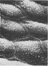
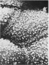
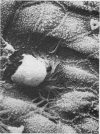
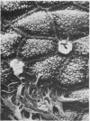
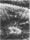
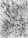
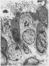
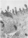
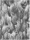
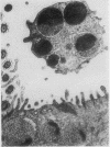
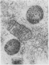
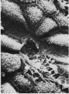
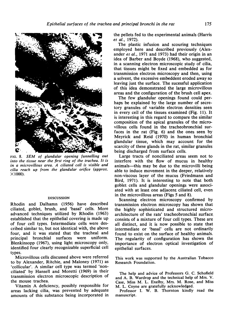
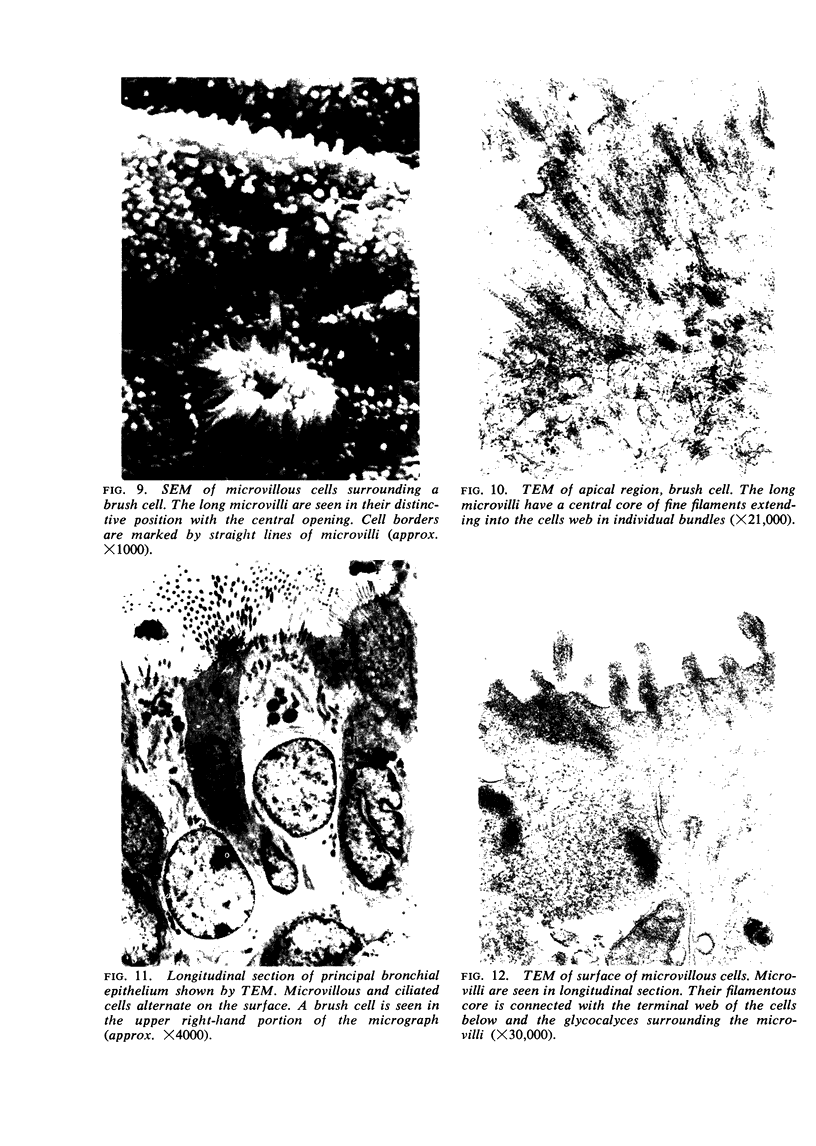
The epithelial surfaces in the trachea and principal bronchi of healthy rats were examined by scanning electron microscopy. A system of four cell types, ciliated, microvillous, brush, and goblet cells, in this order of frequency, were found and intermediate type cells were not seen. An extensive area of the surface examined was covered by densely ciliated epithelium. The presence of other cell types beneath the cilia was confirmed by transmission electron microscopy. Areas up to 1 mm in diameter and randomly distributed were observed where microvillous cells predominated and only occasional ciliated cells were found. Most ciliated cells in these areas were adjacent to glandular openings or goblet cells. The larger microvilli of the brush cells were arranged in a coronal configuration elucidated by the scanning electron microscope. Preparatory techniques recently introduced for the examination of soft tissue in the scanning electron microscope facilitated the confirmation of cell types present and the microarchitecture of the epithelial surface.
Full text
PDF
Images in this article
Selected References
These references are in PubMed. This may not be the complete list of references from this article.
- Alexander I. G., Capicchiano P. M., Ritchie B. C., Maloney J. E. Plastic embedding and surface erosion techniques in soft tissue scanning electron microscopy. J Microsc. 1973 Sep;99(1):69–74. doi: 10.1111/j.1365-2818.1973.tb03854.x. [DOI] [PubMed] [Google Scholar]
- Alexander I. G., Ritchie B. C., Maloney J. E. Scanning electron microscopy of respiratory tissues in the rat. J Anat. 1971 Dec;110(Pt 3):491–492. [PubMed] [Google Scholar]
- Barber V. C., Boyde A. Scanning electron microscopic studies of cilia. Z Zellforsch Mikrosk Anat. 1968;84(2):269–284. doi: 10.1007/BF00330870. [DOI] [PubMed] [Google Scholar]
- Blenkinsopp W. K. Proliferation of respiratory tract epithelium in the rat. Exp Cell Res. 1967 Apr;46(1):144–154. doi: 10.1016/0014-4827(67)90416-8. [DOI] [PubMed] [Google Scholar]
- Ceccarelli B., Clementi F., De Giuli C., Marini D. Aspetto di alcune mucose dell'apparato digerente al microscopio elettronico a scansione. Boll Soc Ital Biol Sper. 1969 May 31;45(10):646–649. [PubMed] [Google Scholar]
- Cireli E. Elektronenmikroskopische Analyse der prä- und postnatalen Differenzierung des Epithels der oberen Luftwege der Ratte. Z Mikrosk Anat Forsch. 1966;74(2):132–178. [PubMed] [Google Scholar]
- Friedmann I., Bird E. S. Ciliary structure, ciliogenesis, microvilli. (Electron microscopy of the mucosa of the upper respiratory tract). Laryngoscope. 1971 Nov;81(11):1852–1868. doi: 10.1288/00005537-197111000-00010. [DOI] [PubMed] [Google Scholar]
- GLAUERT A. M., GLAUERT R. H. Araldite as an embedding medium for electron microscopy. J Biophys Biochem Cytol. 1958 Mar 25;4(2):191–194. doi: 10.1083/jcb.4.2.191. [DOI] [PMC free article] [PubMed] [Google Scholar]
- HUXLEY H. E., ZUBAY G. Preferential staining of nucleic acid-containing structures for electron microscopy. J Biophys Biochem Cytol. 1961 Nov;11:273–296. doi: 10.1083/jcb.11.2.273. [DOI] [PMC free article] [PubMed] [Google Scholar]
- Hansell M. M., Moretti R. L. Ultrastructure of the mouse tracheal epithelium. J Morphol. 1969 Jun;128(2):159–169. doi: 10.1002/jmor.1051280203. [DOI] [PubMed] [Google Scholar]
- Harris C. C., Sporn M. B., Kaufman D. G., Smith J. M., Jackson F. E., Saffiotti U. Histogenesis of squamous metaplasia in the hamster tracheal epithelium caused by vitamin A deficiency or benzo[a]pyrene-Ferric oxide. J Natl Cancer Inst. 1972 Mar;48(3):743–761. [PubMed] [Google Scholar]
- Ito S. The enteric surface coat on cat intestinal microvilli. J Cell Biol. 1965 Dec;27(3):475–491. doi: 10.1083/jcb.27.3.475. [DOI] [PMC free article] [PubMed] [Google Scholar]
- Luciano L., Reale E., Ruska H. Bürstenzellen im Alveolarepithel der Rattenlunge. Z Zellforsch Mikrosk Anat. 1969;95(2):198–201. [PubMed] [Google Scholar]
- Meyrick B., Reid L. The alveolar brush cell in rat lung--a third pneumonocyte. J Ultrastruct Res. 1968 Apr;23(1):71–80. doi: 10.1016/s0022-5320(68)80032-2. [DOI] [PubMed] [Google Scholar]
- REYNOLDS E. S. The use of lead citrate at high pH as an electron-opaque stain in electron microscopy. J Cell Biol. 1963 Apr;17:208–212. doi: 10.1083/jcb.17.1.208. [DOI] [PMC free article] [PubMed] [Google Scholar]
- Silva D. G. The fine structure of multivesicular cells with large microvilli in the epithelium of the mouse colon. J Ultrastruct Res. 1966 Dec;16(5):693–705. doi: 10.1016/s0022-5320(66)80015-1. [DOI] [PubMed] [Google Scholar]


