Abstract
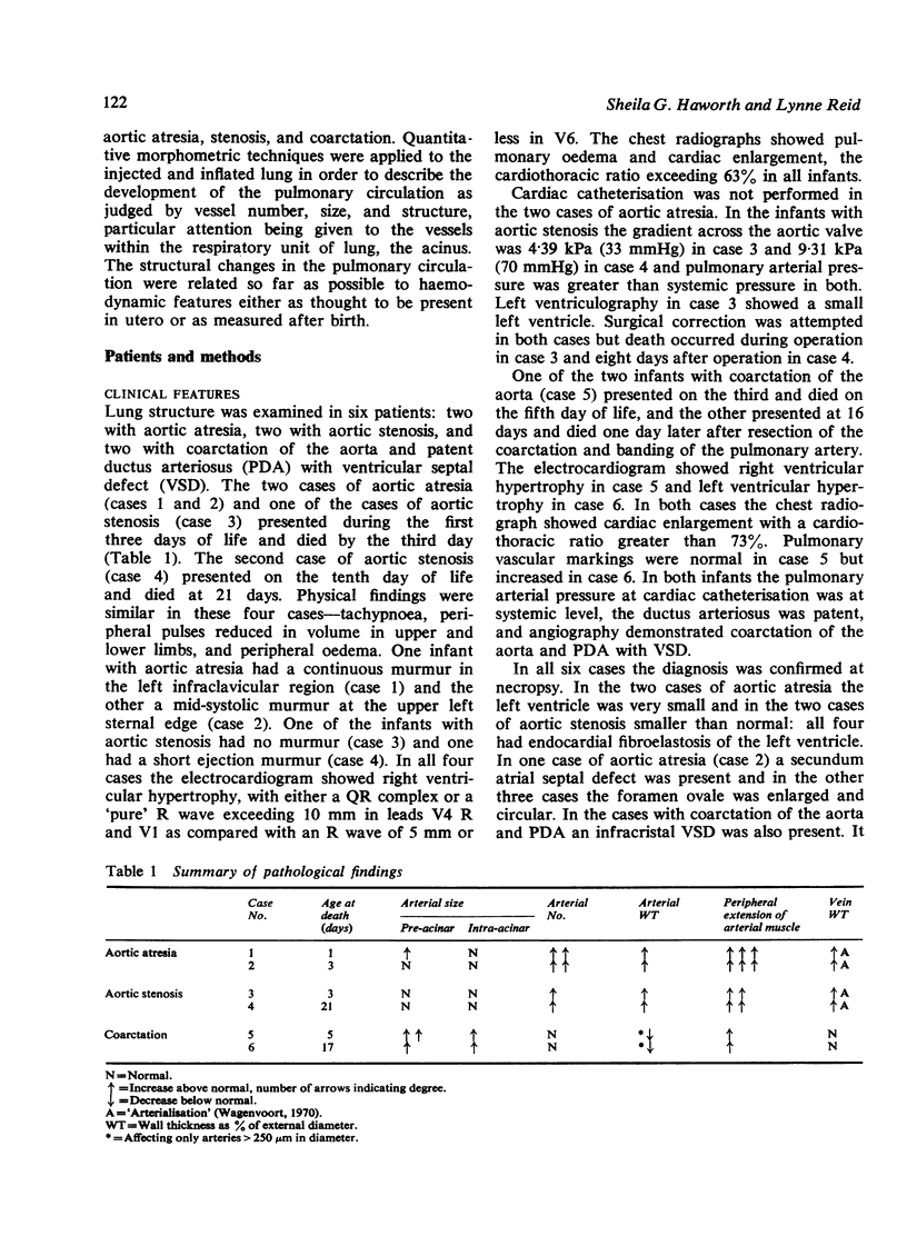
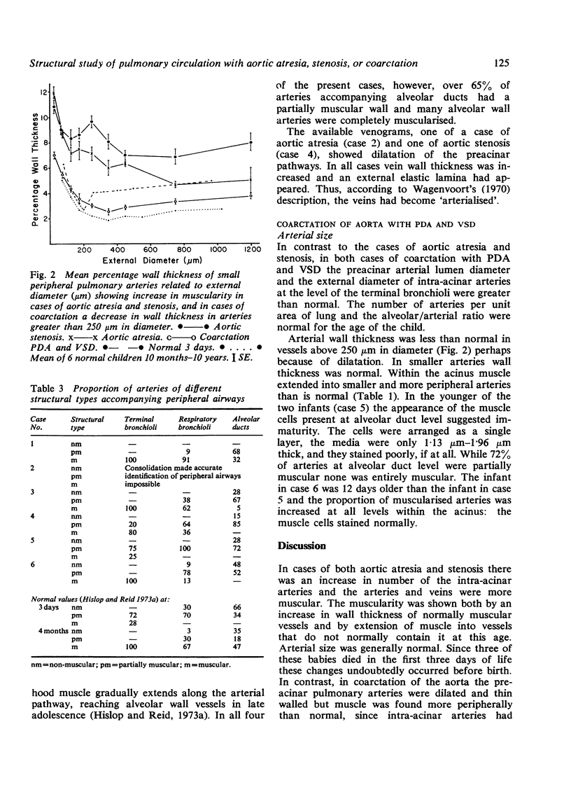
Study of the structural features of the pulmonary circulation in various types of congenital heart disease makes it possible to correlate function and structure in the fetal and newborn lung. We applied quantitative morphometric techniques to the injected and inflated lungs of newborn infants who had died with obstruction to left ventricular outflow from aortic atresia, stenosis, or coarctation. The structure and development of the pulmonary circulation was judged by the number of arteries and veins and their size and wall structure, with particular attention to vessels within the respiratory unit. The study established for the first time that the structure of the pulmonary circulation is modified by the antenatal abnormalities in blood flow that occur through the heart and great vessels in the presence of congenital heart disease. Fetal multiplication of intra-acinar arteries in aortic atresia and stenosis is increased as also is the muscularity of both pre- and intra-acinar arteries and veins, muscle extending into smaller and more peripheral vessels than is normal at birth. When the pulmonary circulation is normal before birth but arterial pressure and flow are abnormally increased at birth, as in coarctation with patent ductus and ventricular septal defect, an increase in arterial diameter and muscularity is apparent within the first week of life.
Full text
PDF
Images in this article
Selected References
These references are in PubMed. This may not be the complete list of references from this article.
- Davies G., Reid L. Growth of the alveoli and pulmonary arteries in childhood. Thorax. 1970 Nov;25(6):669–681. doi: 10.1136/thx.25.6.669. [DOI] [PMC free article] [PubMed] [Google Scholar]
- FERENCZ C., DAMMANN J. F., Jr Significance of the pulmonary vascular bed in congenital heart disease. V. Lesions of the left side of the heart causing obstruction of the pulmonary venous return. Circulation. 1957 Dec;16(6):1046–1056. doi: 10.1161/01.cir.16.6.1046. [DOI] [PubMed] [Google Scholar]
- Haworth S. G., Reid L. Persistent fetal circulation: Newly recognized structural features. J Pediatr. 1976 Apr;88(4 Pt 1):614–620. doi: 10.1016/s0022-3476(76)80021-2. [DOI] [PubMed] [Google Scholar]
- Hislop A., Haworth S. G., Shinebourne E. A., Reid L. Quantitative structural analysis of pulmonary vessels in isolated ventricular septal defect in infancy. Br Heart J. 1975 Oct;37(10):1014–1021. doi: 10.1136/hrt.37.10.1014. [DOI] [PMC free article] [PubMed] [Google Scholar]
- Hislop A., Reid L. Intra-pulmonary arterial development during fetal life-branching pattern and structure. J Anat. 1972 Oct;113(Pt 1):35–48. [PMC free article] [PubMed] [Google Scholar]
- Hislop A., Reid L. New pathological findings in emphysema of childhood. 1. Polyalveolar lobe with emphysema. Thorax. 1970 Nov;25(6):682–690. doi: 10.1136/thx.25.6.682. [DOI] [PMC free article] [PubMed] [Google Scholar]
- Hislop A., Reid L. Pulmonary arterial development during childhood: branching pattern and structure. Thorax. 1973 Mar;28(2):129–135. doi: 10.1136/thx.28.2.129. [DOI] [PMC free article] [PubMed] [Google Scholar]
- Hislop A., Reid L. Structural changes in the pulmonary arteries and veins in tetralogy of Fallot. Br Heart J. 1973 Nov;35(11):1178–1183. doi: 10.1136/hrt.35.11.1178. [DOI] [PMC free article] [PubMed] [Google Scholar]
- Hoffman J. I., Rudolph A. M. The natural history of ventricular septal defects in infancy. Am J Cardiol. 1965 Nov;16(5):634–653. doi: 10.1016/0002-9149(65)90047-0. [DOI] [PubMed] [Google Scholar]
- Maseri A., Caldini P., Harward P., Joshi R. C., Permutt S., Zierler K. L. Determinants of pulmonary vascular volume: recruitment versus distensibility. Circ Res. 1972 Aug;31(2):218–228. doi: 10.1161/01.res.31.2.218. [DOI] [PubMed] [Google Scholar]
- NAEYE R. L. Perinatal vascular changes associated with underdevelopment of the left heart. Am J Pathol. 1962 Sep;41:287–295. [PMC free article] [PubMed] [Google Scholar]
- NAEYE R. L. Perinatal vascular changes in coarctation of the aorta with distal patent ductus arteriosus. Circulation. 1961 Oct;24:754–760. doi: 10.1161/01.cir.24.4.754. [DOI] [PubMed] [Google Scholar]
- Rudolph A. M., Heymann M. A., Spitznas U. Hemodynamic considerations in the development of narrowing of the aorta. Am J Cardiol. 1972 Oct;30(5):514–525. doi: 10.1016/0002-9149(72)90042-2. [DOI] [PubMed] [Google Scholar]
- Samuelson A., Becker A. E., Wagenvoort C. A. A morphometric study of pulmonary veins in normal infants and infants with congenital heart disease. Arch Pathol. 1970 Aug;90(2):112–116. [PubMed] [Google Scholar]
- Wagenvoort C. A. Morphologic changes in intrapulmonary veins. Hum Pathol. 1970 Jun;1(2):205–213. doi: 10.1016/s0046-8177(70)80034-x. [DOI] [PubMed] [Google Scholar]


