

Abstract
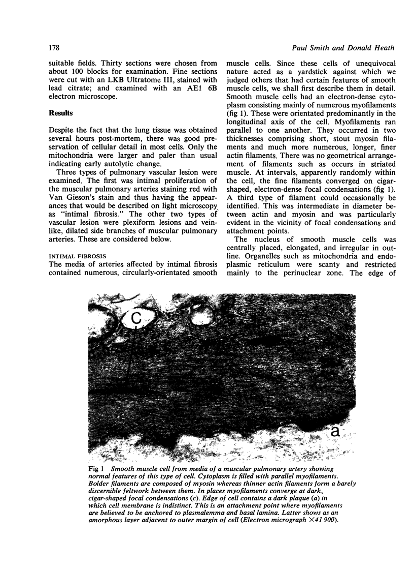
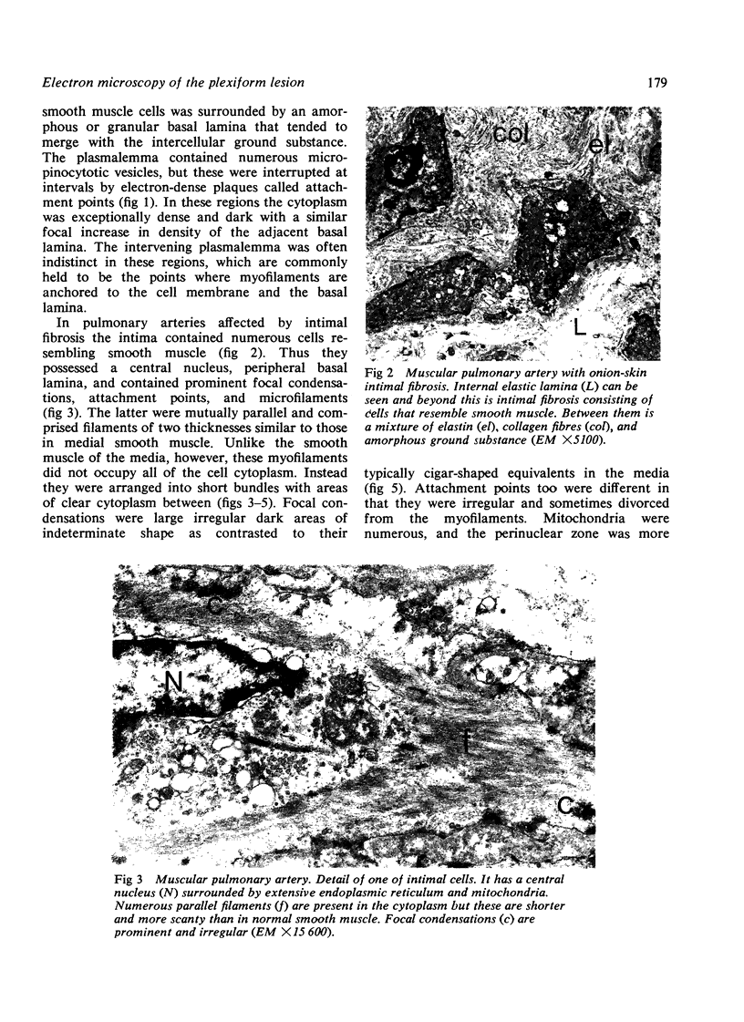
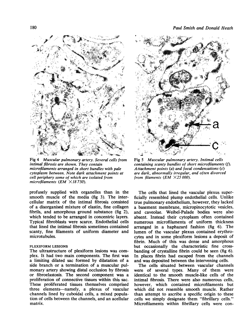
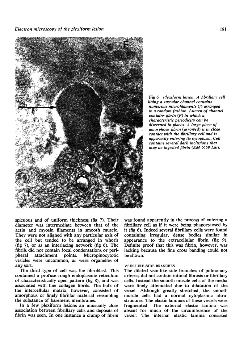
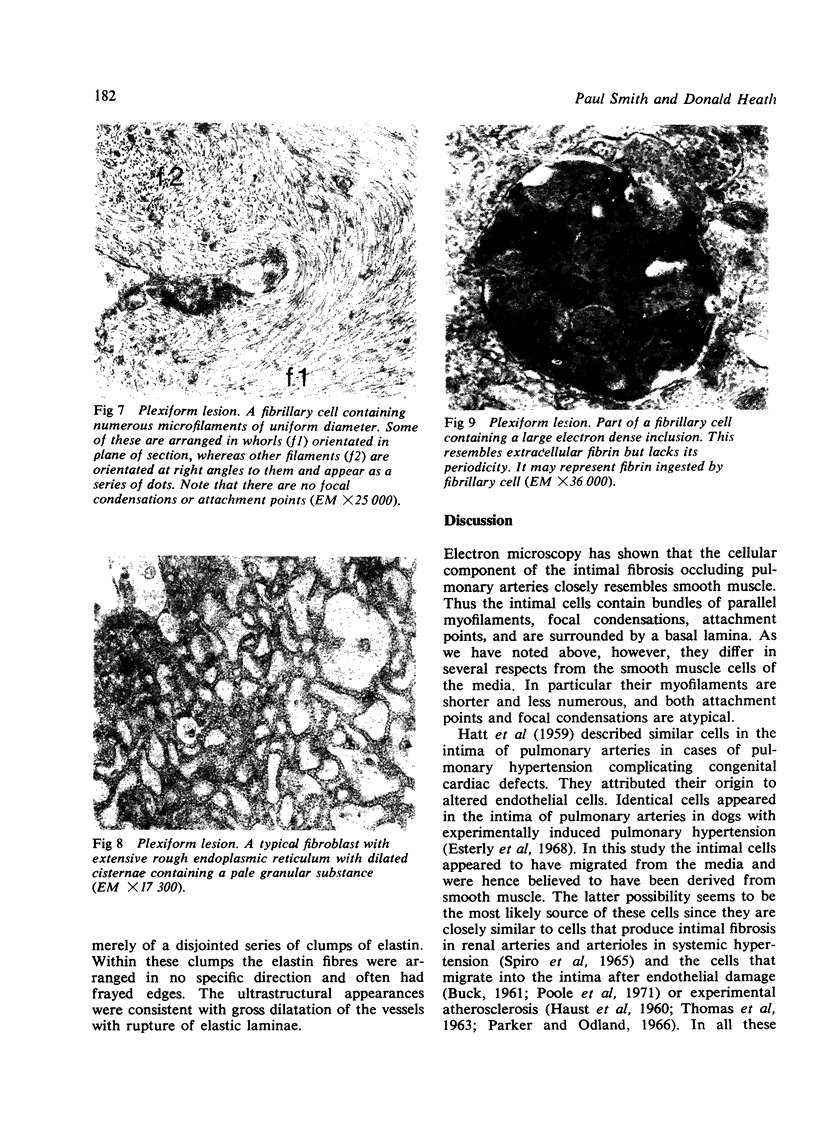
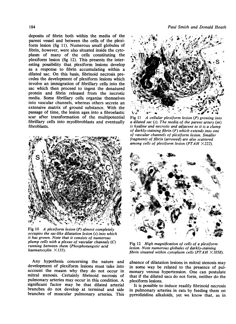
Pulmonary arteries from a case of plexogenic pulmonary arteriopathy were studied with the electron microscope. Many muscular pulmonary arteries showed intimal fibrosis of concentric or "onion-skin" type. The cells embedded within this fibrosis resembled smooth muscle but since they also possessed some features of fibroblasts we refer to them as myofibroblasts. Myofibroblasts also occurred in plexiform lesions together with fibroblasts and "fibrillary cells". These fibrillary cells contained numerous, prominent filaments with a random orientation. They lined the vascular channels of the plexiform lesions as well as being present within the stroma. They appeared to phagocytose fibrin and then to organise the plexiform lesion into a fibrous scar. Fibrillary cells closely resemble vasoformative reserve cells and the cells of the cardiac myxoma and so-called "papillary tumour" of heart valves. They may, therefore, be primitive multipotential cells found throughout the entire cardiovascular system.
Full text
PDF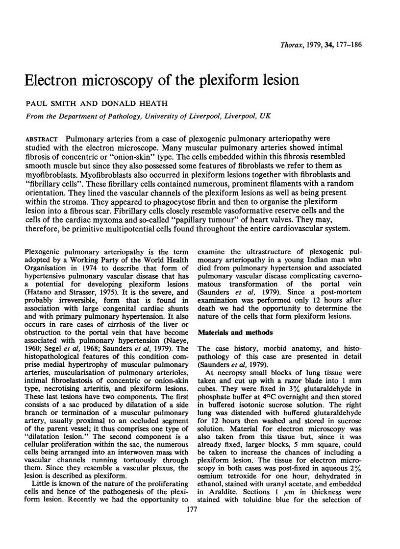
Images in this article


Selected References
These references are in PubMed. This may not be the complete list of references from this article.
- BENSCH K. G., GORDON G. B., MILLER L. FIBRILLAR STRUCTURES RESEMBLING LEIOMYOFIBRILS IN ENDOTHELIAL CELLS OF MAMMALIAN PULMONARY BLOOD VESSELS. Z Zellforsch Mikrosk Anat. 1964 Sep 3;63:759–766. doi: 10.1007/BF00336220. [DOI] [PubMed] [Google Scholar]
- Becker C. G., Murphy G. E. Demonstration of contractile protein in endothelium and cells of the heart valves, endocardium, intima, arteriosclerotic plaques, and Aschoff bodies of rheumatic heart disease. Am J Pathol. 1969 Apr;55(1):1–37. [PMC free article] [PubMed] [Google Scholar]
- Esterly J. A., Glagov S., Ferguson D. J. Morphogenesis of intimal obliterative hyperplasia of small arteries in experimental pulmonary hypertension. An ultrastructural study of the role of smooth-muscle cells. Am J Pathol. 1968 Feb;52(2):325–347. [PMC free article] [PubMed] [Google Scholar]
- Gabbiani G., Badonnel M. C., Rona G. Cytoplasmic contractile apparatus in aortic endothelial cells of hypertensive rats. Lab Invest. 1975 Feb;32(2):227–234. [PubMed] [Google Scholar]
- Gabbiani G., Hirschel B. J., Ryan G. B., Statkov P. R., Majno G. Granulation tissue as a contractile organ. A study of structure and function. J Exp Med. 1972 Apr 1;135(4):719–734. doi: 10.1084/jem.135.4.719. [DOI] [PMC free article] [PubMed] [Google Scholar]
- HAUST M. D., MORE R. H., MOVAT H. Z. The role of smooth muscle cells in the fibrogenesis of arteriosclerosis. Am J Pathol. 1960 Oct;37:377–389. [PMC free article] [PubMed] [Google Scholar]
- Heath D., Smith P. The electron microscopy of "fibrinoid necrosis" in pulmonary arteries. Thorax. 1978 Oct;33(5):579–595. doi: 10.1136/thx.33.5.579. [DOI] [PMC free article] [PubMed] [Google Scholar]
- Heath D., Smith P. The pulmonary endothelial cell. Thorax. 1979 Apr;34(2):200–208. doi: 10.1136/thx.34.2.200. [DOI] [PMC free article] [PubMed] [Google Scholar]
- Issler R. W. The arterial medial cell, smooth muscle or multifunctional mesenchyme? J Atheroscler Res. 1968 Mar-Apr;8(2):201–213. doi: 10.1016/s0368-1319(68)80056-0. [DOI] [PubMed] [Google Scholar]
- NAEYE R. L. "Primary" pulmonary hypertension with coexisting portal hypertension. A retrospective study of six cases. Circulation. 1960 Sep;22:376–384. doi: 10.1161/01.cir.22.3.376. [DOI] [PubMed] [Google Scholar]
- POMERANCE A. Papillary "tumours" of the heart valves. J Pathol Bacteriol. 1961 Jan;81:135–140. doi: 10.1002/path.1700810116. [DOI] [PubMed] [Google Scholar]
- Parker F., Odland G. F. A light microscopic, histochemical and electron microscopic study of experimental atherosclerosis in rabbit coronary artery and a comparison with rabbit aorta atherosclerosis. Am J Pathol. 1966 Mar;48(3):451–481. [PMC free article] [PubMed] [Google Scholar]
- Poole J. C., Cromwell S. B., Benditt E. P. Behavior of smooth muscle cells and formation of extracellular structures in the reaction of arterial walls to injury. Am J Pathol. 1971 Mar;62(3):391–414. [PMC free article] [PubMed] [Google Scholar]
- SPIRO D., LATTES R. G., WIENER J. THE CELLULAR PATHOLOGY OF EXPERIMENTAL HYPERTENSION. I. HYPERPLASTIC ARTERIOLARSCLEROSIS. Am J Pathol. 1965 Jul;47:19–49. [PMC free article] [PubMed] [Google Scholar]
- Saunders J. B., Constable T. J., Heath D., Smith P., Paton A. Pulmonary hypertension complicating portal vein thrombosis. Thorax. 1979 Apr;34(2):281–283. doi: 10.1136/thx.34.2.281. [DOI] [PMC free article] [PubMed] [Google Scholar]
- Segel N., Kay J. M., Bayley T. J., Paton A. Pulmonary hypertension with hepatic cirrhosis. Br Heart J. 1968 Jul;30(4):575–578. doi: 10.1136/hrt.30.4.575. [DOI] [PMC free article] [PubMed] [Google Scholar]
- Smith P., Heath D. Evagination of vascular smooth muscle cells during the early stages of Crotalaria pulmonary hypertension. J Pathol. 1978 Mar;124(3):177–183. doi: 10.1002/path.1711240308. [DOI] [PubMed] [Google Scholar]
- Smith P., Heath D. Ultrastructure of hypoxic hypertensive pulmonary vascular disease. J Pathol. 1977 Feb;121(2):93–100. doi: 10.1002/path.1711210205. [DOI] [PubMed] [Google Scholar]
- Smith P., Kay J. M., Heath D. Hypertensive pulmonary vascular disease in rats after prolonged feeding with Crotalaria spectabilis seeds. J Pathol. 1970 Oct;102(2):97–106. doi: 10.1002/path.1711020205. [DOI] [PubMed] [Google Scholar]
- Stovin P. G., Heath D., Khaliq S. U. Ultrastructure of the cardiac myxoma and the papillary tumour of heart valves. Thorax. 1973 May;28(3):273–285. doi: 10.1136/thx.28.3.273. [DOI] [PMC free article] [PubMed] [Google Scholar]