Abstract
Background
Characterizing the parasite dynamics and population structure provides useful information to understand the dynamic of transmission and to better target control interventions. Despite considerable efforts for its control, vivax malaria remains a major health problem in Peru. In this study, we have explored the population genetics of Plasmodium vivax isolates from Iquitos, the main city in the Peruvian Amazon, and 25 neighbouring peri-urban as well as rural villages along the Iquitos-Nauta Road.
Methodology/ Results
From April to December 2008, 292 P. vivax isolates were collected and successfully genotyped using 14 neutral microsatellites. Analysis of the molecular data revealed a similar proportion of monoclonal and polyclonal infections in urban areas, while in rural areas monoclonal infections were predominant (p = 0.002). Multiplicity of infection was higher in urban (MOI = 1.5–2) compared to rural areas (MOI = 1) (p = 0.003). The level of genetic diversity was similar in all areas (He = 0.66–0.76, p = 0.32) though genetic differentiation between areas was substantial (PHIPT = 0.17, p<0.0001). Principal coordinate analysis showed a marked differentiation between parasites from urban and rural areas. Linkage disequilibrium was detected in all the areas ( = 0.08–0.49, for all p<0.0001). Gene flow among the areas was stablished through Bayesian analysis of migration models. Recent bottleneck events were detected in 4 areas and a recent parasite expansion in one of the isolated areas. In total, 87 unique haplotypes grouped in 2 or 3 genetic clusters described a sub-structured parasite population.
Conclusion/Significance
Our study shows a sub-structured parasite population with clonal propagation, with most of its components recently affected by bottleneck events. Iquitos city is the main source of parasite spreading for all the peripheral study areas. The routes of transmission and gene flow and the reduction of the parasite population described are important from the public health perspective as well for the formulation of future control policies.
Author Summary
We present the population genetics of malaria vivax parasites in a large area of the Peruvian Amazon. Our results showed that the parasite population had a predominant clonal propagation, reproducing themselves with identically or closely related parasites; therefore, the same genetic characteristics are maintained in the offspring. The clonal propagation may favour the higher levels of genetic differentiation among the parasites from isolated areas compared to areas where human migration is common. The patterns of gene flow have been established, finding Iquitos city as a reservoir of parasite genetic variability. Moreover, a recent reduction of the parasite population was observed in areas where recent control activities were performed. This research provides a picture of the nature and dynamics of the parasite population which have a significant impact in the malaria epidemiology; therefore, this knowledge is crucial for the development of efficient control policies.
Introduction
According to the World Health Organization (WHO), Plasmodium vivax caused about 14.2 million malaria cases outside sub-Saharan Africa in 2013 [1]. Despite considerable efforts, Asian and South American countries are still far from achieving malaria elimination [2]. In Peru, the vast majority of malaria cases (76% of 64,673) was reported in the Amazon basin area (Loreto region) for 2014 and about 83% of them are due to P. vivax [3]. Many P. vivax infections are asymptomatic and undetectable by microscopy, providing a potentially important reservoir sustaining local transmission [4–7]. In addition, multiple infections recur even after the administration of the WHO-recommended radical cure treatment against blood- and hepatic-parasite stages (chloroquine and primaquine) [5, 6, 8]. To understand the epidemiology, distribution and transmission dynamics of P. vivax and thus improve its control, it is necessary to unravel the parasite population genetics and dynamics [9, 10]. Such information would be extremely useful for the monitoring and evaluation of control activities, both in the short and long term [10–12].
The extreme genetic variations in the P. vivax populations has already been reported from several endemic areas [10, 13]. In the Peruvian Amazon, the few observations available on P. vivax population genetics were collected in small areas (dispersed villages and communities) and reported heterogeneous and clonal parasite populations [6, 14–16]. Hereby, we report the genetic diversity and population genetics of the P. vivax parasite population from the most important urban city in the Peruvian Amazon and 25 villages located around and along the Iquitos-Nauta road.
Methods
Study sites
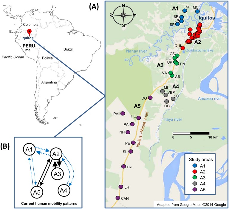
The clinical isolates were collected during an initial screening of the study sites in the Peruvian Amazon (April 2008) followed by active case detection of all fever cases (April to December 2008) during a longitudinal study assessing the efficacy of the recommended radical cure treatment for P. vivax malaria infection (chloroquine 25 mg/kg/day for 3 days and primaquine 0.5 mg//kg/day for 7 days) [17]. The study was conducted both within Iquitos city and in 25 neighbouring villages (Loreto region), some of them along the Iquitos-Nauta Road. Villages were geographically stratified in 5 study areas (A1- A5) (Fig 1A and S1 Table): A1 (3–7 km northwest from Iquitos city and only accessible by boat), A2 (Iquitos city and peripheral villages), A3 (villages situated along the Iquitos-Nauta Road, 11–13 km southwest from Iquitos city), A4 (villages situated 21 km southwest of Iquitos city and 2–9 km far from the Iquitos-Nauta Road) and A5 (villages situated 26–58 km southwest from Iquitos city, most of them along the Iquitos-Nauta road). Participants’ demographics are described in S1 Table.
Fig 1. Study areas and human migration patterns in and around Iquitos city and along the Iquitos-Nauta road (Loreto, Peru).
(A) Number of genotyped P. vivax isolates per area. Overall = 292: A1 = 105 (36.0%), A2 = 70 (24.0%), A3 = 32 (11.0%), A4 = 55 (18.8%) and A5 = 30 (10.3%). For detailed information of the villages that compose each area and frequency of isolates, see S1 Table. (B) The current human migration patterns are described by arrows indicating the direction and the occurrence of transit (thickness). Dotted blue arrows indicate that the transit involves crossing and/or navigating through a river while the solid black arrows indicate transit through the Iquitos Nauta road.
In Fig 1B, we describe the main human mobility patterns in the study areas. Most human mobility occurs around A2 (Iquitos city, economic centre and big markets). Indeed, some people commute every day to A2 but live in A1 or A3, others travel from A5 to A2 during the weekends crossing the Nanay river or come from A4 a couple of times per year navigating along the Itaya river to sell their goods. Some sporadic movement between A1 and A5, crossing the Nanay river, is observed.
The study population consisted primarily of mestizos of low socioeconomic status. Villages located along Iquitos-Nauta road or next to the river had no electricity and in most cases drinking water was taken from the river or natural springs. Malaria transmission is perennial with peaks from November to May (rainy season)[18], and the majority of malaria cases are due to P. vivax. Recurrent sub-patent and asymptomatic infections are frequent [5, 6]. Anopheles darlingi is the main anthropophilic and exo/endophilic vector [19]. Malaria prevention and control activities were conducted before the sample collection in Loreto region including all our study areas but in A4 (S1 Table) as part of PAMAFRO (Malaria Control Program in Andean-country Border Regions) which started in 2005 [20].
Sample collection
Patients were examined daily during the treatment and followed up weekly with blood sampling according to WHO guidelines for P. vivax drug efficacy during the first 28 days [WHO, 2009]; and systematic monthly follow-up was carried out thereafter. For the purpose of the current study, all D0 (before-) and D1 (after treatment) isolates were analysed, including a blood sample for microscopy (thick and thick film) and a blood spot on filter paper (BSFP) (Whatman grade 3, Whatman, Springfield Mill, USA).
Ethics statement
The study was approved by the Ethical Boards of Universidad Peruana Cayetano Heredia, Peru (Project PVIVAX-UPCH, SIDISI code: 053256), the institutional review board of the Institute of Tropical Medicine Antwerp and the University Hospital of Antwerp, Belgium. Adult participants provided informed written consent, and a parent or guardian of any child participant provided informed written consent on their behalf.
Laboratory methods
All slides were read by microscopy (thin and thick smear) to confirm P. vivax infection and estimate the parasite density (number of asexual parasites for 200 white blood cells (WBC) assuming 8000 WBC/μl) [6, 21]. Parasite and human DNA was extracted using the Chelex method [22]. P. vivax mono-infections were confirmed by species-specific PCR (ssPCR) [23] and genotyped using a panel of 14 well-described microsatellite markers (MS), namely MS1, MS2, MS3, MS4, MS5, MS6, MS7, MS8, MS9, MS10, MS12, MS15, MS16 and MS20 [24, 25]. Briefly, the DNA extracted from each sample was used to perform a separate PCR for each MS. The forward primer of each MS was labelled with a fluorophore to identify the size of the amplicon through capillary electrophoresis in a 3730 XL ABI sequencer (Applied Biosystems, Foster City, CA, USA) [6]. MS PCR was repeated for those isolates with no MS PCR amplification. Some isolates from A1 and A4 were previously analysed and already published [6, 14].
Data analysis
The allele fragment sizes recovered from the capillary electrophoresis were determined using Genemapper (Applied Biosystems, Foster City, CA, USA). Only fragments with ≥100 relative fluorescence units (RFU) were considered as ‘real’ alleles. In case of the presence of two or more alleles, only alleles with RFU ≥30% of the dominant allele RFU were considered for further analysis. The genetic and statistical analysis were performed mainly at study areas level but whenever feasible also at the village level to assess the influence of individual villages on the areas. SPSS for Windows v.20 (IBM Corp., NY) was used to perform non-genetic statistical analysis.
Multiplicity of infection
Infections were classified as monoclonal (1 allele per loci) or polyclonal (≥2 alleles in at least one locus). The haplotypes (unique combination of 14-MS alleles) were determined using GenAlEx v.6.5 [26, 27]. The predominant haplotypes were recovered from polyclonal infections based on the highest peak of the alleles [28, 29]. The multiplicity of infection (MOI, minimal number of distinct “clones” or haplotypes within a isolate) was estimated by taking the locus with the highest number of alleles as a proxy [6]. The proportions of polyclonal infections were compared between areas with Pearson χ2 test and the average MOI compared using the Kruskal-Wallis test.
Genetic diversity and parasite differentiation
The mean number of alleles and the number of private alleles (alleles unique to a single area) were determined for each area using GenAlEx. The allelic richness, a non-biased-by-sample size measure of the number of alleles, was determined using FSTAT v.2.9.3 [30]. The expected heterozygosity (He) was determined per locus and per study area using GenAlEx. The He represents the probability of finding a different allele for a given locus in any pair of samples randomly drawn from the same population. He varies from no diversity at all (0) to highly diverse (1). Overall monoclonal/polyclonal infections with complete/incomplete MS allele data were included for the calculations of these metrics. The He and the allelic richness were compared among study areas using Kruskal-Wallis Test.
The 2-level hierarchical genetic variation of the parasites between and within study areas was analysed by the Analysis of Molecular Variance (AMOVA) using GenAlEx [26, 27], and the PHIPT statistic, an estimate of the proportion of the parasites’ genetic variance between areas relative to the total variance, was calculated. When a third hierarchical level (villages) was included the proportion of variance between the areas (PHIRT), among the villages (PHIPR) and within the villages (PHIPT) were calculated. Probabilities for the AMOVA indexes were calculated based on individual randomizations (9999 permutations) [26, 27]. Pairwise comparisons of the genetic differentiation between areas or villages were further evaluated calculating the pairwise PHIPT values. PHIPT, analogue of the fixation index Fst, suppresses the within-population variance and ranges from 0 (no differentiation) to 1 (full differentiation). PHIPT was determined only for isolates with full haplotype data [26, 27].
Principal Coordinate Analysis (PCoA), which plots the major relationships within a multivariate dataset, was generated with GenAlEx by intercomparison of geographic areas and villages using the PHIPT matrix obtained via AMOVA and the individual-by-individual binary genetic distance matrix (for later genetic relatedness analysis) [26, 27]. Only isolates with full allele data were included on the PCoA. Mantel test for Matrix Correspondence was performed using GenAlEx to test the occurrence of a positive correlation (Rxy>0) between the genetic PHIPT matrix and geographic distances, so called the Isolation-by-distance (IBD) hypothesis. Geographic distances were computed using geospatial data for each village and converting UTM to km with GenAlEx [26, 27].
Linkage disequilibrium
The presence of linkage disequilibrium (LD), the non-random association of alleles from different loci, was evaluated by determining the standardized Index of Association () as a measure of multilocus linkage using LIAN [31]. Bias due to equivocal assignment of haplotypes in polyclonal infections was assessed by determining the linkage in (i) both monoclonal and polyclonal infections, and (ii) in monoclonal infections only [32]. LD was also assessed on infections with unique haplotypes in order to differentiate between clonal propagation and epidemic expansion [29, 33]. Pairwise-loci LD was evaluated using FSTAT to discard physical linkage between loci located within the same contig (MS4-MS5, MS7-MS8 and MS12-MS15) [30].
Genetic structure. The genetic structure was explored using three different approaches using eBURST, STRUCTURE and GENODIVE. The software eBURST v.3 assigned the haplotypes to genetic clusters (‘haplogroups’) of parasites that shared the same alleles in at least 7–13 loci [34]. Haplotypes not complying with the clustering criteria were defined as singletons. The software STRUCTURE v.2.3.3 was used to elucidate the genetic structure and the most likely number of clusters (K)[35]. STRUCTURE runs were performed exploring K from 1 to 15 (10 iterations each), consisting of a burn-in period of 50,000 iterations followed by 150,000 Markov Chain Monte Carlo (MCMC) iterations, assuming a mixture model and correlated allele frequencies. The most likely K was defined according to Evanno et al.[35] by calculating the rate of change of K, ΔK, using STRUCTURE HARVESTER v0.6.94 [36]. The haplotypes with >85% ancestry were assigned to a specific cluster for later cluster differentiation analysis. Hierarchical structure were analysed within the identified clusters using sampling location as priors (LOCPRIOR option) and increasing the up to 20 iterations for each K, burn-in 50,000 iteration followed by 500,000 MCMC iterations [37]. Additional data parsing and formatting of the STRUCTURE results were subsequently done using CLUMMP [38] and DISTRUCT [39]. CLUMMP aligns the cluster assignment across replicate analyses while DISTRUCT performs a graphical display of the aligned cluster assignments. Genetic differentiation among the predicted genetic clusters was evaluated through AMOVA and PCoA. In order to confirm the genetic clustering of the parasites, an AMOVA-based K-means clustering method implemented in the software GENODIVE v.2.0b23 (OS X 10.6 operating system) was used [40]. This method combines the K-means clustering which divides a number of individuals into an a priori assigned number of groups (K) in such a way that the within-group diversity is minimized and the between-group is maximized [41]. In order to determine the optimal clustering, the pseudo-F statistic (the highest value = optimal clustering) was calculated using the simulated annealing with 150,000 steps as convergence method and the number of algorithm repeats set to 50. GENODIVE retrieves the Rho index (analogue of Fst ranging from 0 to 1) as a measure of genetic differentiation between the genetic clusters [40]. Only isolates with full haplotype data were included for eBURST, STRUCTURE and GENODIVE analyses, as well as isolates with up to 3 loci with missing data for the two last analyses. In order to verify the presence of independent genetic clusters, He, pairwise PHIPT and multilocus and pairwise-loci LD were assessed as described above.
Network and gene glow analysis
PHYLOViZ v1.0 was used to display the phylogenetic relationship through minimum spanning trees (MST) among P. vivax haplotypes [42]. MST’s were constructed to explore the haplotypes’ genetic relationship in function to its geographic origin (areas differentially coloured) and to check the genetic clustering retrieved from STRUCTURE (clusters differentially coloured).
The gene flow was inferred through the assessment of 13 migration models using a Bayesian approach (MCMC analysis) based on the coalescence theory and implemented in MIGRATE-N v3.6 [43]. MIGRATE-N estimates the posterior probability distribution of the migration parameters: mutation-scaled migration rates (M) and population sizes (Θ); and calculates marginal log-likelihoods (log mL) for each model using a thermodynamic integration. The log mL’s were used to calculate the log Bayes factors (LBF, mL ratios) and probabilities [44]. The best model is the one with the highest log mL and the model ranking is made by comparison of the LBF’s and probabilities with respect to the best model. The models were built considering the genetic structuring gathered on the previous steps and on the knowledge on the people’s current mobility patterns in and around Iquitos (Fig 1B). Each migration model was conducted in MIGRATE-N deriving the starting migration parameters from Fst-like calculations and through a long chain of 1x10⁶ recorded genealogies at a sampling increment of 100 iterations after discarding the first 10,000 as burn-in for each locus. To improve the MCMC searches, static heating scheme with 4 chains with temperatures 1, 1.5, 3 and 1’000,000 was set. Uniform-distributed priors for the migration parameters were set to range 0–100. Brownian motion approximation to the ladder model was selected as microsatellite data model for faster calculations [45]. The number of immigrants per generation (Nm) was calculated as the product of M by Θ(recipient). The effective population size (Ne) was also calculated: Ne = Θ/2μ; where μ = mutation rate per generation per locus, μ only described for P. falciparum = 1.59 x 10⁻⁴ (CI 95% 3.7x10⁻⁴–6.98x10⁻⁵) [46].
Bottleneck analysis
We tested the deviation from the mutation-drift equilibrium due to recent bottleneck or rapid expansion events in the parasite population using the software BOTTLENECK v1.2.02 [47, 48]. After a recent reduction of the effective population size, the extent of the reduction of the number of alleles (rare alleles mainly) would be higher compared to the reduction of genetic diversity (He). In contrast, the deficiency of He may indicate a recent population expansion. Using the Wilcoxon rank test, BOTTLENECK determines if a significant number of microsatellites exhibits excess or deficiency of He under the most strict mutational models: Infinite Allele Model (IAM) and the Stepwise Mutational Model (SMM) and also the Two-phase model (TPM) which allows to fractionate the proportion of mutational models [47, 49]. The test was performed initially grouping the isolates by their geographic origin using only 10 polymorphic microsatellites (MS1, MS3, MS5 and MS7 were not used for this analysis, average He<0.5). Since the microsatellites we used may not follow the same mutational model we also performed the analysis separately for perfect 3-4-bp repeats microsatellites (MS4, MS9, MS15, MS20) and imperfect microsatellites (MS2, MS6, MS8, MS10, MS12, MS16) which may tend to follow the SSM and IAM, respectively [47, 50]. The mutational models were simulated by 10,000 replications, and specifically for TPM the proportion of SSM was fractionated in 5–95% a variance among multiple steps of 30.
Results
Multiplicity of infection
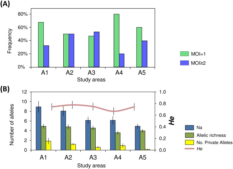
Overall we have analysed 292 P. vivax isolates (out of 302) with genotyping success on 56.2% (out of 292 isolates) with all 14 loci, 62.3% with 11–13 loci and 81.5% with 10 or less loci (efficiency of PCR amplification for each locus tabulated in S2 Table). Most of the isolates, 62.7% (183/292) were monoclonal infections and the polyclonal infections carried a minimum of 2 or 3 different haplotypes (34.6% and 2.7%, respectively). The proportion of mono/polyclonal infections significantly differed between areas (p = 0.002), i.e. A1 and A4 presented higher frequency of monoclonal infections while in A2 and A3 the proportions of mono/polyclonal infections were similar (Fig 2A). The median MOI was higher in A2 (MOI = 1.5) and A3 (MOI = 2) compared to the other areas (MOI = 1) (p = 0.003).
Fig 2. Distribution of monoclonal and polyclonal infections and allelic patterns between the study areas.
All samples were considered for the analysis (A1 = 105, A2 = 70, A3 = 32, A4 = 55, A5 = 30). (A) Frequency of monoclonal (MOI = 1) and polyclonal infections (MOI≥2) per area. (B) Allelic patterns per area: Na = No. of different alleles, No. Private alleles = Number of alleles unique to a single area, He = Expected heterozygosity.
Genetic diversity and parasite differentiation
The overall genetic diversity estimates among the areas described a median He = 0.74 (range 0.66–0.76), median allelic richness = 4.6 alleles (range 3.6–4.9), and did not differ significantly between areas (He: p = 0.32); allelic richness: p = 0.17), Fig 2B). Private alleles were less in A3 and almost absent in A5 than in the other areas (Fig 2B). The level of polymorphism (He) of each locus is presented in S2 Table. The AMOVA revealed that most of the genetic variation of the parasite population relied within areas (83%) (Table 1), though genetic differentiation between areas was also observed (PHIPT = 0.17, p = 0.0001).
Table 1. Summary of hierarchical AMOVA for different geographical groupings and genetic clustering.
| Grouping | Source of variation | df | Sum of squares | Estimated variation (%) | Statistic | p-value | |
|---|---|---|---|---|---|---|---|
| Geographic locations | By areas1 | Between areas | 4 | 93.9 | 17% | PHIPT = 0.17 | 0.0001 |
| Within areas | 123 | 525.9 | 83% | ||||
| By villages2 | Between areas | 4 | 136.8 | 22% | PHIRT = 0.22 | 0.0002 | |
| Between villages within areas | 7 | 43.6 | 4% | PHIPR = 0.06 | 0.0036 | ||
| Within villages | 116 | 482.3 | 73% | PHIPT = 0.27 | 0.0001 | ||
| Genetic clusters3 | K = 2 | Between clusters | 1 | 73 | 37% | PHIPT = 0.37 | 0.0001 |
| Within clusters | 87 | 355.3 | 63% | ||||
| K = 3* | Between clusters | 1 | 53.3 | 26% | PHIPT = 0.26 | 0.0001 | |
| Within clusters | 79 | 338.7 | 74% | ||||
| K = 7 | Between clusters | 6 | 224.9 | 59% | PHIPT = 0.59 | 0.0001 | |
| Within clusters | 80 | 167.2 | 41% | ||||
At 2-level hierarchical AMOVA: PHIPT = estimate of the proportion of the parasites’ genetic variance between areas/clusters relative to the total variance. At 3-level hierarchical AMOVA: PHIRT, PHIPR and PHIPT = proportion of variance between the areas, between villages and within villages, respectively.
1Number of isolates: A1 (n = 60), A2 (n = 16), A3 (n = 10), A4 (n = 38), A5 (n = 4)
2Number of isolates: A1 (MN = 20, FM = 7, SR = 20, LP = 7, SP = 6), A2 (VS = 4, others A2 = 12), A3 (VA = 7, others A3 = 3), A4 (SC = 36, VBP = 2), A5 = 4.
3Genetic clustering assignment performed using STRUCTURE. Parasites classified as admixed were not considered for AMOVA. When K = 2 the clusters contained: 71 and 18 isolates; when K = 3: 58 and 23 isolates; and when K = 7: 23, 18, 15, 11, 10, 7 and 3 isolates.
* For K = 3, only two clusters were considered since no isolate was assigned as full member of the third cluster.
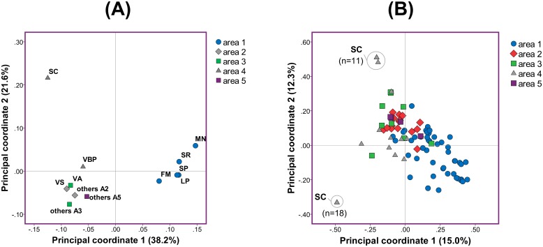
Pairwise calculations of the genetic differentiation and PCoA between areas and/or villages showed that parasites in the geographically isolated areas A1 and A4 were differentiated compared to those circulating in the other three areas with direct access to the Iquitos-Nauta road (Table 2 and Fig 3A). The first two coordinates of the PCoA explained 59.8% of the total variance pointing out clustering of parasites in villages from A1 (median PHIPT 0.02 within A1), presence of related parasites in A2, A3 and A5 and genetic differentiation of parasites from San Carlos village (A4) (Fig 3A).
Table 2. PHIPT values obtained from pairwise comparison between study areas (A1–A5) and between villages within each area.
| A1 | A2 | A3 | A4 | |||||||||||||
|---|---|---|---|---|---|---|---|---|---|---|---|---|---|---|---|---|
| A1 | MN | FM | SR | LP | SP | A2 | VS | others A2 | A3 | VA | others A3 | A4 | SC | VBP | ||
| A1 | ||||||||||||||||
| FM | 0.07 | |||||||||||||||
| SR | 0.03 | 0.01 | ||||||||||||||
| A1 | LP | 0.08 | 0 | 0.01 | ||||||||||||
| SP | 0.05 | 0 | 0.02 | 0 | ||||||||||||
| A2 | 0.11 | |||||||||||||||
| A2 | VS | 0.33 | 0.26 | 0.28 | 0.29 | 0.29 | ||||||||||
| others A2 | 0.23 | 0.12 | 0.17 | 0.16 | 0.15 | 0.09 | ||||||||||
| A3 | A3 | 0.14 | 0.06 | |||||||||||||
| VA | 0.3 | 0.18 | 0.23 | 0.26 | 0.26 | 0.34 | 0.15 | |||||||||
| others A3 | 0.3 | 0.10 | 0.21 | 0.2 | 0.18 | 0.26 | 0.10 | 0.10 | ||||||||
| A4 | 0.23 | 0.18 | 0.19 | |||||||||||||
| A4 | SC | 0.36 | 0.32 | 0.33 | 0.37 | 0.36 | 0.37 | 0.28 | 0.31 | 0.32 | ||||||
| VBP | 0.26 | 0.10 | 0.14 | 0.11 | 0.12 | 0.23 | 0.09 | 0.21 | 0.06 | 0.18 | ||||||
| A5 | A5 | 0.09 | 0.26 | 0.10 | 0.17 | 0.13 | 0.10 | -0.02 | 0.25 | 0.05 | 0.03 | 0.17 | 0.06 | 0.19 | 0.29 | 0.04 |
Fig 3. PCoA of the parasite population at village and individual haplotype level.
(A) Principal coordinate analysis of the study areas resulted from intercomparison of individual villages. Percentages in the parenthesis indicate the proportion of total variation explained by each principal coordinate. The term “others…” was used to group villages within specific areas that had less than 2 isolates with known haplotype. (B) PCoA among the individual haplotypes within the areas. Areas are defined in Fig 1A and village abbreviations are detailed in S1 Table.
When considering parasite populations per village, most of the genetic variation actually lied within villages (73% of the total genetic variation) with a high differentiation among the parasites within the villages (PHIPT = 0.27, p = 0.0001), while little differentiation was found between the villages (PHIPR = 0.06, p = 0.004) (Table 1). The PCoA at the individual haplotype level confirmed genetic differentiation among parasites within the same areas even within the same village: i.e. two different groups of haplotypes in San Carlos village (Fig 3B). The genetic distance of the parasite population was not correlated with geographic distance between villages (using PHIPT matrix Rxy = -0.41 p = 0.17) (scatterplot of the genetic and geographic distances in S1 Fig).
Linkage disequilibrium
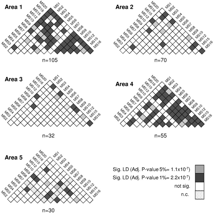
In all study areas except for A5, multilocus linkage disequilibrium (LD) was found when all isolates (MOI≥1) or only monoclonal (MOI = 1) isolates were analysed ( = 0.08–0.49, for all areas except A5: p<0.0001) (Table 3). LD remained in those areas when only unique haplotypes were considered for the analysis ( = 0.08–0.17, p<0.0001). The number of isolates decreased drastically when only monoclonal infections were considered (in A3, from 10 polyclonal isolates to 3 monoclonal isolates). Further LD analysis was performed at village-level only for those villages with more than four isolates. LD was found in villages within A1 (MN, SR and LP, = 0.18–0.21 p<0.0001); A2 (VS, = 0.38 p<2.0x10-04); A3 (VA, = 0.39 p<1.0x10-05) and A4 (SC, = 0.52 p<0.0002). In contrast to the multilocus LD analysis, the pairwise LD analysis was performed by including isolates with missing alleles and showed presence of LD in all areas. The pairwise LD was found mainly between loci located in different contigs therefore the LD found among loci within the same contigs did not alter the outcome (Fig 4).
Table 3.
Multilocus linkage disequilibrium assessed using the Index of association ().
| Area | All isolates | MOI = 1 | Unique haplotypes | ||||||
|---|---|---|---|---|---|---|---|---|---|
| # isolates | p-value | # isolates | p-value | # isolates | p-value | ||||
| A1 | 60 | 0.15 | <0.0001 | 59 | 0.19 | <0.0001 | 46 | 0.10 | <0.0001 |
| A2 | 16 | 0.08 | <0.0001 | 10 | 0.05 | 0.0078 | 16 | 0.08 | <0.0001 |
| A3 | 10 | 0.24 | <0.0001 | 3 | 0.83 | 0.0002 | 9 | 0.17 | <0.0001 |
| A4 | 38 | 0.49 | <0.0001 | 36 | 0.49 | <0.0001 | 12 | 0.14 | <0.0001 |
| A5 | 4 | 0.01 | 0.58 | 3 | 0.27 | 0.11 | 4 | 0.01 | 0.58 |
| All areas | 128 | 0.12 | <0.0001 | 111 | 0.17 | <0.0001 | 87 | 0.06 | <0.0001 |
LD analysis for each area at three levels: all isolates, monoclonal isolates only (MOI = 1) and unique haplotypes only. p-Values to test the null hypothesis of linkage equilibrium. Isolates with missing alleles were excluded from the analysis.
Fig 4. Pairwise-locus linkage disequilibrium analysis by study areas.
Significant association between alleles at pairs of loci in each isolate (LD) was tested using FSTAT. p-Values were adjusted after Bonferroni corrections and depicted by a colour as described in the legend. Isolates with missing alleles where included in the analysis.
Genetic structure
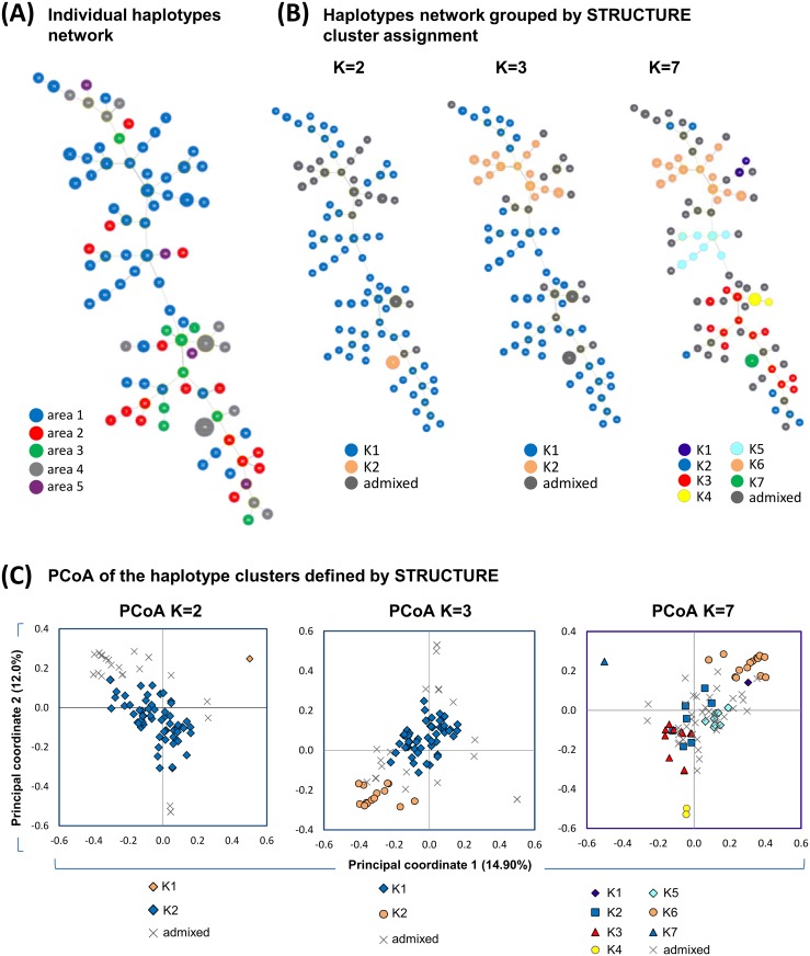
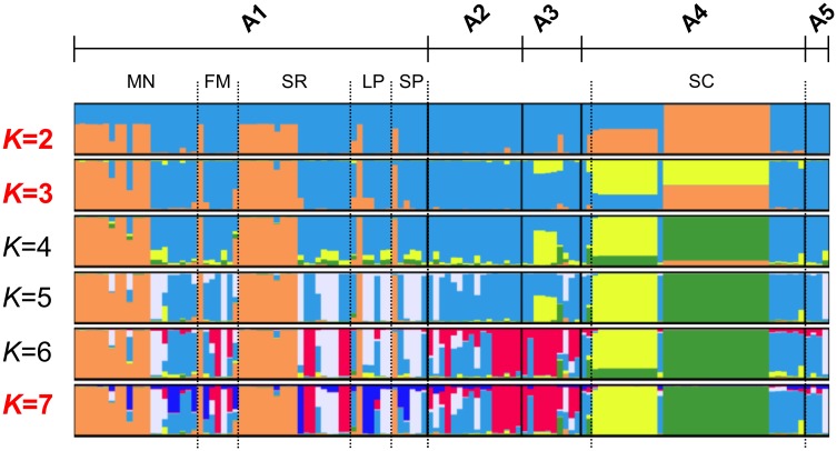
87 unique haplotypes were found (52.9% of them in A1) in 128 isolates without allelic missing data (Fig 5A; list of haplotypes in S3 Table). The two most frequent haplotypes were found 18 and 11 times (both in A4), followed by 11 haplotypes found 2–4 times (10 from A1 and 1 from A3) and 74 haplotypes were present only once (Fig 5A). Haplotypes were not shared between areas and only shared within the villages of A1. The haplotypes were grouped into 9 to 13 genetic clusters when the eBURST criteria was set to 8 to 13 loci with identical alleles. When the criteria was set to 7 loci, eBURST assigned the parasite population into 3 genetic clusters (PHIPT among clusters = 0.25, p = 0.001) and 6 singletons, where one cluster accounted for 91% (116/128) of the isolates. The K-means clustering divided the population into 2 and 3 genetic clusters and significant genetic differentiation between clusters was found (Rho 0.35 and 0.23, respectively). STRUCTURE analysis was performed to infer cluster assignment including only isolates with up to 3 missing alleles and with known haplotypes. Using STRUCTURE results HARVESTER predicted the most likely number of clusters being K = 2, followed by K = 3, and K = 7 (Fig 6). Using a threshold of 85% for the assignment of group representatives to each cluster, 70–63% of the isolates were assigned to clusters for K = 2, K = 3 and K = 7 and the remaining isolates were assigned as admixed parasites (Fig 5B). The AMOVA analysis revealed high differentiation between these genetic clusters (PHIPT = 0.26–0.59) (Table 1) which were graphically displayed using PCoA (Fig 5C). When the number of clusters was set to K = 2, parasites from all areas belonged to cluster 2 (K2: He = 0.76; = 0.06 p<0.0001) while 18 isolates from San Carlos village belonged to cluster 1 (K1: all the isolates shared the same haplotype) (Fig 5B). Hierarchical structure was subsequently found within cluster 2 where two sub-clusters were found (PHIPT = 0.20, p<0.0001), A1 contained one cluster and hybrid samples while the other cluster was present in all the other areas (S2 Fig). When K = 3, the former 18 isolates from San Carlos and 11 additional isolates from San Carlos were classified as admixed parasites where their genetic composition shared part of the cluster 1 or 2 with a third cluster. No parasites with >85% ancestry belonging to the third cluster were found within our study. Cluster 1 and 2 contained parasites from all areas (He = 0.77 and 0.35; = 0.05 and 0.11, for both p<0.0001). The extent of genetic diversity and LD within the clusters decreased when K = 7 (He varied from no diversity to 0.74, median He = 0.35) and linkage disequilibrium was found for all the clusters (p<0.01).
Fig 5. Phylogenetic relationship and PCoA of the P. vivax haplotypes arranged by the geographic origin and the genetic clustering.
(A) Network analysis displaying the phylogenetic relationship of the P. vivax haplotypes. The haplotypes are coloured following the area colour code displayed in the legend. The size of the nodes denotes the number of haplotypes transformed in logarithmic scale. (B) Phylogenetic relationships and PCoA of the isolates assigned into 2, 3 and 7 genetic clusters following STRUCTURE criteria. The nodes were coloured to discriminate their assignment to a genetic cluster (K1, K2…) or as an admixed haplotype and the node’s size varied as described previously. When K = 3 only 2 clusters are described (K1 and K2) since no isolate achieved effective assignment to K3. (C) The genetic distances among the haplotypes are described by the PCoA graphs and coloured according to the assignment to a specific genetic cluster or as admixed haplotypes. Percentages in the parentheses indicate the proportion of total variation explained by each principal coordinate.
Fig 6. Genetic clustering analysis by STRUCTURE.
The graph depicts the clustering models when the isolates were assigned into 2 to 7 clusters (K = 2, 3 and 7 being the best models). Each isolate is represented by a single vertical line broken into K coloured segments, with lengths proportional to each of the K inferred clusters.
Network and gene flow
The minimum spanning tree in Fig 5A displayed the phylogenetic relationship among the parasites coloured by its geographic origin. Besides most A1’s parasites were related to other A1’s parasite (probably due to its larger sample size), all the rest of A1 parasites shared a phylogenetic relationship with parasites from other areas. Among A2 and A3 haplotypes, most A2 haplotypes diverged from A3 haplotypes but almost none from A2 to A3. By the way, the two largest clusters of haplotypes from A4 have diverged from parasites similar to the ones found in A3 while all A5 parasites have diverged from parasites from other areas.
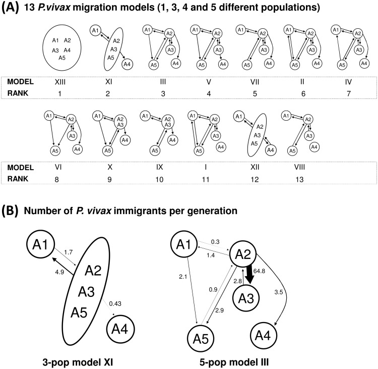
The gene flow was assessed evaluating parasite migration models which relied on the combined knowledge of the genetic structuring of the parasite population and the known human mobility patterns (Figs 1B and 7A). Thirteen migration models were evaluated through Bayesian analysis (marginal likelihoods and LBF of all the models tabulated in S4 Table). The high gene flow rates among all areas denoted the model XIII as the best model (log mL = -47467.4, prob>0.99), which describes a single panmictic population (random mating among the parasites from all the five areas) with an effective population size of 6,891 haplotypes (credibility interval 95% 4,144–9,640). Considering the genetic substructuring and asymmetric human mobilization observed for most of the areas which contrast with the panmictic model, the 2nd and 3rd best models which consider 3 and 5 populations with asymmetric migration were also explored in detail (Fig 7B). The 3-population model XI suggests A2, A3 and A5 as a panmictic unit with unequal migration among A1 to A2/3/5 and few parasite moving from A2/3/5 to A4 (Nm<1). The 5-population model III adds unidirectional gene flow from A1 to A5 which is in line with the phylogenetic results (Figs 5A and 7B). For Model III the highest rate of gene flow was found between the A2 and A3, areas which contain parasites with common genetic characteristics (PCoA and cluster analysis), have the highest multiplicity of infection rates compared to the other areas and is consistent with the current people’s mobilization patterns (Figs 1B, 2A, 3A and 7B). Other models were evaluated were A4 was treated as a fully isolated area or with bidirectional gene flow but these models had the lowest probabilities (Fig 7A and S4 Table). However, model V described unidirectional gene flow from A3 to A4 with a high number of migrants (Nm = 5.6) probably due to related parasite among A3 and A4.
Fig 7. Comparison of 13 parasite migration models and summary estimates of gene flow.
(A) The study areas were pooled as an unique population and in 3–5 populations. The arrows represent the direction of migration and the models are displayed by its rank order. The marginal log-likelihoods and LBF used to rank the models are tabulated in S4 Table. The model XIII (panmixia) has the highest model probability (>0.99) while all the rest have near zero probability. (B) The estimated number of immigrants per generation are described for the models XI and III. The thicknesses of the arrows are proportional to the number of immigrants. The Θ and M estimates for the models XIII, XI and III are tabulated in S5 Table.
Bottleneck
BOTTLENECK analysis using 10 polymorphic markers showed a significant number of microsatellites had an excess of He in areas A1, A2 and A3 under IAM (p<0.002) and TPM (only for A2 and A3, p<0.04), indicating a recent bottleneck event and a deficiency of He for A4 under SMM (p = 0.002), possibly indicating a rapid expansion (S3 Fig). However, A1 also presented contrasting He deficiency under SMM (p = 0.002). A5 was not included in the analysis (n = 4). Similar results were obtained when the analysis was performed separately for perfect and imperfect microsatellites as shown in S3 Fig The presence of excess and deficiency of He in A1 was further investigated by grouping the isolates by village (whenever the sample size was sufficiently large): excess of He in all villages was found but in addition deficit was also found in Manacamiri village (MA: IAM for imperfect MS: excess p = 0.008 and deficit p = 0.04; SMM for perfect MS: excess p = 0.06 and deficit p = 0.03).
Discussion
The present study provides a very comprehensive dataset on population genetics and gene flow analysis of P. vivax parasites circulating in the Peruvian Amazon. We determined and compared the genetic diversity and multiplicity of infection in five areas in and around Iquitos. We further unravelled the population structure by assessing the LD, genetic differentiation and determined the most likely number of genetic clusters and the genetic relationships among parasites. Multiple gene flow models were assessed determining the parasite migration patterns that may affect the genetic structuring. Moreover the occurrence of recent bottleneck events as result of recent malaria control programs were also explored. The findings support a sub-structured parasite population with a predominant clonal propagation and revealed that Iquitos city (A2) is the source of parasite spreading for all the other areas due to socio-economic patterns. Recent bottleneck events were found only in areas where intervention control programs were carried before we started the sample collection.
Multiplicity of infection
MOI, the average number of distinct parasites infecting an individual in a specific area, has been used as a proxy of malaria transmission [12], and it provides information about the configuration of the parasite population [10]. Overall, monoclonal P. vivax infections were the most frequent, confirming previous studies from the Peruvian Amazon [6, 14, 51]. Areas with some degree of isolation, such as areas A1 and A4, had mainly monoclonal infections, whereas the areas close to the Iquitos-Nauta road (A2 and A3) had polyclonal infections with 2 or 3 different clones and a similar proportion of monoclonal and polyclonal infections. This difference may be explained by a higher mobility rate of the people from A2 and A3 leading to a higher probability of being infected with distinct parasites. In areas with limited gene flow, A1 and A4, frequent mating between genetically identical or very related parasites may increase the odds that a person is re-infected with the same “clone”, which would result in a low MOI [52]. Moreover, the MOI will be affected by the patterns of hypnozoite activation where the probability of having a homologous activation may be higher in areas with one or few circulating clones compared to areas where unrelated clones are circulating.
Genetic diversity
Previously, varying levels of genetic diversity have been reported in endemic settings of the Peruvian Amazon revealing different transmission patterns [6, 14, 15, 51]. Multivariate analysis (AMOVA) indicated that the major source of the genetic variation was due to variation within villages instead of between areas. The greater genetic variation within the villages may be explained by the coexistence of different haplotypes within the villages as results of gene flow, genetic drift and/or a large hidden P. vivax reservoir but also by the high mutability of the microsatellites [53] or overestimation of the number of different haplotypes. The degree of polymorphism of the microsatellites or genotyping errors due to technical artefacts (false alleles) may influence the accuracy of defining the presence of one or more haplotypes within an infection [54].
In our study the levels of genetic diversity remained at intermediate levels with no substantial differences between areas. The high rates of gene flow found may have increased the levels of He. In the present study we reported coexistence of LD and extensive diversity also reported in other studies [55–57]. In the case of repeat-sequence in tandems like the microsatellites, the level of genetic polymorphism (‘genetic diversity’) may be maintained or increased due to the appearance of new alleles by mutational events during replication in the host cells without affecting the LD [55, 57]. Considering long-term P. vivax infections in the Peruvian Amazon the rate of allele mutations occurring within the host needs to be further explored. On the other hand, the extent of LD could be overestimated due to some of the recruited vivax patients were living in the same household [57]. The presence of LD and relatively high genetic diversity may be also favoured by a scenario where inbreeding of few sympatric divergent parasites is frequent [58].
LD and predominant clonal propagation
Similarly to other P. vivax populations from South America [11, 53, 58–62], the presence of LD and low MOI indicated a clonal propagation type in our study population. The low malaria transmission and/or the long-term vivax infections in the Peruvian Amazon may also favour the predominant clonal propagation [6]. Passive clonality due to restrained diversity and low gene flow, a different scenario to what we found in the present study, may not be the only type of clonal propagation occurring in the study areas. Tibayrenc and Ayala (2014) coined the ‘in-built, active clonality’ whereas P. falciparum despite the possibility of recombination with sympatric unrelated clones P. falciparum would prefer self-fertilization in order to gain biological and evolutionary adaptations to its environment [52]. The high rate of gene flow and high variability within the study villages drive us to consider that P. vivax may have also in-built, active clonal behaviour in order to take advantage during its adaptation to the environment/hosts. Further study is needed to verify the in-built clonal behaviour and its impact on the epidemiology.
Genetic relatedness
Genetic clustering approaches revealed the presence of at least two or three independent parasite clusters in our study population. We rerun STRUCTURE within one cluster of the cluster when K = 2 to: (1) look for hierarchical structure and; (2) to avoid misleading cluster assignment [37]. Sub-structured parasite population was confirmed in areas A1 and A4 and possibly misleading initial STRUCTURE assignment of the clusters (this is also graphically detected in Fig 5C). The cluster 1, which contained 18 isolates carrying the same haplotype, may influence the misleading assignment: STRUCTURE tends to group strongly related samples into one cluster and the rest of samples are assigned in a large cluster [37].
Little to moderate differentiation among the parasites was found in the urban areas A2, A3 and A5 (“urban cluster”) where continuous human mobilization and parasite gene flow on the Iquitos-Nauta Road occur. Conversely, different clusters of parasites were found circulating in the area A1 and A4 increasing the levels of genetic differentiation. In the San Carlos village (A4) was detected a group of parasites circulating only within this village (Fig 6). Major divergence of these parasites may have occurred due to the limited gene flow, genetic drift, bottleneck events, selection and/or recombination with imported parasites not being sampled in this study. The LD and the deficiency of He detected in A4 confirmed the rapid expansion of these clones with clustered transmission previously described after 2-year follow up [6].
P. vivax transmission in the Peruvian Amazon
Determining the transmission patterns is a priority for the implementation of control and elimination programs [63]. Our initial analysis revealed no genetic isolation of the study areas despite the geographic distance indicating that exist gene flow. Overall, few private alleles were found among areas where A3 and A5 had the lowest numbers supporting gene flow among the areas where A3 and A5 are beneficiated with more immigrants (parasites). However due to sample size bias on the calculation of private alleles we further used a Bayes approach where our sample size was not anymore an issue. We evaluated 13 migration models, using a Bayes approach based on the coalescence theory, which were proposed under the assumptions of current human mobilization and the genetic structuring data. The panmictic model was inferred as the best model, which is in line with the AMOVA results (less genetic differentiation among areas compared to within the areas). However, due to geographic constrains it is unlikely that people mobilizes to every area while parasites mate randomly. Possibly our genetic data was not strong enough to distinguish the best model: i.e. recent bottleneck events may affect the analysis [45]; however still we were able to recover two models that proposed a better explanation regarding the transmission dynamics and genetic structuring. Both models (XI and III) agreed that area A2 (for model XI: A2/3/5) is the source of parasites spreading for all the other areas: A2 comprises Iquitos city, place where people from A1 and A3 transmutes to every day, crossing the Nanay river by boat or going by car/bus/motorbike through the Iquitos-Nauta Road, for economic activities (Figs 1A and 1B and 7B). The influx of people from A1 to A2 is drives most of the parasite influx among these areas where people from A1 may be infected in A2 then importing parasites from A1 to A2 when they return to their households. Model XI described a panmixia between A2, A3 and A while the model III showed that most of migration occurs between A2 and A3, favoured by the proximity and the road (no geographic barrier) in line with the shared genetic parasite characteristics found previously. In line to the phylogenetic analysis, the model III described that A5 have influx of parasites from A2 which could occur during the visit of people from A5 to A1 on weekends to sell their products in the markets of Iquitos. Model III also described influx of parasites from A1 to A5 despite the Nanay river isolate both areas. High rates of parasite migration from A2 to A3 and A5 is in line with low (or none) number of private alleles recorded for A3 and A5. Regarding to area A4, is it known that occasionally people from A4 (especially from San Carlos) travels through the Itaya river to the A2 to sell their products and that may explain the importation of parasites from A2 to A4. As mentioned before, some parasites from A4 are related to parasites from A3, which in turn are highly related to A2 parasites. The relatedness of A4 and A3 parasites (Network analysis) and migration of parasites from A3 to A4 (referred only in model IV) may have occur by unknown human migration patterns or events where the vector mobilizes among these areas or genetic divergence of A3 parasites into the current A4 parasites. We have documented parasite transmission from A2 to the other areas but not significant immigration of parasites towards A2 which explains why A2 has the smallest population size (number of haplotypes) compared to the other areas (S5 Table) and why we still found significant pairwise genetic differentiation against A1 and A4. In addition to the low rate of immigrants to A2, recent bottleneck events may have also negatively affected the effective population size in A2.
Bottleneck events after intervention program
By describing the parasite structure, genetic diversity and dynamics, population genetics can also contribute in assessing the impact of an intervention [10]. Before our sample collection in 2008, the PAMAFRO project which involved campaigns of malaria prevention and control program with active case detection and treatment as well as distribution of insecticide-treated mosquito nets was carried out in Loreto (including all our study areas except for area A4), resulting in a 49% drop of the incidence of clinical vivax malaria from 2005 to 2008 [20]. The expected impact of the intervention on the parasite population besides lowering the malaria incidence would be a reduction of the effective parasite population size, the so called bottleneck effect. Since no data prior to the intervention on the effective parasite population size were available, we performed a retrospective analysis looking for recent bottleneck effect. A parasite population having experienced a recent bottleneck shows a faster decline in the number of alleles compared to a He reduction because rare alleles will be lost with little influence on the He [47]. The predominant clonal propagation found in this study did not affect the Bottleneck analysis since moderate He and significant intra-area genetic variation were found. Only areas with n>15 isolates (S3 Fig) were used for the analysis to increase the resolution power as described by Luikart et al. [64]. In this study, bottleneck events were detected in all areas where control interventions were implemented. San Carlos village did not benefit of any control activity before the sample collection, and no bottleneck but rather a rapid clonal expansion was observed. This is the first report of bottleneck events for P. vivax population in the Peruvian Amazon following the implementation of prevention and control activities. Noteworthy that the reduction of malaria cases in Peru lasted only until 2011 coinciding with the finalization of the PAMAFRO project and since then there has been an steady increase of malaria cases: i.e. Loreto region reported 11,779 vivax malaria cases in 2011 and 60,566 in 2014 [3, 65]. Further and continuous monitoring of the population structure and dynamics of the parasite population is necessary to understand the factors that are involved in the evolution of malaria in the Peruvian Amazon. The detection of recent bottlenecks in the parasite population could be used as complementary tool to measure the efficacy and impact of malaria control programs.
In conclusion, we have elucidated the population genetics of Plasmodium vivax in a large geographical area in and around Iquitos, the main socio-economic capital city of the Peruvian Amazon. We have shown the use of a Bayes approach to infer the gene flow pattern among our study areas and the detection of the reduction of the population size as a result of a control program. The knowledge about the routes of malaria transmission (gene flow) and the effect of control policies on the parasite population is a priority from the public health perspective as well for the formulation of future control policies and assessment of current control/elimination strategies.
Supporting Information
. The number of isolates is described for each village.
(PDF)
The allelic size range is described in base pairs (bp). In this table, the level of polymorphism of each locus was described as the expected heterozygosity (He) calculated for each marker using pooled isolates from all areas. The efficiency of amplification was calculated as the percentage of isolates with positive PCR amplification for a specific locus over the total number of analysed isolates (n = 292).
(PDF)
(PDF)
The models were constructed considering 5, 3 and 1 populations evaluating from 1 to 14 combined parameters (Θ and M) using MIGRATE-N.
(PDF)
(PDF)
Mantel test for Matrix Correspondence was performed using GenAlEx (1) to test the occurrence of a positive correlation (Rxy>0) between the genetic PHIPT matrix and geographic distances, so called the Isolation-by-distance (IBD) hypothesis. The genetic and geographic distance were not correlated (Rxy = -0.41 p = 0.17).
(PDF)
(PDF)
(PDF)
Acknowledgments
We thank all P. vivax patients for their enthusiastic participation in the study as well as all field workers for their dedication in the cohort follow-up. We also thank the journal editor and three anonymous reviewers for their critical comments of the text. We also acknowledge the generous support of Peter Beerli (University of Florida) providing active coaching for the MIGRATE-N nalysis during the MBL course held Woods Hole, MA, US in July 2015 and later on.
Data Availability
All relevant data are within the paper and its Supporting Information files.
Funding Statement
The sample collection was funded by the Directorate General for Development Cooperation (DGCD) of the Belgian Government (framework agreement 2 and 3; project 95501 and 95502 respectively): http://diplomatie.belgium.be/en/policy/development_cooperation/. The funders had no role in study design, data collection and analysis, decision to publish, or preparation of the manuscript.
References
- 1.WHO. World Malaria Report 2014. Switzerland: World Health Organization, 2014. [Google Scholar]
- 2.Gething PW, Elyazar IR, Moyes CL, Smith DL, Battle KE, Guerra CA, et al. A long neglected world malaria map: Plasmodium vivax endemicity in 2010. PLoS Negl Trop Dis. 2012;6(9):e1814 10.1371/journal.pntd.0001814 [DOI] [PMC free article] [PubMed] [Google Scholar]
- 3.Chapilliquén F. Situación epidemiológica de la malaria (a la SE 53), Perú 2014. Lima, Peru: Ministery of Health of Peru, 2015. [Google Scholar]
- 4.Branch O, Casapia WM, Gamboa DV, Hernandez JN, Alava FF, Roncal N, et al. Clustered local transmission and asymptomatic Plasmodium falciparum and Plasmodium vivax malaria infections in a recently emerged, hypoendemic Peruvian Amazon community. Malar J. 2005;4:27 Epub 2005/06/25. [DOI] [PMC free article] [PubMed] [Google Scholar]
- 5.Van den Eede P, Soto-Calle VE, Delgado C, Gamboa D, Grande T, Rodriguez H, et al. Plasmodium vivax sub-patent infections after radical treatment are common in Peruvian patients: results of a 1-year prospective cohort study. PLoS One. 2011;6(1):e16257 Epub 2011/02/08. 10.1371/journal.pone.0016257 [DOI] [PMC free article] [PubMed] [Google Scholar]
- 6.Delgado-Ratto C, Soto-Calle V, Van den Eede P, Gamboa D, Rosas A, Abatih E, et al. Population structure and spatio-temporal transmission dynamics of Plasmodium vivax after radical cure treatment in a rural village of the Peruvian Amazon. Malaria Journal. 2014;13(1):8. [DOI] [PMC free article] [PubMed] [Google Scholar]
- 7.Griffing SM, Gamboa D, Udhayakumar V. The history of 20th century malaria control in Peru. Malar J. 2013;12:303 10.1186/1475-2875-12-303 [DOI] [PMC free article] [PubMed] [Google Scholar]
- 8.Graf PC, Durand S, Alvarez Antonio C, Montalvan C, Galves Montoya M, Green MD, et al. Failure of Supervised Chloroquine and Primaquine Regimen for the Treatment of Plasmodium vivax in the Peruvian Amazon. Malaria research and treatment. 2012;2012:936067 10.1155/2012/936067 [DOI] [PMC free article] [PubMed] [Google Scholar]
- 9.Volkman SK, Ndiaye D, Diakite M, Koita OA, Nwakanma D, Daniels RF, et al. Application of genomics to field investigations of malaria by the international centers of excellence for malaria research. Acta Trop. 2012;121(3):324–32. 10.1016/j.actatropica.2011.12.002 [DOI] [PMC free article] [PubMed] [Google Scholar]
- 10.Arnott A, Barry AE, Reeder JC. Understanding the population genetics of Plasmodium vivax is essential for malaria control and elimination. Malar J. 2012;11:14 10.1186/1475-2875-11-14 [DOI] [PMC free article] [PubMed] [Google Scholar]
- 11.Chenet SM, Schneider KA, Villegas L, Escalante AA. Local population structure of Plasmodium: impact on malaria control and elimination. Malar J. 2012;11(1):412. Epub 2012/12/13. [DOI] [PMC free article] [PubMed] [Google Scholar]
- 12.Volkman SK, Neafsey DE, Schaffner SF, Park DJ, Wirth DF. Harnessing genomics and genome biology to understand malaria biology. Nat Rev Genet. 2012;13(5):315–28. 10.1038/nrg3187 [DOI] [PubMed] [Google Scholar]
- 13.Carlton JM, Das A, Escalante AA. Genomics, population genetics and evolutionary history of Plasmodium vivax. Adv Parasitol. 2013;81:203–22. 10.1016/B978-0-12-407826-0.00005-9 [DOI] [PubMed] [Google Scholar]
- 14.Van den Eede P, Van der Auwera G, Delgado C, Huyse T, Soto-Calle VE, Gamboa D, et al. Multilocus genotyping reveals high heterogeneity and strong local population structure of the Plasmodium vivax population in the Peruvian Amazon. Malar J. 2010;9:151 Epub 2010/06/08. 10.1186/1475-2875-9-151 [DOI] [PMC free article] [PubMed] [Google Scholar]
- 15.Kosek M, Yori PP, Gilman RH, Calderon M, Zimic M, Chuquiyauri R, et al. High degree of Plasmodium vivax diversity in the Peruvian Amazon demonstrated by tandem repeat polymorphism analysis. Am J Trop Med Hyg. 2012;86(4):580–6. 10.4269/ajtmh.2012.11-0627 [DOI] [PMC free article] [PubMed] [Google Scholar]
- 16.McCollum AM, Soberon V, Salas CJ, Santolalla ML, Udhayakumar V, Escalante AA, et al. Genetic variation and recurrent parasitaemia in Peruvian Plasmodium vivax populations. Malar J. 2014;13(1):67. [DOI] [PMC free article] [PubMed] [Google Scholar]
- 17.Erhart A, Sotto-Calle V, Delgado-Ratto C, Contreras-Mancilla J, Torres E, Sanchez-Martinez L, et al. Plasmodium vivax morbidity after radical cure: a two-year cohort study in the Peruvian Amazon 63th Annual meeting American Society of Tropical Medicine and Hygiene; Philadelphia, Pennsylvania, USA: 2014. p. 291. [Google Scholar]
- 18.Aramburu Guarda J, Ramal Asayag C, Witzig R. Malaria reemergence in the Peruvian Amazon region. Emerg Infect Dis. 1999;5(2):209–15. Epub 1999/04/30. [DOI] [PMC free article] [PubMed] [Google Scholar]
- 19.Roshanravan B, Kari E, Gilman RH, Cabrera L, Lee E, Metcalfe J, et al. Endemic malaria in the Peruvian Amazon region of Iquitos. Am J Trop Med Hyg. 2003;69(1):45–52. Epub 2003/08/23. [PubMed] [Google Scholar]
- 20.ORAS-CONHU. Compartiendo Lecciones Aprendidas. Proyecto control de la malaria en las zonas fronterizas de la region andina: Un enfoque comunitario-PAMAFRO. Lima, Peru: Organismo Andino de la Salud—Convenio Hipólito Unanue, 2009. [Google Scholar]
- 21.Gutierrez S, Arróspide N. Manual de Procedimientos de Laboratorio para el diagnóstico de Malaria. Ministerio de Salud del Perú—Instituto Nacional de Salud; 2003. p. 7–14. [Google Scholar]
- 22.Plowe CV, Djimde A, Bouare M, Doumbo O, Wellems TE. Pyrimethamine and proguanil resistance-conferring mutations in Plasmodium falciparum dihydrofolate reductase: polymerase chain reaction methods for surveillance in Africa. Am J Trop Med Hyg. 1995;52(6):565–8. Epub 1995/06/01. [DOI] [PubMed] [Google Scholar]
- 23.Rubio JM, Post RJ, van Leeuwen WM, Henry MC, Lindergard G, Hommel M. Alternative polymerase chain reaction method to identify Plasmodium species in human blood samples: the semi-nested multiplex malaria PCR (SnM-PCR). Trans R Soc Trop Med Hyg. 2002;96 Suppl 1:S199–204. Epub 2002/06/12.. [DOI] [PubMed] [Google Scholar]
- 24.Karunaweera ND, Ferreira MU, Hartl DL, Wirth DF. Fourteen polymorphic microsatellite DNA markers for the human malaria parasite Plasmodium vivax. Molecular Ecology Notes. 2007;7(1):172–5. [Google Scholar]
- 25.Van den Eede P, Erhart A, Van der Auwera G, Van Overmeir C, Thang ND, Hung le X, et al. High complexity of Plasmodium vivax infections in symptomatic patients from a rural community in central Vietnam detected by microsatellite genotyping. Am J Trop Med Hyg. 2010;82(2):223–7. Epub 2010/02/06. 10.4269/ajtmh.2010.09-0458 [DOI] [PMC free article] [PubMed] [Google Scholar]
- 26.Peakall ROD, Smouse PE. genalex 6: genetic analysis in Excel. Population genetic software for teaching and research. Molecular Ecology Notes. 2006;6(1):288–95. [DOI] [PMC free article] [PubMed] [Google Scholar]
- 27.Peakall R, Smouse P. GenAlEx 6.5: Genetic analysis in Excel. Population genetic software for teaching and research–an update. Bioinformatics. 2012. [DOI] [PMC free article] [PubMed] [Google Scholar]
- 28.Anderson TJ, Su XZ, Bockarie M, Lagog M, Day KP. Twelve microsatellite markers for characterization of Plasmodium falciparum from finger-prick blood samples. Parasitology. 1999;119 (Pt 2):113–25. Epub 1999/08/31. [DOI] [PubMed] [Google Scholar]
- 29.Orjuela-Sanchez P, Sa JM, Brandi MC, Rodrigues PT, Bastos MS, Amaratunga C, et al. Higher microsatellite diversity in Plasmodium vivax than in sympatric Plasmodium falciparum populations in Pursat, Western Cambodia. Exp Parasitol. 2013;134(3):318–26. 10.1016/j.exppara.2013.03.029 [DOI] [PMC free article] [PubMed] [Google Scholar]
- 30.Goudet J. FSTAT (Version 1.2): A Computer Program to Calculate F-Statistics. J Hered. 1995;86(6):485–6. [Google Scholar]
- 31.Haubold B, Hudson RR. LIAN 3.0: detecting linkage disequilibrium in multilocus data. Linkage Analysis. Bioinformatics. 2000;16(9):847–8. Epub 2000/12/08. [DOI] [PubMed] [Google Scholar]
- 32.Thanapongpichat S, McGready R, Luxemburger C, Day N, White N, Nosten F, et al. Microsatellite genotyping of Plasmodium vivax infections and their relapses in pregnant and non-pregnant patients on the Thai-Myanmar border. Malaria Journal. 2013;12(1):275. [DOI] [PMC free article] [PubMed] [Google Scholar]
- 33.Smith JM, Smith NH, O'Rourke M, Spratt BG. How clonal are bacteria? Proceedings of the National Academy of Sciences. 1993;90(10):4384–8. [DOI] [PMC free article] [PubMed] [Google Scholar]
- 34.Feil EJ, Li BC, Aanensen DM, Hanage WP, Spratt BG. eBURST: inferring patterns of evolutionary descent among clusters of related bacterial genotypes from multilocus sequence typing data. J Bacteriol. 2004;186(5):1518–30. Epub 2004/02/20. [DOI] [PMC free article] [PubMed] [Google Scholar]
- 35.Evanno G, Regnaut S, Goudet J. Detecting the number of clusters of individuals using the software STRUCTURE: a simulation study. Mol Ecol. 2005;14(8):2611–20. Epub 2005/06/23. [DOI] [PubMed] [Google Scholar]
- 36.Earl D, vonHoldt B. STRUCTURE HARVESTER: a website and program for visualizing STRUCTURE output and implementing the Evanno method. Conservation Genetics Resources. 2012;4(2):359–61. [Google Scholar]
- 37.Kalinowski ST. The computer program STRUCTURE does not reliably identify the main genetic clusters within species: simulations and implications for human population structure. Heredity (Edinb). 2011;106(4):625–32. [DOI] [PMC free article] [PubMed] [Google Scholar]
- 38.Jakobsson M, Rosenberg NA. CLUMPP: a cluster matching and permutation program for dealing with label switching and multimodality in analysis of population structure. Bioinformatics. 2007;23(14):1801–6. [DOI] [PubMed] [Google Scholar]
- 39.Rosenberg NA. DISTRUCT: a program for the graphical display of population structure. Molecular Ecology Notes. 2004;4(1):137–8. [Google Scholar]
- 40.Meirmans PG, Van Tienderen PH. GENOTYPE and GENODIVE: two programs for the analysis of genetic diversity of asexual organisms. Molecular Ecology Notes. 2004;4(4):792–4. [Google Scholar]
- 41.Meirmans PG. AMOVA-Based Clustering of Population Genetic Data. Journal of Heredity. 2012;103(5):744–50. 10.1093/jhered/ess047 [DOI] [PubMed] [Google Scholar]
- 42.Francisco A, Vaz C, Monteiro P, Melo-Cristino J, Ramirez M, Carrico J. PHYLOViZ: phylogenetic inference and data visualization for sequence based typing methods. BMC Bioinformatics. 2012;13(1):87. [DOI] [PMC free article] [PubMed] [Google Scholar]
- 43.Beerli P. Comparison of Bayesian and maximum-likelihood inference of population genetic parameters. Bioinformatics. 2006;22(3):341–5. [DOI] [PubMed] [Google Scholar]
- 44.Beerli P, Palczewski M. Unified framework to evaluate panmixia and migration direction among multiple sampling locations. Genetics. 2010;185(1):313–26. 10.1534/genetics.109.112532 [DOI] [PMC free article] [PubMed] [Google Scholar]
- 45.Beerli P, Beerli P. How to use MIGRATE or why are Markov chain Monte Carlo programs difficult to use? Population Genetics for Animal Conservation: Cambridge University Press; 2009. [Google Scholar]
- 46.Anderson TJ, Su XZ, Roddam A, Day KP. Complex mutations in a high proportion of microsatellite loci from the protozoan parasite Plasmodium falciparum. Mol Ecol. 2000;9(10):1599–608. Epub 2000/10/26. [DOI] [PubMed] [Google Scholar]
- 47.Cornuet JM, Luikart G. Description and power analysis of two tests for detecting recent population bottlenecks from allele frequency data. Genetics. 1996;144(4):2001–14. [DOI] [PMC free article] [PubMed] [Google Scholar]
- 48.Piry S, Luikart G, Cornuet J-M. BOTTLENECK: A Computer Program for Detecting Recent Reductions in the Effective Population Size Using Allele Frequency Data. 1999. [Google Scholar]
- 49.Luikart G, Cornuet JM. Empirical evaluation of a test for identifying recently bottlenecked populations from allele frequency data. Conserv Biol. 1998;12(1):228–37. [Google Scholar]
- 50.Cristescu R, Sherwin WB, Handasyde K, Cahill V, Cooper DW. Detecting bottlenecks using BOTTLENECK 1.2.02 in wild populations: the importance of the microsatellite structure. Conservation Genetics. 2009;11(3):1043–9. [Google Scholar]
- 51.Sutton PL, Neyra V, Hernandez JN, Branch OH. Plasmodium falciparum and Plasmodium vivax infections in the Peruvian Amazon: propagation of complex, multiple allele-type infections without super-infection. Am J Trop Med Hyg. 2009;81(6):950–60. Epub 2009/12/10. 10.4269/ajtmh.2009.09-0132 [DOI] [PMC free article] [PubMed] [Google Scholar]
- 52.Tibayrenc M, Ayala FJ. New insights into clonality and panmixia in Plasmodium and toxoplasma. Adv Parasitol. 2014;84:253–68. 10.1016/B978-0-12-800099-1.00005-3 [DOI] [PubMed] [Google Scholar]
- 53.Batista CL, Barbosa S, DASB M, Viana SA, Ferreira MU. Genetic diversity of Plasmodium vivax over time and space: a community-based study in rural Amazonia. Parasitology. 2014:1–11. [DOI] [PubMed] [Google Scholar]
- 54.Sutton PL. A call to arms: on refining Plasmodium vivax microsatellite marker panels for comparing global diversity. Malar J. 2013;12:447 10.1186/1475-2875-12-447 [DOI] [PMC free article] [PubMed] [Google Scholar]
- 55.Karunaweera ND, Ferreira MU, Munasinghe A, Barnwell JW, Collins WE, King CL, et al. Extensive microsatellite diversity in the human malaria parasite Plasmodium vivax. Gene. 2008;410(1):105–12. Epub 2008/01/30. 10.1016/j.gene.2007.11.022 [DOI] [PubMed] [Google Scholar]
- 56.Orjuela-Sanchez P, Karunaweera ND, da Silva-Nunes M, da Silva NS, Scopel KK, Goncalves RM, et al. Single-nucleotide polymorphism, linkage disequilibrium and geographic structure in the malaria parasite Plasmodium vivax: prospects for genome-wide association studies. BMC genetics. 2010;11:65 10.1186/1471-2156-11-65 [DOI] [PMC free article] [PubMed] [Google Scholar]
- 57.Gunawardena S, Ferreira MU, Kapilananda GM, Wirth DF, Karunaweera ND. The Sri Lankan paradox: high genetic diversity in Plasmodium vivax populations despite decreasing levels of malaria transmission. Parasitology. 2014;141(7):880–90. 10.1017/S0031182013002278 [DOI] [PMC free article] [PubMed] [Google Scholar]
- 58.Ferreira MU, Karunaweera ND, da Silva-Nunes M, da Silva NS, Wirth DF, Hartl DL. Population structure and transmission dynamics of Plasmodium vivax in rural Amazonia. J Infect Dis. 2007;195(8):1218–26. Epub 2007/03/16. [DOI] [PubMed] [Google Scholar]
- 59.Orjuela-Sanchez P, da Silva NS, da Silva-Nunes M, Ferreira MU. Recurrent parasitemias and population dynamics of Plasmodium vivax polymorphisms in rural Amazonia. Am J Trop Med Hyg. 2009;81(6):961–8. Epub 2009/12/10. 10.4269/ajtmh.2009.09-0337 [DOI] [PubMed] [Google Scholar]
- 60.Koepfli C, Rodrigues PT, Antao T, Orjuela-Sanchez P, Van den Eede P, Gamboa D, et al. Plasmodium vivax Diversity and Population Structure across Four Continents. PLoS Negl Trop Dis. 2015;9(6):e0003872 10.1371/journal.pntd.0003872 [DOI] [PMC free article] [PubMed] [Google Scholar]
- 61.Menegon M, Durand P, Menard D, Legrand E, Picot S, Nour B, et al. Genetic diversity and population structure of Plasmodium vivax isolates from Sudan, Madagascar, French Guiana and Armenia. Infect Genet Evol. 2014;27C:244–9. [DOI] [PubMed] [Google Scholar]
- 62.Imwong M, Nair S, Pukrittayakamee S, Sudimack D, Williams JT, Mayxay M, et al. Contrasting genetic structure in Plasmodium vivax populations from Asia and South America. Int J Parasitol. 2007;37(8–9):1013–22. Epub 2007/04/20. [DOI] [PubMed] [Google Scholar]
- 63.Barry AE, Waltmann A, Koepfli C, Barnadas C, Mueller I. Uncovering the transmission dynamics of Plasmodium vivax using population genetics. Pathogens and global health. 2015;109(3):142–52. 10.1179/2047773215Y.0000000012 [DOI] [PMC free article] [PubMed] [Google Scholar]
- 64.Luikart G, Sherwin WB, Steele BM, Allendorf FW. Usefulness of molecular markers for detecting population bottlenecks via monitoring genetic change. Mol Ecol. 1998;7(8):963–74. [DOI] [PubMed] [Google Scholar]
- 65.Mateo Lizarbe S. ¿Re-emergencia de la malaria en el Perú?: Reflexiones sobre el control de la malaria. Lima, Peru: Ministery of Health of Peru, 2013. [Google Scholar]
Associated Data
This section collects any data citations, data availability statements, or supplementary materials included in this article.
Supplementary Materials
. The number of isolates is described for each village.
(PDF)
The allelic size range is described in base pairs (bp). In this table, the level of polymorphism of each locus was described as the expected heterozygosity (He) calculated for each marker using pooled isolates from all areas. The efficiency of amplification was calculated as the percentage of isolates with positive PCR amplification for a specific locus over the total number of analysed isolates (n = 292).
(PDF)
(PDF)
The models were constructed considering 5, 3 and 1 populations evaluating from 1 to 14 combined parameters (Θ and M) using MIGRATE-N.
(PDF)
(PDF)
Mantel test for Matrix Correspondence was performed using GenAlEx (1) to test the occurrence of a positive correlation (Rxy>0) between the genetic PHIPT matrix and geographic distances, so called the Isolation-by-distance (IBD) hypothesis. The genetic and geographic distance were not correlated (Rxy = -0.41 p = 0.17).
(PDF)
(PDF)
(PDF)
Data Availability Statement
All relevant data are within the paper and its Supporting Information files.


