

Abstract
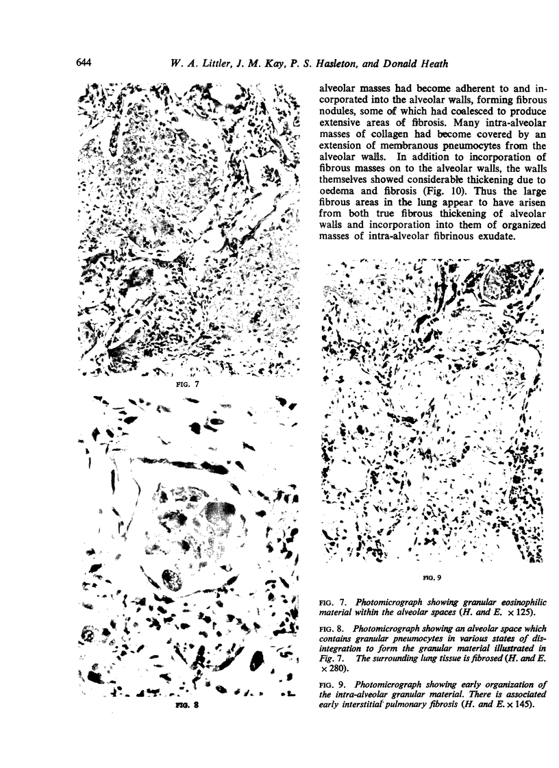
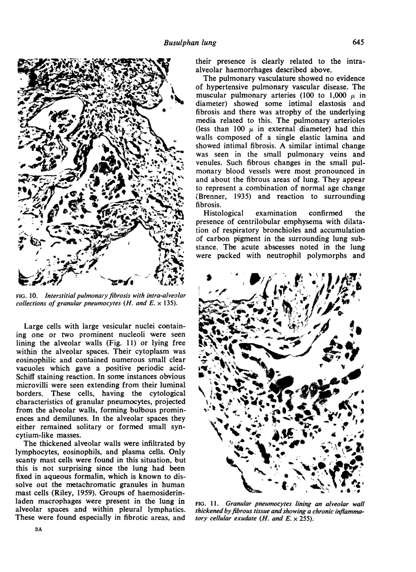
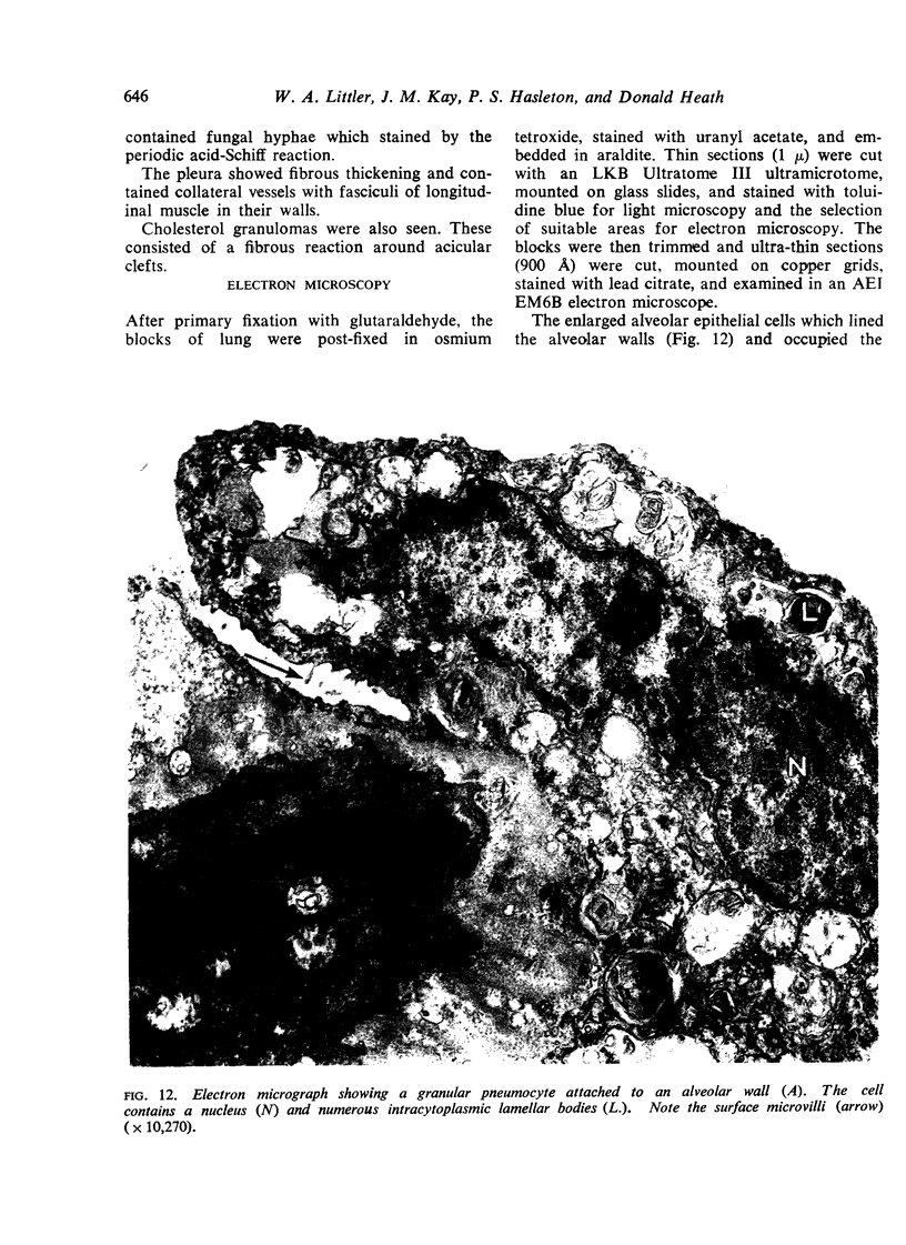
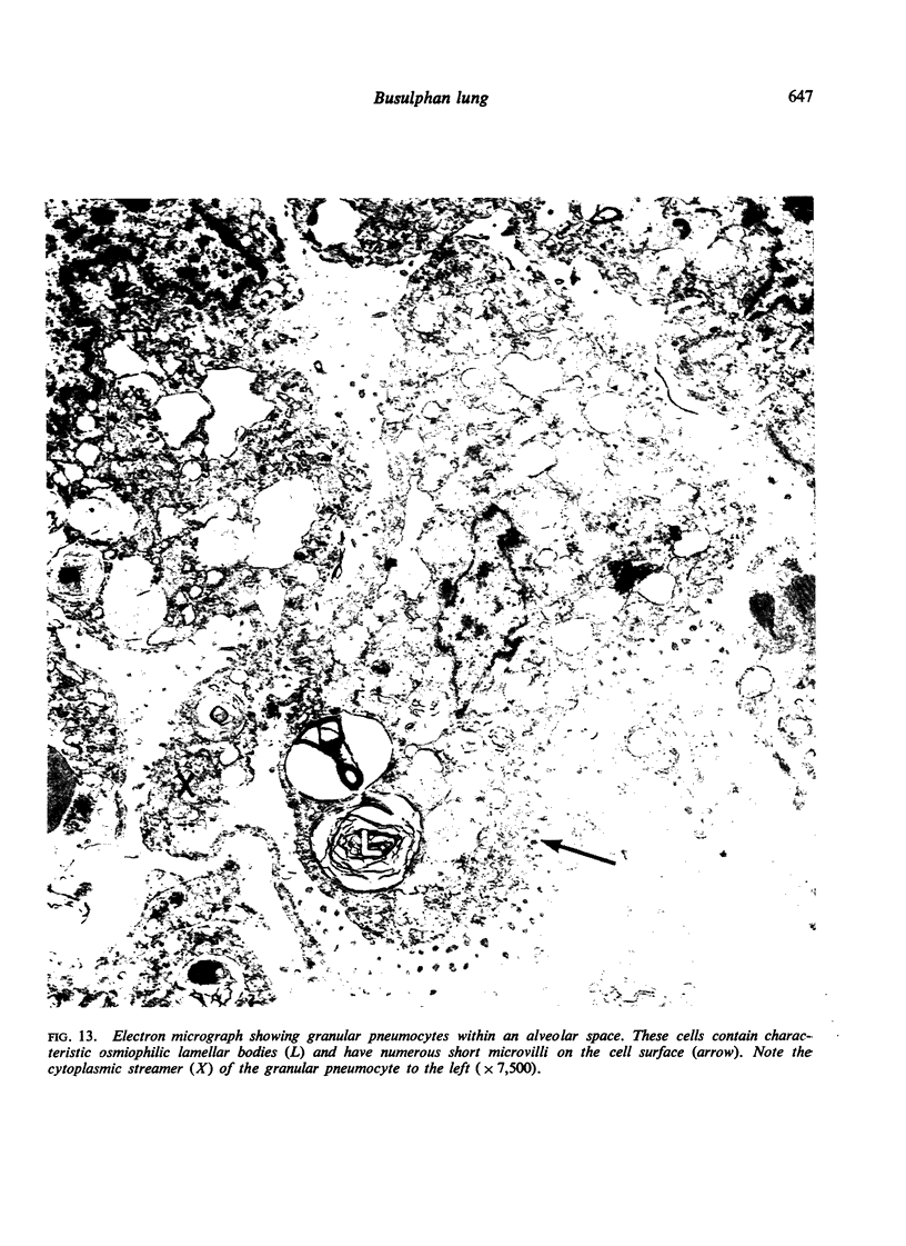
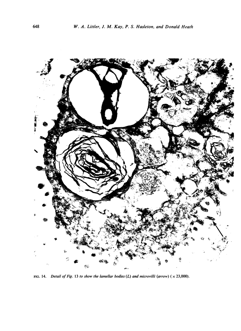
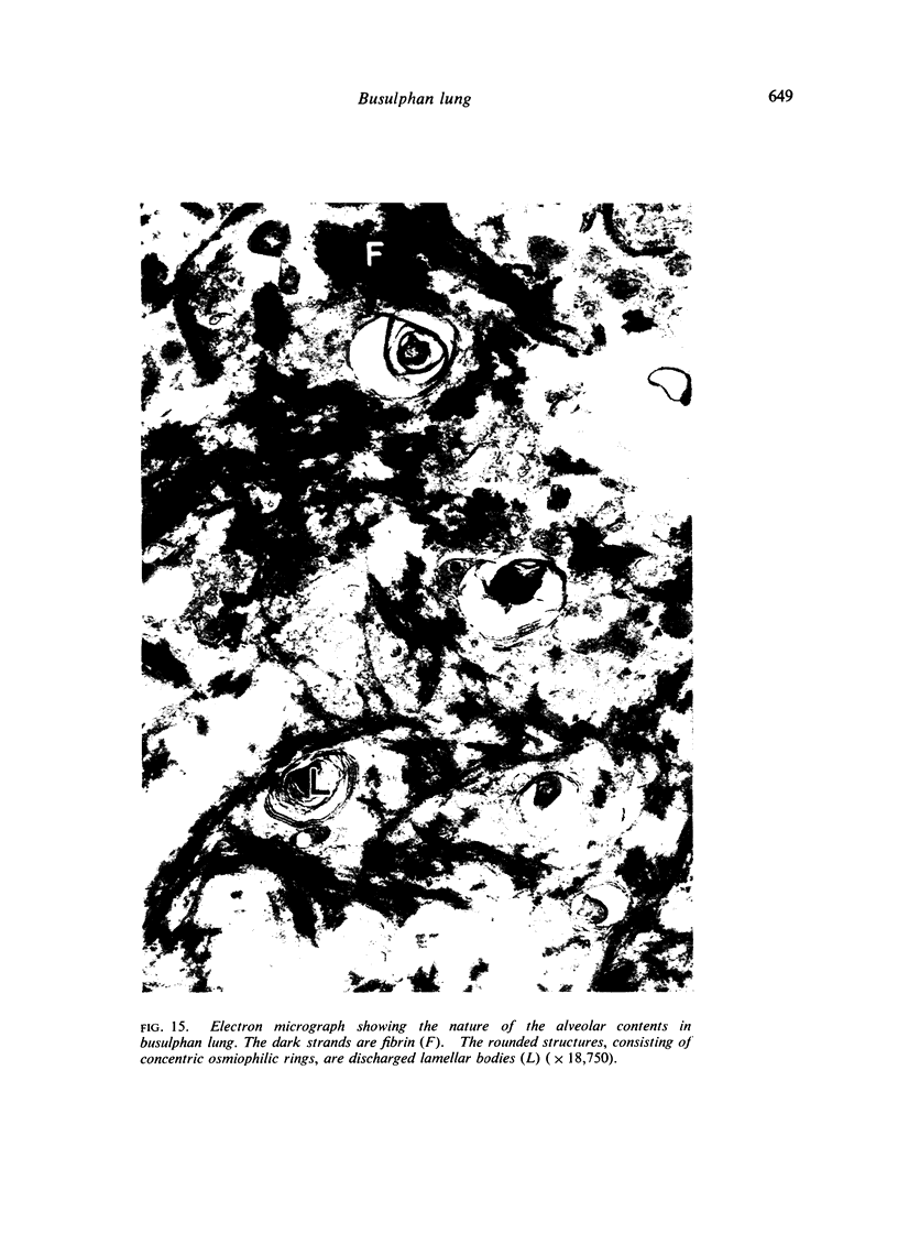
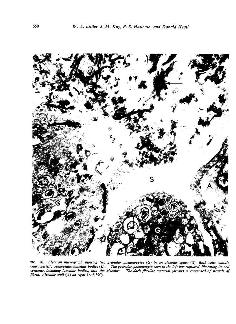
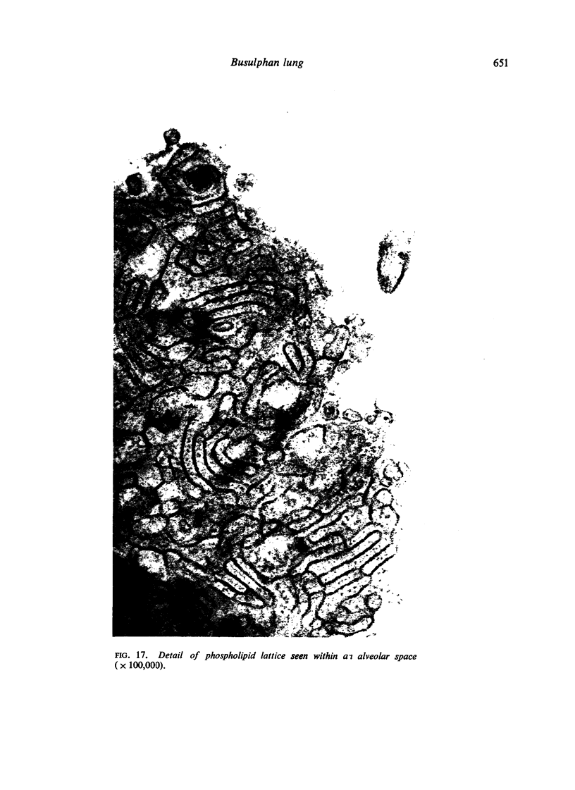
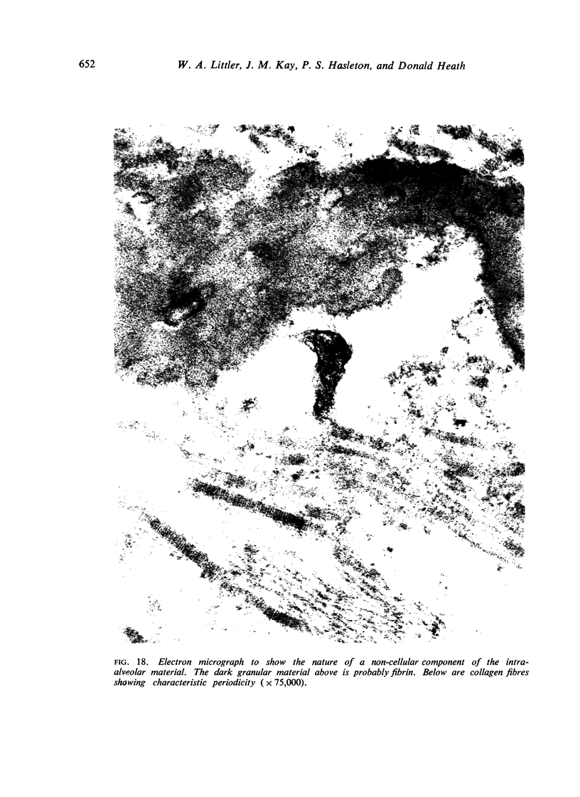
A 61-year-old man with chronic myeloid leukaemia was treated with busulphan (Myleran). After receiving 1 g. of this drug over a period of 20 months he became dyspnoeic and developed crepitations in the lungs. Two months later radiographs of the chest revealed peri-hilar infiltrates and subsequently diffuse mottling throughout both lungs. Lung function tests showed a gross impairment of the transfer factor to a quarter of the predicted normal. At necropsy the lungs showed a striking proliferation of granular pneumocytes, many of which had disintegrated to produce intra-alveolar debris, some of which showed organization by fibrous tissue. There was associated interstitial pulmonary fibrosis. Electron microscopy confirmed the desquamated alveolar cells to be type II (granular) pneumocytes containing characteristic lamellar bodies. Many of these osmiophilic bodies, believed to be the source of pulmonary surfactant, had been liberated into the alveolar spaces, with the formation of phospholipid myelin figures and lattices. We think that the basic pathology of busulphan lung is a chemically induced alveolitis with proliferation of granular pneumocytes followed by fibrosis of alveolar walls and intra-alveolar contents.
Full text
PDF
Images in this article


Selected References
These references are in PubMed. This may not be the complete list of references from this article.
- BLAKEMORE W. S., FORSTER R. E., MORTON J. W., OGILVIE C. M. A standardized breath holding technique for the clinical measurement of the diffusing capacity of the lung for carbon monoxide. J Clin Invest. 1957 Jan;36(1 Pt 1):1–17. doi: 10.1172/JCI103402. [DOI] [PMC free article] [PubMed] [Google Scholar]
- Brewer D. B., Heath D., Asquith P. Electron microscopy of desquamative interstitial pneumonia. J Pathol. 1969 Feb;97(2):317–323. doi: 10.1002/path.1710970217. [DOI] [PubMed] [Google Scholar]
- FULTON R. M., HUTCHINSON E. C., JONES A. M. Ventricular weight in cardiac hypertrophy. Br Heart J. 1952 Jul;14(3):413–420. doi: 10.1136/hrt.14.3.413. [DOI] [PMC free article] [PubMed] [Google Scholar]
- Glancy D. L., Frazier P. D., Roberts W. C. Pulmonary parenchymal cholesterol-ester granulomas in patients with pulmonary hypertension. Am J Med. 1968 Aug;45(2):198–210. doi: 10.1016/0002-9343(68)90038-7. [DOI] [PubMed] [Google Scholar]
- Heard B. E., Cooke R. A. Busulphan lung. Thorax. 1968 Mar;23(2):187–193. doi: 10.1136/thx.23.2.187. [DOI] [PMC free article] [PubMed] [Google Scholar]
- Kay J. M., Smith P., Heath D. Electron microscopy of Crotalaria pulmonary hypertension. Thorax. 1969 Sep;24(5):511–526. doi: 10.1136/thx.24.5.511. [DOI] [PMC free article] [PubMed] [Google Scholar]
- LEAKE E., SMITH W. G. DIFFUSE INTERSTITIAL PULMONARY FIBROSIS AFTER BUSULPHAN THERAPY. Lancet. 1963 Aug 31;2(7305):432–434. doi: 10.1016/s0140-6736(63)92173-1. [DOI] [PubMed] [Google Scholar]
- LIEBOW A. A., STEER A., BILLINGSLEY J. G. DESQUAMATIVE INTERSTITIAL PNEUMONIA. Am J Med. 1965 Sep;39:369–404. doi: 10.1016/0002-9343(65)90206-8. [DOI] [PubMed] [Google Scholar]
- LUCY J. A., GLAUERT A. M. STRUCTURE AND ASSEMBLY OF MACROMOLECULAR LIPID COMPLEXES COMPOSED OF GLOBULAR MICELLES. J Mol Biol. 1964 May;8:727–748. doi: 10.1016/s0022-2836(64)80121-2. [DOI] [PubMed] [Google Scholar]
- OLINER H., SCHWARTZ R., RUBIO F., DAMESHEK W. Interstitial pulmonary fibrosis following busulfan therapy. Am J Med. 1961 Jul;31:134–139. doi: 10.1016/0002-9343(61)90229-7. [DOI] [PubMed] [Google Scholar]
- Smalley R. V., Wall R. L. Two cases of busulfan toxicity. Ann Intern Med. 1966 Jan;64(1):154–164. doi: 10.7326/0003-4819-64-1-154. [DOI] [PubMed] [Google Scholar]