Abstract
Since alveolar and airway growth have previously been studied in greater detail than has arterial growth, the present study establishes the growth pattern of the arteries and relates this to that of the alveoli and airways.
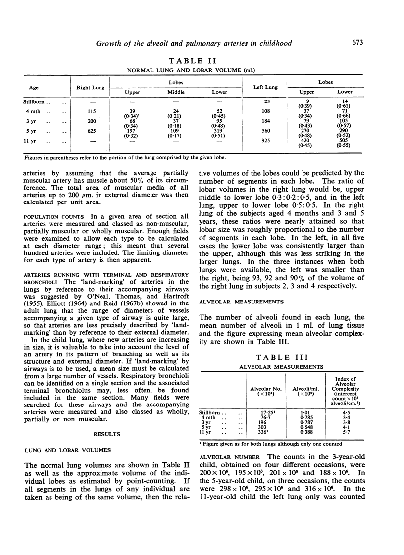
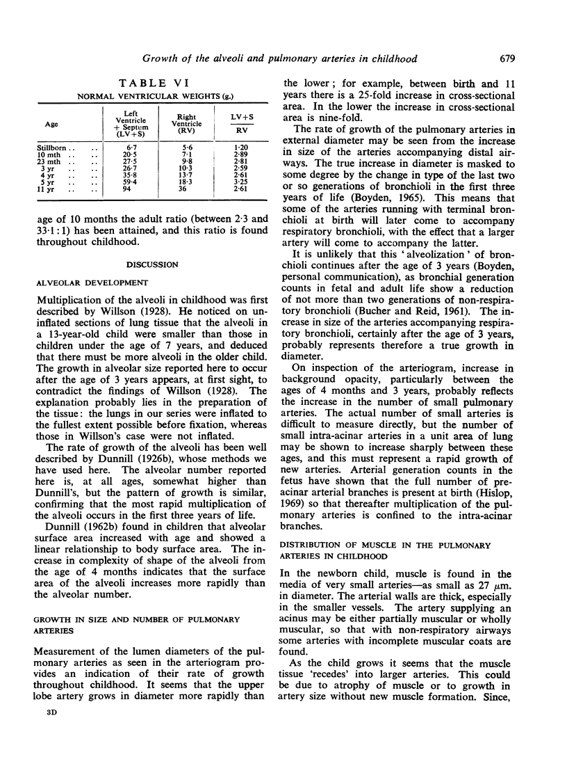
The pattern of post-natal alveolar multiplication found is similar to that reported by Dunnill, but for all ages about 10% above his figure. Alveolar size hardly changes in the first three or four years of life but thereafter increases steadily. The complexity of alveolar shape increases from the age of 4 months.
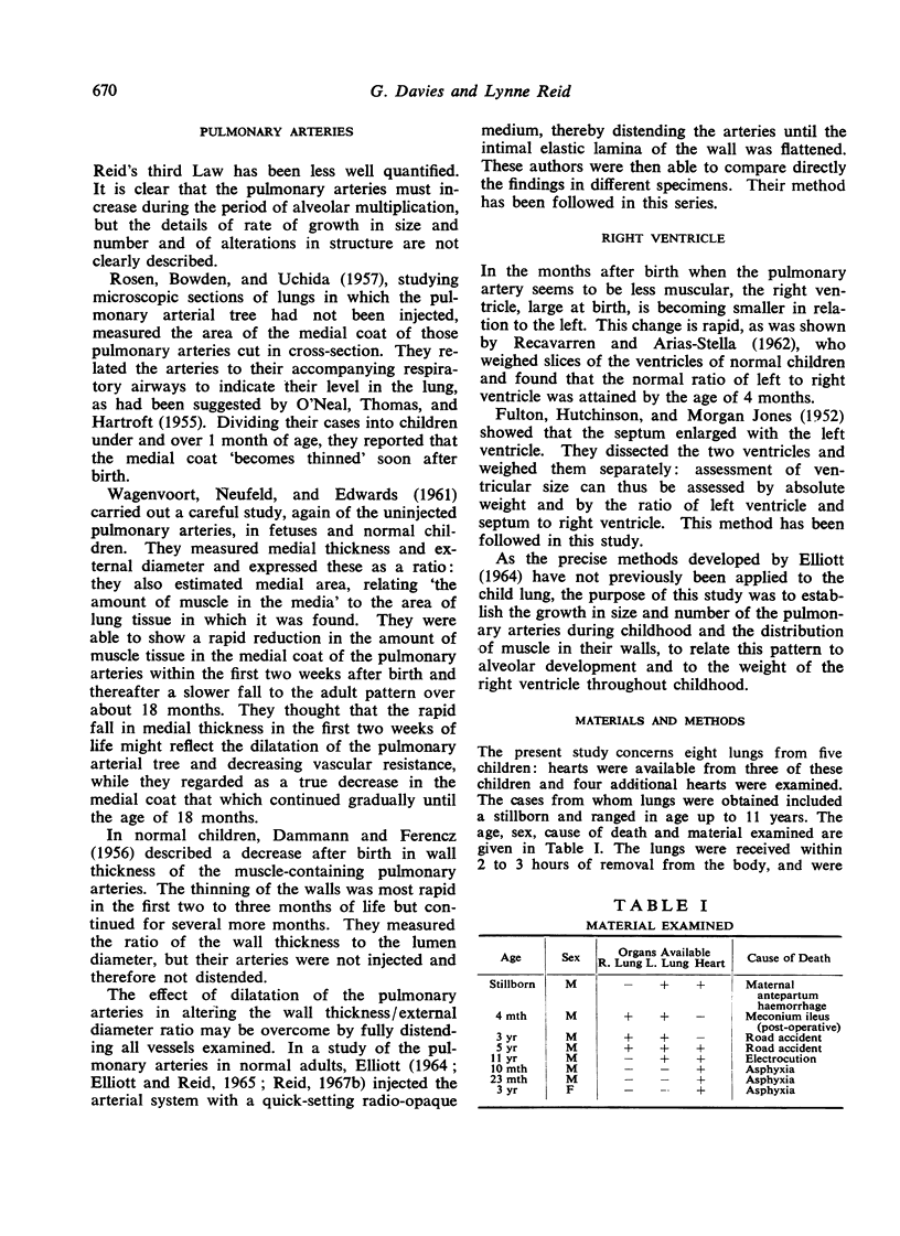
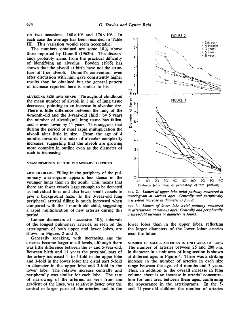
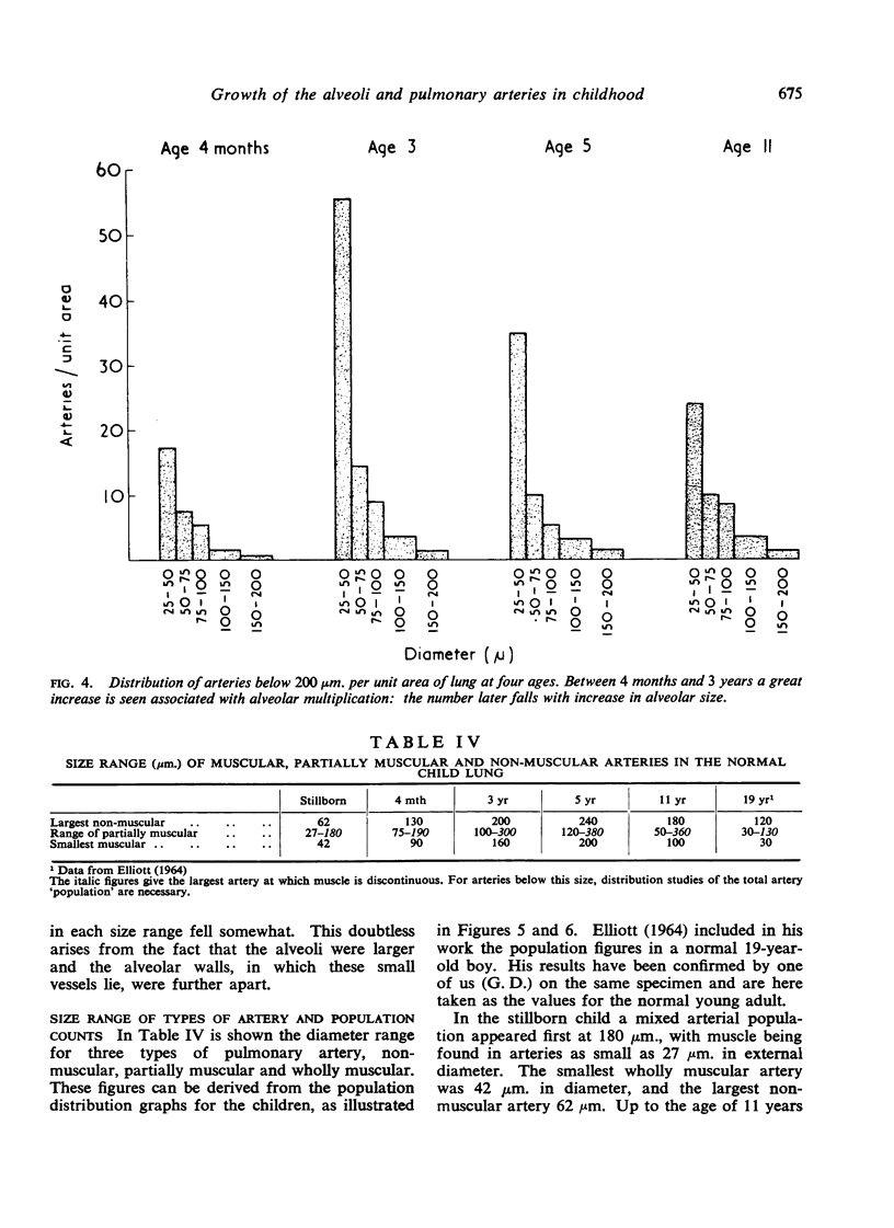
In arteriograms the arterial lumen, both centrally and peripherally, increases 4- to 5-fold in the upper lobe, 3-fold in the lower. Microscopic examination shows a striking increase between 4 months and 3 years in the number of arteries below 200 μm. per unit area of lung tissue; after 5 years this number falls slightly, probably reflecting an increase in alveolar size.
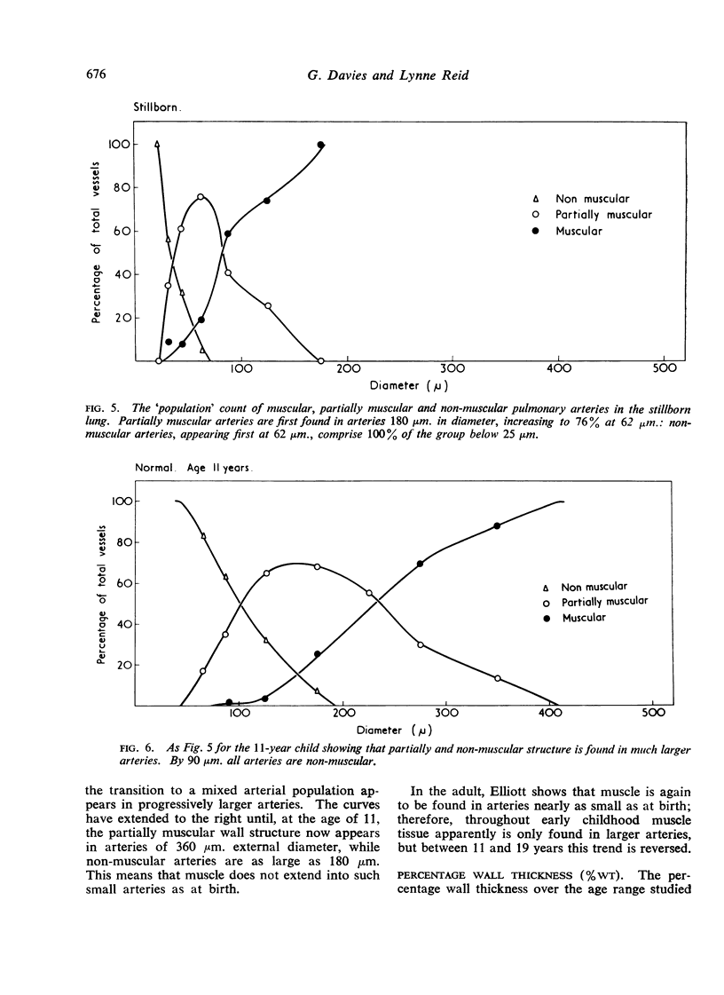
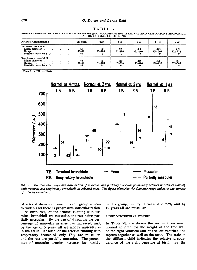
Muscular, partially muscular and non-muscular arteries show a similar distribution over the same size range in the newborn as in the adult. During childhood, however, the arteries over this size range are free of muscle and much larger arteries show this mixed `population'. Between 11 and 19 years the adult pattern forms by progressive penetration of muscle into the acinus.
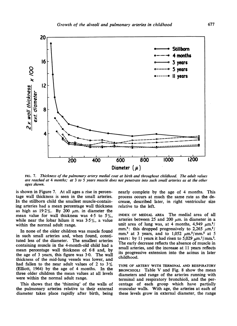
The thickness of the muscle coat of pulmonary artery branches falls to adult levels by the age of 4 months. Between birth and 5 years the medial area of arteries between 25 and 200 μm. in diameter per unit area of lung falls, but has risen again by 11 years.
Full text
PDF

Images in this article
Selected References
These references are in PubMed. This may not be the complete list of references from this article.
- BOYDEN E. A. THE TERMINAL AIR SACS AND THEIR BLOOD SUPPLY IN A 37-DAY INFANT LUNG. Am J Anat. 1965 Mar;116:413–428. doi: 10.1002/aja.1001160207. [DOI] [PubMed] [Google Scholar]
- BUCHER U., REID L. Development of the intrasegmental bronchial tree: the pattern of branching and development of cartilage at various stages of intra-uterine life. Thorax. 1961 Sep;16:207–218. doi: 10.1136/thx.16.3.207. [DOI] [PMC free article] [PubMed] [Google Scholar]
- DAMMANN J. F., Jr, FERENCZ C. The significance of the pulmonary vascular bed in congenital heart disease. I. Normal lungs. II. Malformations of the heart in which there is pulmonary stenosis. Am Heart J. 1956 Jul;52(1):7–17. doi: 10.1016/0002-8703(56)90115-6. [DOI] [PubMed] [Google Scholar]
- ELLIOTT F. M., REID L. SOME NEW FACTS ABOUT THE PULMONARY ARTERY AND ITS BRANCHING PATTERN. Clin Radiol. 1965 Jul;16:193–198. doi: 10.1016/s0009-9260(65)80042-3. [DOI] [PubMed] [Google Scholar]
- FULTON R. M., HUTCHINSON E. C., JONES A. M. Ventricular weight in cardiac hypertrophy. Br Heart J. 1952 Jul;14(3):413–420. doi: 10.1136/hrt.14.3.413. [DOI] [PMC free article] [PubMed] [Google Scholar]
- O'NEAL R. M., THOMAS W. A., HARTROFT P. M. The media of small muscular pulmonary arteries in mitral stenosis. AMA Arch Pathol. 1955 Sep;60(3):267–270. [PubMed] [Google Scholar]
- RECAVARREN S., ARIAS-STELLA J. Topography of right ventricular hypertrophy in children native to high altitude. Am J Pathol. 1962 Oct;41:467–475. [PMC free article] [PubMed] [Google Scholar]
- WAGENVOORT C. A. Vasoconstriction and medial hypertrophy in pulmonary hypertension. Circulation. 1960 Oct;22:535–546. doi: 10.1161/01.cir.22.4.535. [DOI] [PubMed] [Google Scholar]
- WEIBEL E. R., GOMEZ D. M. A principle for counting tissue structures on random sections. J Appl Physiol. 1962 Mar;17:343–348. doi: 10.1152/jappl.1962.17.2.343. [DOI] [PubMed] [Google Scholar]


