Abstract
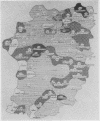
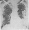
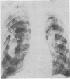
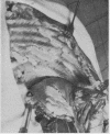
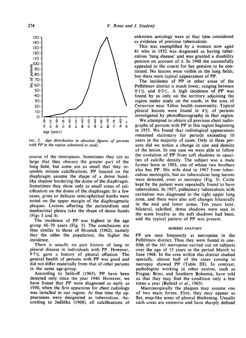
Pleural plaques were found in 644 (6·6%) of 9,760 photofluorograms taken in 1965 in a region of Pelhřimov district; the incidence was highest in the age group 66-70 years. The advanced age of those affected may be explained by the greater frequency of the causative agent in the past. The disorder was known in Pelhřimov district as early as 1930; it was then thought to be posttuberculous. The past history of the cases was uninformative; as a rule, the only common previous disease was pleurisy with effusion, occurring in 9·7%. The general condition of those affected was excellent; only 8% were aware of the fact that pleural lesions were present. The disorder was found mainly in farmers, familial incidence was common, and if two generations of one family suffered from the condition, the older generation was affected in 100%.
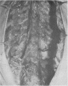
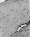
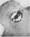
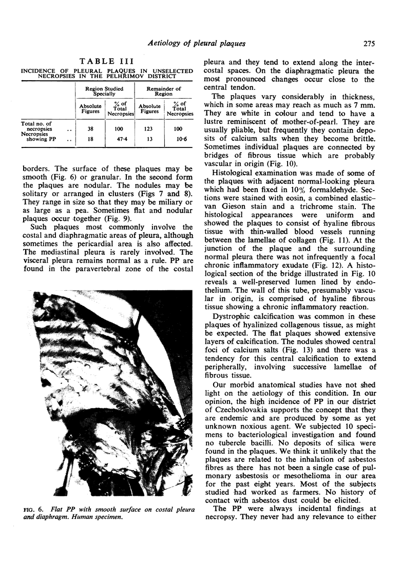
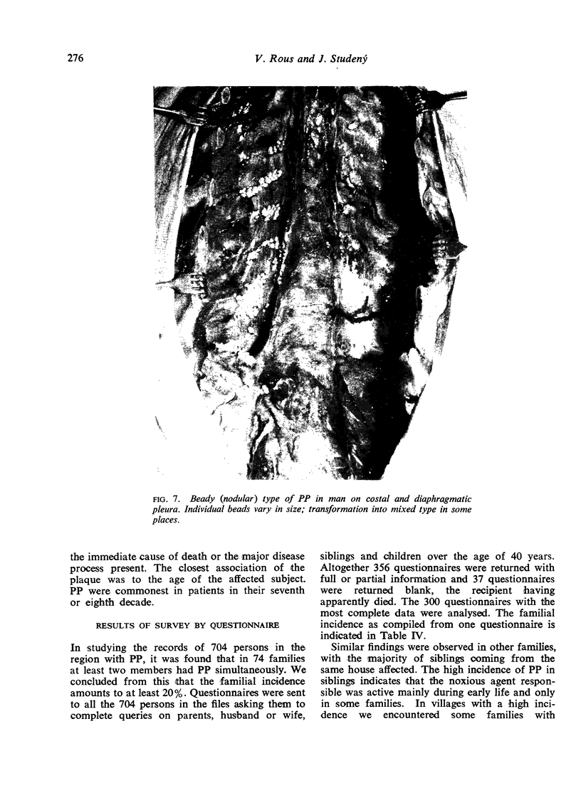
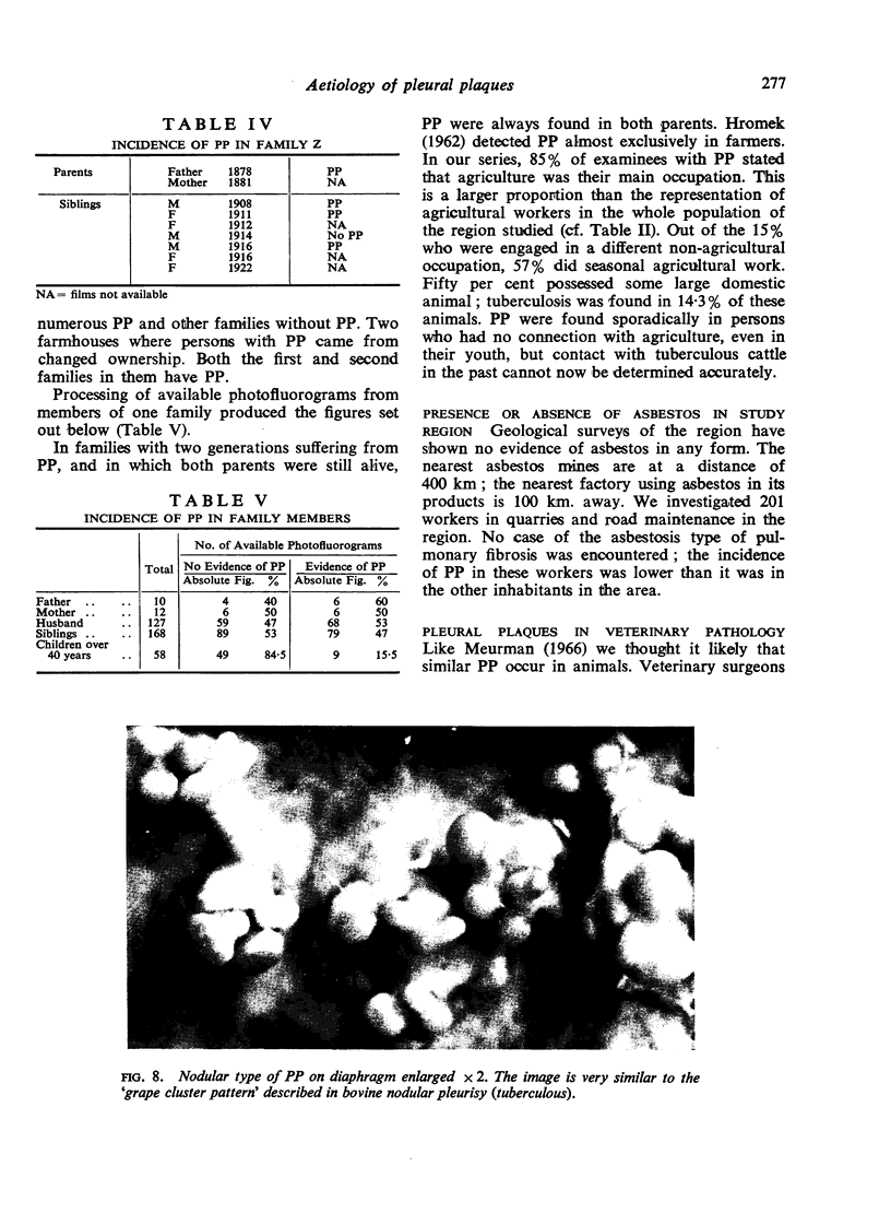
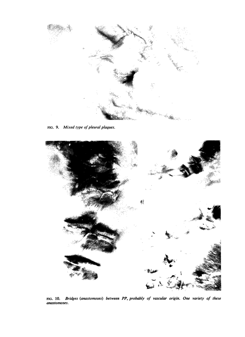
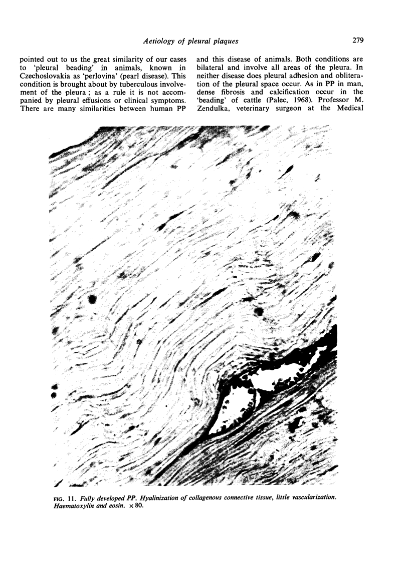
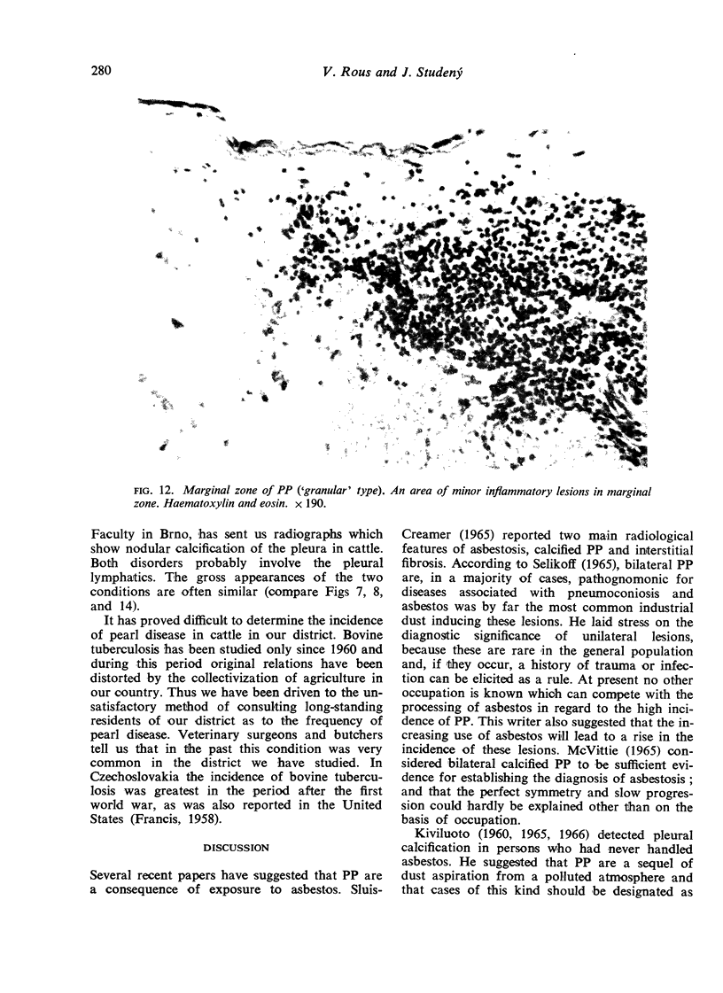
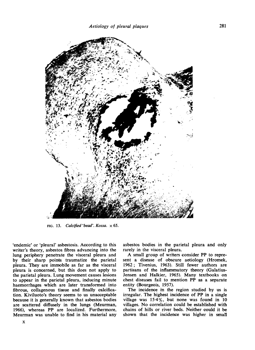
Pleural plaques consist morphologically of limited areas of hyalinized collagenous connective tissue with calcium salt deposits. Tubercle bacilli could not be cultivated from the lesions. Mineralological analysis showed no evidence of silicates in the pleural plaques and a normal content in the lungs.
The aetiological factor responsible for the development of pleural plaques in Pelhřimov district is not known, but asbestos cannot be implicated. The unknown noxious agent is carried to the pleura by the lymph and blood stream. Pleural plaques are an endemic disorder. The traditional view that lesions are post-tuberculous appears, in the region submitted to this study, to be a possible explanation.
Full text
PDF
Images in this article
Selected References
These references are in PubMed. This may not be the complete list of references from this article.
- ANSPACH M. [Are pleural calcifications pathognomic for an asbestosis?]. Int Arch Gewerbepathol Gewerbehyg. 1962;19:108–120. [PubMed] [Google Scholar]
- Galatius-Jensen F., Halkier E. Radiological aspects of pleural hyalo-serositis. Br J Radiol. 1965 Dec;38(456):944–946. doi: 10.1259/0007-1285-38-456-944. [DOI] [PubMed] [Google Scholar]
- Hourihane D. O., Lessof L., Richardson P. C. Hyaline and calcified pleural plaques as an index of exposure to asbestos. Br Med J. 1966 Apr 30;1(5495):1069–1074. doi: 10.1136/bmj.1.5495.1069. [DOI] [PMC free article] [PubMed] [Google Scholar]
- MARSOVA D. BEITRAG ZUR ATIOLOGIE DER PLEURAVERKALKUNGEN. Z Tuberk Erkr Thoraxorg. 1964 Jun;121:329–334. [PubMed] [Google Scholar]
- McVittie J. C. Asbestosis in Great Britain. Ann N Y Acad Sci. 1965 Dec 31;132(1):128–138. doi: 10.1111/j.1749-6632.1965.tb41096.x. [DOI] [PubMed] [Google Scholar]
- Selikoff I. J. The occurrence of pleural calcification among asbestos insulation workers. Ann N Y Acad Sci. 1965 Dec 31;132(1):351–367. doi: 10.1111/j.1749-6632.1965.tb41116.x. [DOI] [PubMed] [Google Scholar]
- Sluis-Cremer G. K. Asbestosis in South Africa--certain geographical and environmental considerations. Ann N Y Acad Sci. 1965 Dec 31;132(1):215–234. doi: 10.1111/j.1749-6632.1965.tb41103.x. [DOI] [PubMed] [Google Scholar]
- TIVENIUS L. Benign pleural lesions simulating tumour. Thorax. 1963 Mar;18:39–44. doi: 10.1136/thx.18.1.39. [DOI] [PMC free article] [PubMed] [Google Scholar]


