Abstract
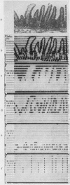
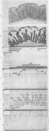
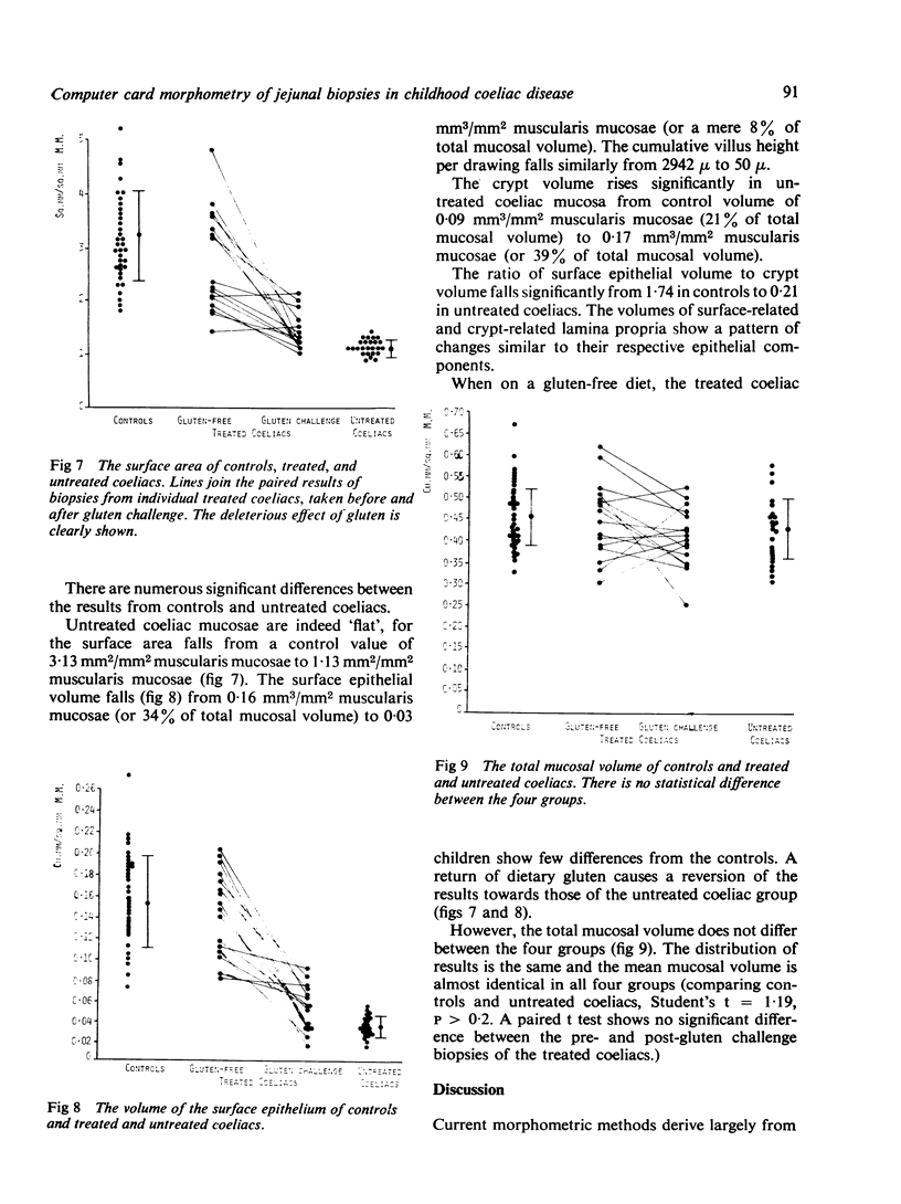
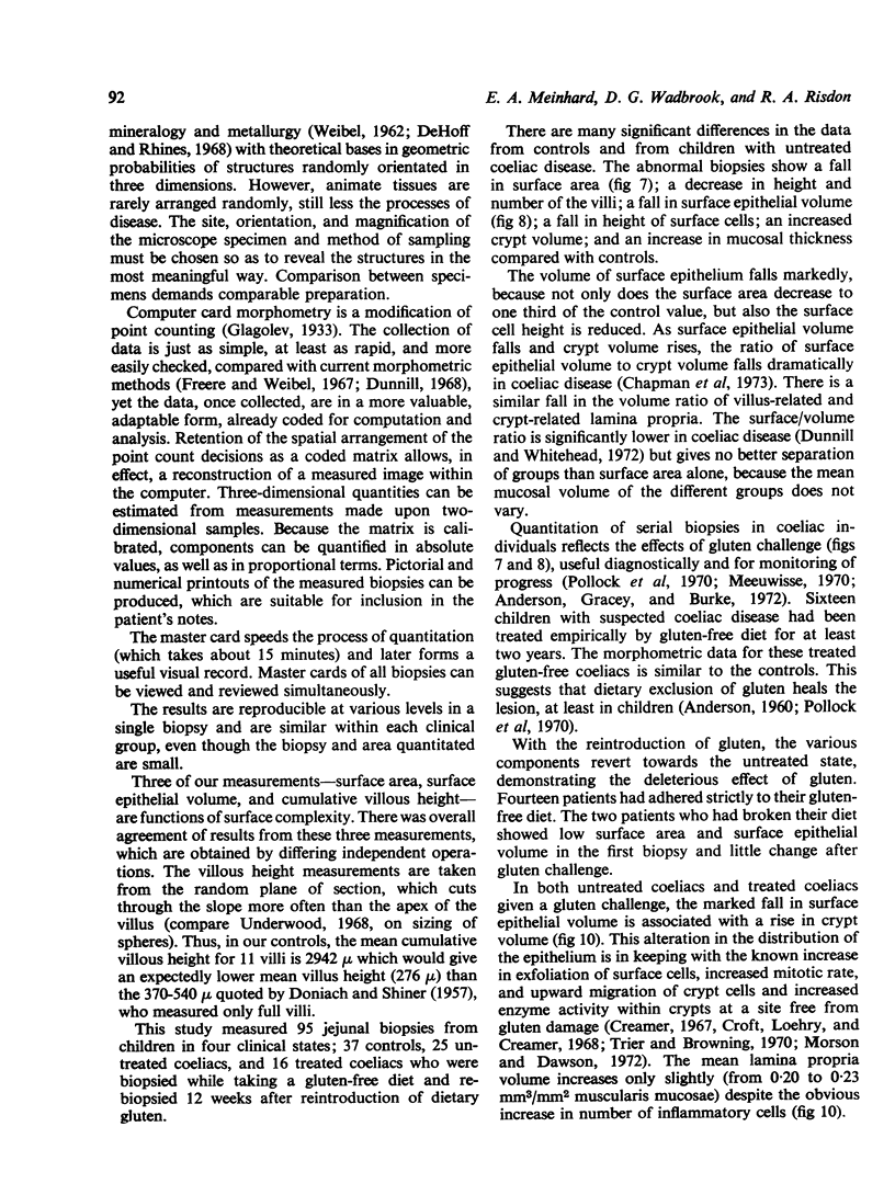
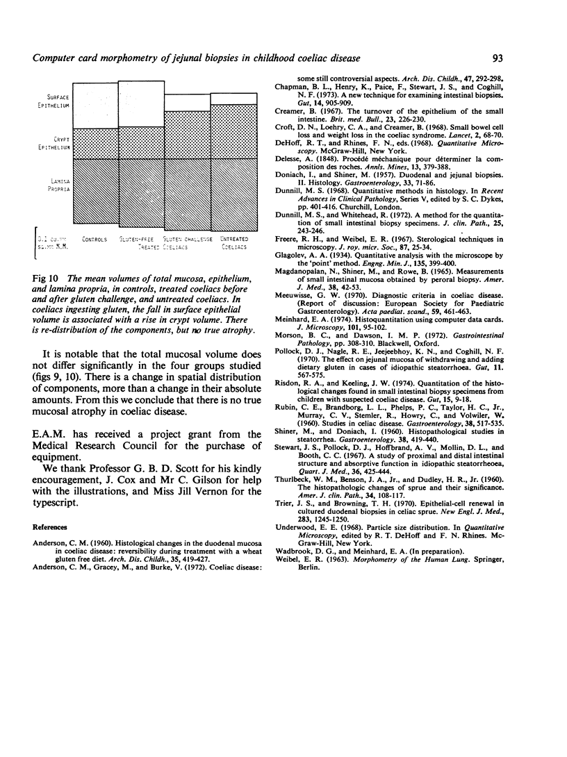
The histological changes in 95 jejunal biopsy specimens from children have been analyzed by a new mporphometric technique. The microscope image of the specimen is traced directly onto computer data cards. A simple sketch records accurate quantitative data in a matrix of 840 points, retaining the spatial arrangement of the tissue components. The data are fed via an optical mark data card reader, into a mini-computer. FORTRAN IV programs allow calculation of surface area, villous heights, and component volumes in metric units, and of volume proportions, volume-to-volume ratios, and surface-to-volume ratios. Pictorial and numerical printouts are produced, which are suitable for inclusion in the patient's notes. Jejunal biopsies from 37 controls and 26 untreated coeliac patients were clearly distinguished morphometrically. Sixteen pairs of biopsies from coeliac patients on long-term gluten-free diets before, and 12 weeks after, the reintroduction of dietary gluten significantly reflected the effects of gluten challenge. Comparison of control and abnormal biopsies showed a spatial redistribution of the components, more than a change in their absolute amounts. There was no significant differences in the total epithelial volumes in controls, treated or untreated patients, suggesting that the mucosal lesion in coeliac disease is not a true atrophy.
Full text
PDF
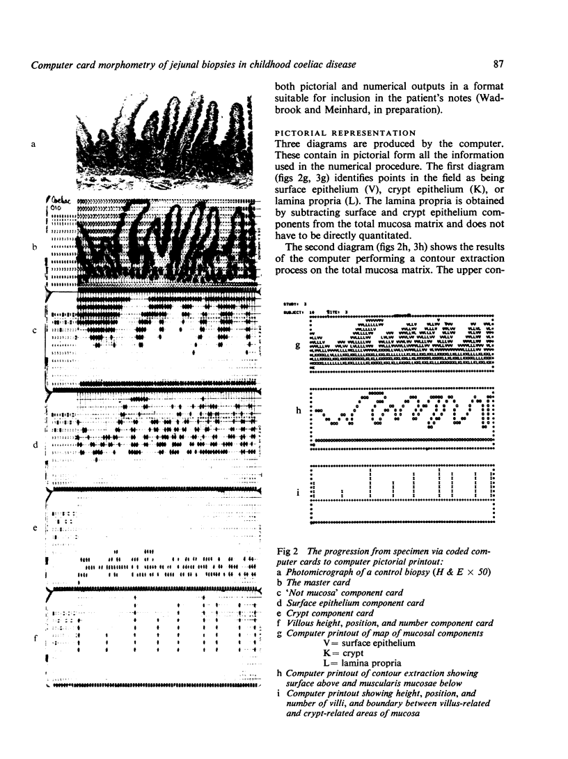
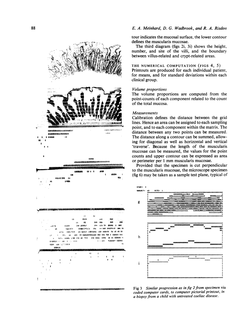
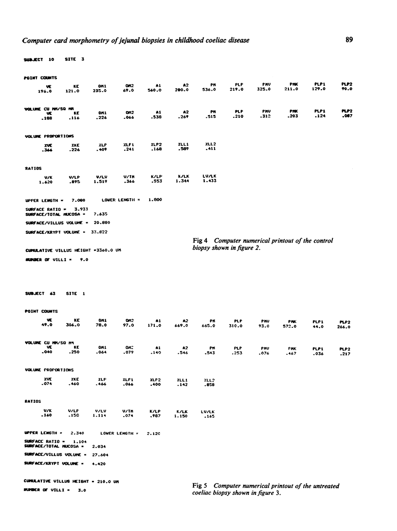
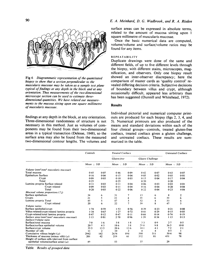
Images in this article
Selected References
These references are in PubMed. This may not be the complete list of references from this article.
- ANDERSON C. M. Histological changes in the duodenal mucosa in coeliac disease. Reversibility during treatment with a wheat gluten free diet. Arch Dis Child. 1960 Oct;35:419–427. doi: 10.1136/adc.35.183.419. [DOI] [PMC free article] [PubMed] [Google Scholar]
- Anderson C. M., Gracey M., Burke V. Coeliac disease. Some still controversial aspects. Arch Dis Child. 1972 Apr;47(252):292–298. doi: 10.1136/adc.47.252.292. [DOI] [PMC free article] [PubMed] [Google Scholar]
- Chapman B. L., Henry K., Paice F., Stewart J. S., Coghill N. F. A new technique for examining intestinal biopsies. Gut. 1973 Nov;14(11):905–909. doi: 10.1136/gut.14.11.905. [DOI] [PMC free article] [PubMed] [Google Scholar]
- Creamer B. The turnover of the epithelium of the small intestine. Br Med Bull. 1967 Sep;23(3):226–230. doi: 10.1093/oxfordjournals.bmb.a070561. [DOI] [PubMed] [Google Scholar]
- DONIACH I., SHINER M. Duodenal and jejunal biopsies. II. Histology. Gastroenterology. 1957 Jul;33(1):71–86. [PubMed] [Google Scholar]
- Dunnill M. S., Whitehead R. A method for the quantitation of small intestinal biopsy specimens. J Clin Pathol. 1972 Mar;25(3):243–246. doi: 10.1136/jcp.25.3.243. [DOI] [PMC free article] [PubMed] [Google Scholar]
- Hfreere R. H., Weibel E. R. Stereologic techniques in microscopy. J R Microsc Soc. 1967;87(1):25–34. doi: 10.1111/j.1365-2818.1967.tb04489.x. [DOI] [PubMed] [Google Scholar]
- MADANAGOPALAN N., SHINER M., ROWE B. MEASUREMENTS OF SMALL INTESTINAL MUCOSA OBTAINED BY PERORAL BIOPSY. Am J Med. 1965 Jan;38:42–53. doi: 10.1016/0002-9343(65)90158-0. [DOI] [PubMed] [Google Scholar]
- Meinhard E. Histoquantitation using computer data cards. J Microsc. 1974 May;101(1):95–102. doi: 10.1111/j.1365-2818.1974.tb03870.x. [DOI] [PubMed] [Google Scholar]
- Pollock D. J., Nagle R. E., Jeejeebhoy K. N., Coghill N. F. The effect on jejunal mucosa of withdrawing and adding dietary gluten in cases of idiopathic steatorrhoea. Gut. 1970 Jul;11(7):567–575. doi: 10.1136/gut.11.7.567. [DOI] [PMC free article] [PubMed] [Google Scholar]
- RUBIN C. E., BRANDBORG L. L., PHELPS P. C., TAYLOR H. C., Jr, MURRAY C. V., STEMLER R., HOWRY C., VOLWILER W. Studies of celiac disease. II. The apparent irreversibility of the proximal intestinal pathology in celiac disease. Gastroenterology. 1960 Apr;38:517–532. [PubMed] [Google Scholar]
- Risdon R. A., Keeling J. W. Quantitation of the histological changes found in small intestinal biopsy specimens from children with suspected coeliac disease. Gut. 1974 Jan;15(1):9–18. doi: 10.1136/gut.15.1.9. [DOI] [PMC free article] [PubMed] [Google Scholar]
- SHINER M., DONIACH I. Histopathologic studies in steatorrhea. Gastroenterology. 1960 Mar;38:419–440. [PubMed] [Google Scholar]
- Stewart J. S., Pollock D. J., Hoffbrand A. V., Mollin D. L., Booth C. C. A study of proximal and distal intestinal structure and absorptive function in idiopathic steatorrhoea. Q J Med. 1967 Jul;36(143):425–444. [PubMed] [Google Scholar]
- THURLBECK W. M., BENSON J. A., Jr, DUDLEY H. R., Jr The histopathologic changes of sprue and their significance. Am J Clin Pathol. 1960 Aug;34:108–117. doi: 10.1093/ajcp/34.2.108. [DOI] [PubMed] [Google Scholar]
- Trier J. S., Browning T. H. Epithelial-cell renewal in cultured duodenal biopsies in celiac sprue. N Engl J Med. 1970 Dec 3;283(23):1245–1250. doi: 10.1056/NEJM197012032832302. [DOI] [PubMed] [Google Scholar]


