Abstract
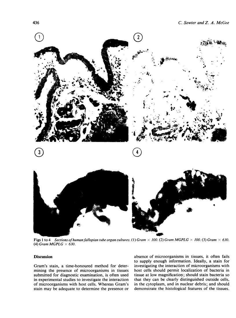
A method (Gram-MGPLG) for demonstrating micro-organisms was compared with Gram and four other known methods. Each method was tested on tissue infected with Staphylococcus aureus, Pseudomonas aeruginosa or Neisseria gonorrhoeae, which were then fixed in Bouin's formol saline, formol sublimate, or Van de Grift solutions. Gram-positive organisms in tissues were easily seen even at low magnification when stained by several of the methods tested. Gram-negative organisms, however, are very difficult to locate when stained by Gram's method because tissue components and the organisms are all shades of red, whereas the Gram-MGPLG provided easier location of organisms because these are stained red while the nuclei are blue and connective tissue is green. All methods are markedly affected by fixation; better preservation of cytological detail and improved staining reactions were produced by fixative containing mercuric chloride.
Full text
PDF
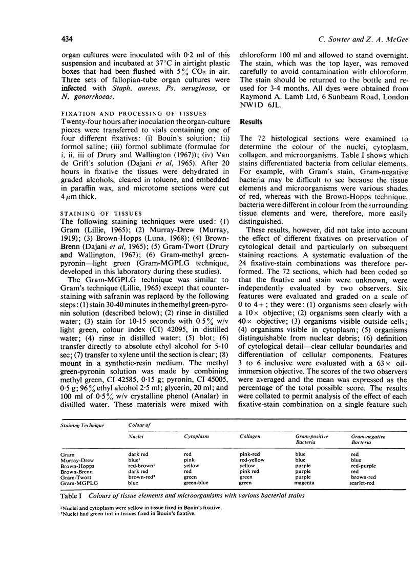
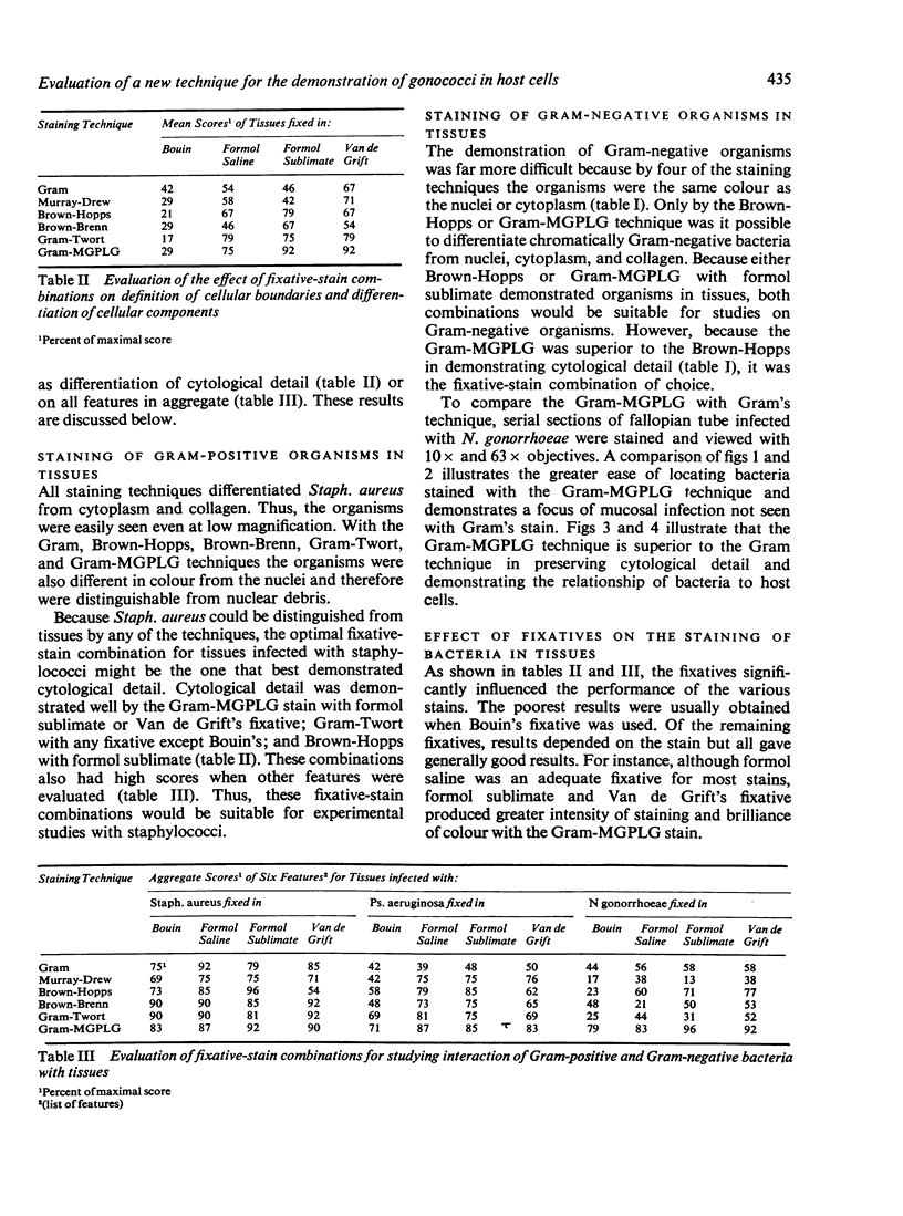

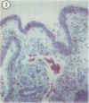
Images in this article
Selected References
These references are in PubMed. This may not be the complete list of references from this article.
- DAJANI A. S., CLYDE W. A., Jr, DENNY F. W. EXPERIMENTAL INFECTION WITH MYCOPLASMA PNEUMONIAE (EATON'S AGENT). J Exp Med. 1965 Jun 1;121:1071–1086. doi: 10.1084/jem.121.6.1071. [DOI] [PMC free article] [PubMed] [Google Scholar]


