

Abstract
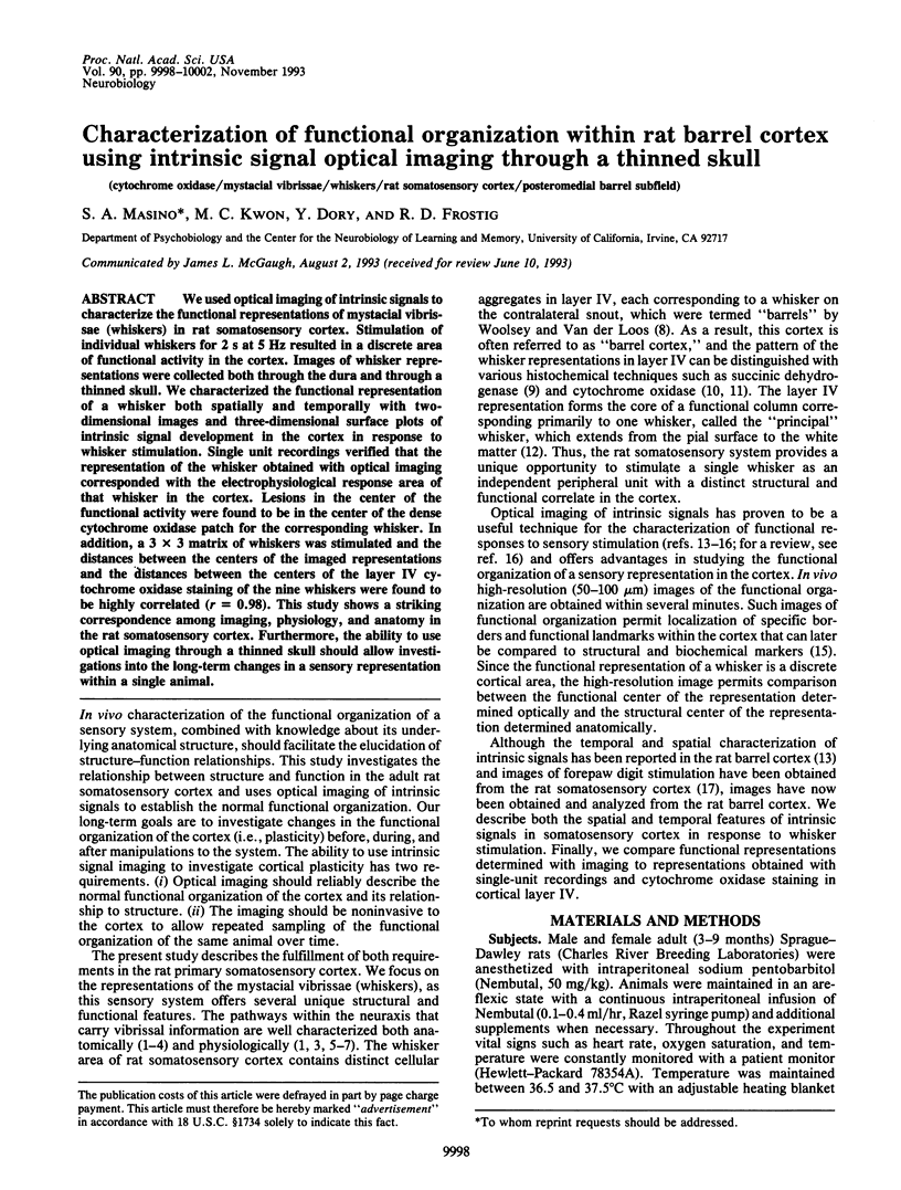
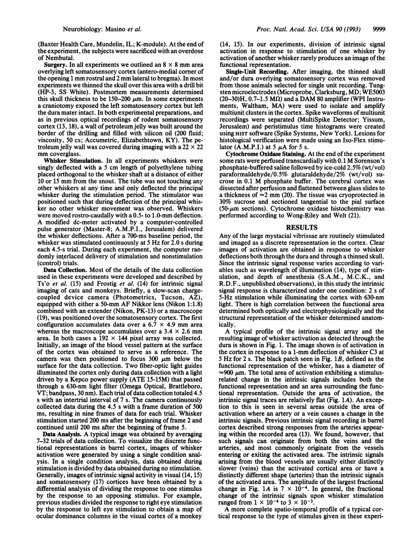
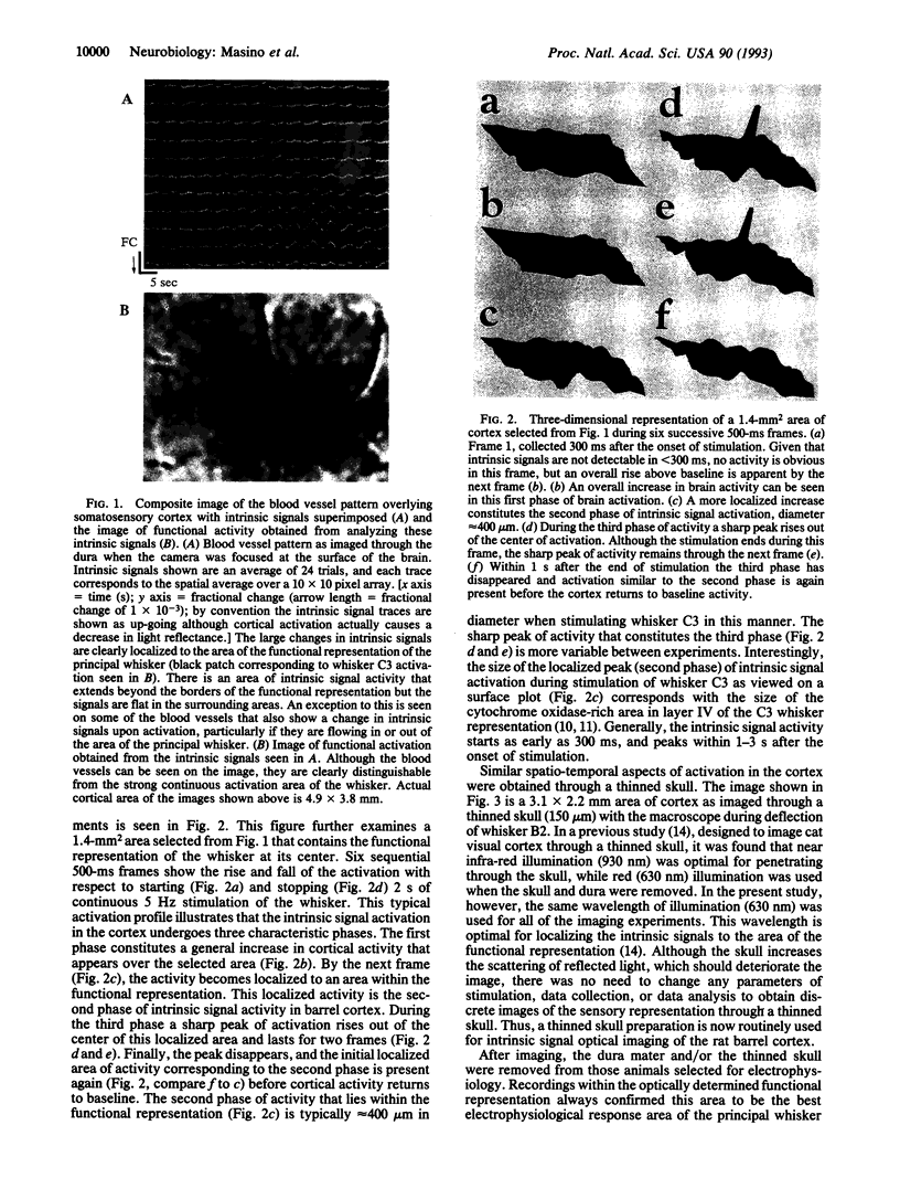
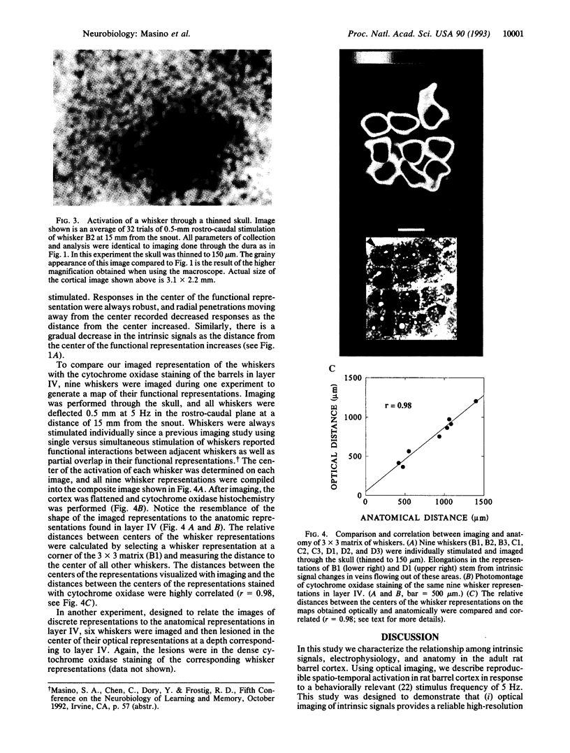
We used optical imaging of intrinsic signals to characterize the functional representations of mystacial vibrissae (whiskers) in rat somatosensory cortex. Stimulation of individual whiskers for 2 s at 5 Hz resulted in a discrete area of functional activity in the cortex. Images of whisker representations were collected both through the dura and through a thinned skull. We characterized the functional representation of a whisker both spatially and temporally with two-dimensional images and three-dimensional surface plots of intrinsic signal development in the cortex in response to whisker stimulation. Single unit recordings verified that the representation of the whisker obtained with optical imaging corresponded with the electrophysiological response area of that whisker in the cortex. Lesions in the center of the functional activity were found to be in the center of the dense cytochrome oxidase patch for the corresponding whisker. In addition, a 3 x 3 matrix of whiskers was stimulated and the distances between the centers of the imaged representations and the distances between the centers of the layer IV cytochrome oxidase staining of the nine whiskers were found to be highly correlated (r = 0.98). This study shows a striking correspondence among imaging, physiology, and anatomy in the rat somatosensory cortex. Furthermore, the ability to use optical imaging through a thinned skull should allow investigations into the long-term changes in a sensory representation within a single animal.
Full text
PDF
Images in this article
Selected References
These references are in PubMed. This may not be the complete list of references from this article.
- Armstrong-James M., Fox K., Das-Gupta A. Flow of excitation within rat barrel cortex on striking a single vibrissa. J Neurophysiol. 1992 Oct;68(4):1345–1358. doi: 10.1152/jn.1992.68.4.1345. [DOI] [PubMed] [Google Scholar]
- Armstrong-James M., Fox K. Spatiotemporal convergence and divergence in the rat S1 "barrel" cortex. J Comp Neurol. 1987 Sep 8;263(2):265–281. doi: 10.1002/cne.902630209. [DOI] [PubMed] [Google Scholar]
- Bartfeld E., Grinvald A. Relationships between orientation-preference pinwheels, cytochrome oxidase blobs, and ocular-dominance columns in primate striate cortex. Proc Natl Acad Sci U S A. 1992 Dec 15;89(24):11905–11909. doi: 10.1073/pnas.89.24.11905. [DOI] [PMC free article] [PubMed] [Google Scholar]
- Bonhoeffer T., Grinvald A. Iso-orientation domains in cat visual cortex are arranged in pinwheel-like patterns. Nature. 1991 Oct 3;353(6343):429–431. doi: 10.1038/353429a0. [DOI] [PubMed] [Google Scholar]
- Chiaia N. L., Rhoades R. W., Bennett-Clarke C. A., Fish S. E., Killackey H. P. Thalamic processing of vibrissal information in the rat. I. Afferent input to the medial ventral posterior and posterior nuclei. J Comp Neurol. 1991 Dec 8;314(2):201–216. doi: 10.1002/cne.903140202. [DOI] [PubMed] [Google Scholar]
- Chmielowska J., Carvell G. E., Simons D. J. Spatial organization of thalamocortical and corticothalamic projection systems in the rat SmI barrel cortex. J Comp Neurol. 1989 Jul 15;285(3):325–338. doi: 10.1002/cne.902850304. [DOI] [PubMed] [Google Scholar]
- Dawson D. R., Killackey H. P. The organization and mutability of the forepaw and hindpaw representations in the somatosensory cortex of the neonatal rat. J Comp Neurol. 1987 Feb 8;256(2):246–256. doi: 10.1002/cne.902560205. [DOI] [PubMed] [Google Scholar]
- Frostig R. D., Lieke E. E., Ts'o D. Y., Grinvald A. Cortical functional architecture and local coupling between neuronal activity and the microcirculation revealed by in vivo high-resolution optical imaging of intrinsic signals. Proc Natl Acad Sci U S A. 1990 Aug;87(16):6082–6086. doi: 10.1073/pnas.87.16.6082. [DOI] [PMC free article] [PubMed] [Google Scholar]
- Gochin P. M., Bedenbaugh P., Gelfand J. J., Gross C. G., Gerstein G. L. Intrinsic signal optical imaging in the forepaw area of rat somatosensory cortex. Proc Natl Acad Sci U S A. 1992 Sep 1;89(17):8381–8383. doi: 10.1073/pnas.89.17.8381. [DOI] [PMC free article] [PubMed] [Google Scholar]
- Grinvald A., Lieke E., Frostig R. D., Gilbert C. D., Wiesel T. N. Functional architecture of cortex revealed by optical imaging of intrinsic signals. 1986 Nov 27-Dec 3Nature. 324(6095):361–364. doi: 10.1038/324361a0. [DOI] [PubMed] [Google Scholar]
- Jacquin M. F., Renehan W. E., Mooney R. D., Rhoades R. W. Structure-function relationships in rat medullary and cervical dorsal horns. I. Trigeminal primary afferents. J Neurophysiol. 1986 Jun;55(6):1153–1186. doi: 10.1152/jn.1986.55.6.1153. [DOI] [PubMed] [Google Scholar]
- Jensen K. F., Killackey H. P. Terminal arbors of axons projecting to the somatosensory cortex of the adult rat. I. The normal morphology of specific thalamocortical afferents. J Neurosci. 1987 Nov;7(11):3529–3543. doi: 10.1523/JNEUROSCI.07-11-03529.1987. [DOI] [PMC free article] [PubMed] [Google Scholar]
- Killackey H. P., Belford G. R. The formation of afferent patterns in the somatosensory cortex of the neonatal rat. J Comp Neurol. 1979 Jan 15;183(2):285–303. doi: 10.1002/cne.901830206. [DOI] [PubMed] [Google Scholar]
- Kossut M., Hand P. J., Greenberg J., Hand C. L. Single vibrissal cortical column in SI cortex of rat and its alterations in neonatal and adult vibrissa-deafferented animals: a quantitative 2DG study. J Neurophysiol. 1988 Aug;60(2):829–852. doi: 10.1152/jn.1988.60.2.829. [DOI] [PubMed] [Google Scholar]
- Land P. W., Simons D. J. Cytochrome oxidase staining in the rat SmI barrel cortex. J Comp Neurol. 1985 Aug 8;238(2):225–235. doi: 10.1002/cne.902380209. [DOI] [PubMed] [Google Scholar]
- Ratzlaff E. H., Grinvald A. A tandem-lens epifluorescence macroscope: hundred-fold brightness advantage for wide-field imaging. J Neurosci Methods. 1991 Feb;36(2-3):127–137. doi: 10.1016/0165-0270(91)90038-2. [DOI] [PubMed] [Google Scholar]
- Simons D. J., Carvell G. E. Thalamocortical response transformation in the rat vibrissa/barrel system. J Neurophysiol. 1989 Feb;61(2):311–330. doi: 10.1152/jn.1989.61.2.311. [DOI] [PubMed] [Google Scholar]
- Simons D. J. Response properties of vibrissa units in rat SI somatosensory neocortex. J Neurophysiol. 1978 May;41(3):798–820. doi: 10.1152/jn.1978.41.3.798. [DOI] [PubMed] [Google Scholar]
- Ts'o D. Y., Frostig R. D., Lieke E. E., Grinvald A. Functional organization of primate visual cortex revealed by high resolution optical imaging. Science. 1990 Jul 27;249(4967):417–420. doi: 10.1126/science.2165630. [DOI] [PubMed] [Google Scholar]
- Welker C. Receptive fields of barrels in the somatosensory neocortex of the rat. J Comp Neurol. 1976 Mar 15;166(2):173–189. doi: 10.1002/cne.901660205. [DOI] [PubMed] [Google Scholar]
- Wong-Riley M. T., Welt C. Histochemical changes in cytochrome oxidase of cortical barrels after vibrissal removal in neonatal and adult mice. Proc Natl Acad Sci U S A. 1980 Apr;77(4):2333–2337. doi: 10.1073/pnas.77.4.2333. [DOI] [PMC free article] [PubMed] [Google Scholar]
- Woolsey T. A., Van der Loos H. The structural organization of layer IV in the somatosensory region (SI) of mouse cerebral cortex. The description of a cortical field composed of discrete cytoarchitectonic units. Brain Res. 1970 Jan 20;17(2):205–242. doi: 10.1016/0006-8993(70)90079-x. [DOI] [PubMed] [Google Scholar]