Abstract
MOB1 protein is a core component of the Hippo signaling pathway in animals where it is involved in controlling tissue growth and tumor suppression. Plant MOB1 proteins display high sequence homology to animal MOB1 proteins, but little is known regarding their role in plant growth and development. Herein we report the critical roles of Arabidopsis MOB1 (AtMOB1A) in auxin-mediated development in Arabidopsis. We found that loss-of-function mutations in AtMOB1A completely eliminated the formation of cotyledons when combined with mutations in PINOID (PID), which encodes a Ser/Thr protein kinase that participates in auxin signaling and transport. We showed that atmob1a was fully rescued by its Drosophila counterpart, suggesting functional conservation. The atmob1a pid double mutants phenocopied several well-characterized mutant combinations that are defective in auxin biosynthesis or transport. Moreover, we demonstrated that atmob1a greatly enhanced several other known auxin mutants, suggesting that AtMOB1A plays a key role in auxin-mediated plant development. The atmob1a single mutant displayed defects in early embryogenesis and had shorter root and smaller flowers than wild type plants. AtMOB1A is uniformly expressed in embryos and suspensor cells during embryogenesis, consistent with its role in embryo development. AtMOB1A protein is localized to nucleus, cytoplasm, and associated to plasma membrane, suggesting that it plays roles in these subcellular localizations. Furthermore, we showed that disruption of AtMOB1A led to a reduced sensitivity to exogenous auxin. Our results demonstrated that AtMOB1A plays an important role in Arabidopsis development by promoting auxin signaling.
Author Summary
MOB1 protein is a key component of the Hippo signaling pathway in animals, and it plays critical roles in organ size control. The plant hormone auxin regulates many aspects of plant growth and development including organogenesis. In this work, we showed that AtMOB1A, which is highly homologous to animal MOB1 proteins, plays an important role in plant organogenesis. Furthermore, we demonstrated that AtMOB1A synergistically interacts with auxin biosynthesis, transport, and signaling pathways to regulate Arabidopsis development. We further showed that AtMOB1A likely controls plant development by promoting auxin signaling. This work identified a new player in auxin-mediated plant development and lays a foundation for further dissection of the mechanisms by which auxin regulates organogenesis.
Introduction
In recent years, the Hippo signaling pathway has emerged as a very important pathway for animal development [1]. This highly conserved pathway was initially identified in Drosophila as a key pathway controlling organ size, and later was shown to play a role in controlling cell fate and pattern formation in mammals [2–5]. The core part of the pathway is a phosphorylation cascade composed of four key components in mammals and Drosophila: a Ste20-like Ser/Thr protein kinase Mst1/2 [Hippo (Hpo) in Drosophila] [6,7], an NDR-family protein kinase Lats1/2 [Warts (Wts) in Drosophila] [8,9], and two kinase regulatory components, Sav and MOB1 (Sav and Mats in Drosophila) [10,11] (S1 Fig). Mst1/2 phosphorylates MOB1 and Lats1/2, and activates Lats1/2. MOB1 can bind to Lats1/2 and potentiate its intrinsic kinase activity. The activated Lats1/2 in turn phosphorylates and inactivates a transcriptional co-activator YAP/TAZ (Yorkie in Drosophila) [12]. YAP/TAZ is an effector of the Hippo pathway. Phosphorylation of YAP/TAZ results in its cytoplasmic retention, largely by facilitating its interaction with 14-3-3 proteins. Dephosphorylation of YAP/TAZ promotes its nuclear localization where it interacts with transcription factors and regulates gene expression. Drosophila mutants of core components in this pathway, such as hpo, wts, mats, sav, showed larger organs. In mammals, Hippo signaling controls patterning and differentiation of airway epithelial progenitors, mammary gland differentiation, intestinal fate, cardiovascular, liver, pancreas, central nervous system, and lymphocyte development [2]. It also regulates stem cell self-renewal and cell polarity in animals [2,13,14]. Recently, it was reported that the Arabidopsis thaliana MOB1A gene is required for tissue patterning of the root tip [15] and the development of both sporophyte and gametophyte [16]. MOB1 proteins in plants and animals share high sequence homology [11]. It is tempting to hypothesize that the Hippo pathway may also function in plants. However, very little is known regarding how the hypothesized Hippo pathway may regulate plant growth and development.
The plant hormone auxin plays critical roles in plant growth and development. Local auxin biosynthesis, polar transport, and auxin signaling all contribute to proper plant growth and development. The best characterized tryptophan-dependent auxin biosynthesis pathway is the indole-3-pyruvate pathway, in which tryptophan is converted into indole-3-pyruvate by TAA/TAR family of amino transferases. Indole-3-pyruvate is then converted into IAA by YUC family of flavin-containing monooxygenases [17–21]. Auxin biosynthesis is temporally and spatially regulated [22,23]. Auxin transport is carried out by auxin influx carriers AUX1/LAXs, auxin efflux carriers PINs, and ABCB transporters [24]. Both local auxin biosynthesis and polar transport are important for generating auxin gradients and maxima, which are perceived by auxin receptors. The best characterized auxin receptor is TIR1/AFBs and Aux/IAA co-receptor complexes [25,26]. Disruption of auxin biosynthesis, polar transport or signal transduction pathways leads to defects in almost every aspect of developmental processes, such as flower, embryo, root, and leaf development [22,27,28]. For example, auxin biosynthetic mutants yuc1/4/10/11 quadruple mutants are defective in embryogenesis, and auxin signaling mutants such as mp fail to develop normal hypocotyls and roots [23]. Auxin transport mutant pin1 develops pin-like inflorescences, which was also observed in auxin signaling mutant mp and npy mutants [29,30]. Although it has been well documented that auxin plays essential roles in plant development, little is actually understood regarding how auxin gradients are translated into guiding proper developmental events.
In this paper, we provide evidence that links AtMOB1A, which is homologous to a key component of the animal Hippo pathway, to auxin-mediated plant organogenesis and development. We conducted a genetic screen for mutants that could enhance the phenotypes of pid, which is defective in auxin signaling and transport [31,32]. One of the pid enhancers, ncp1 (no-cotyledon in pid 1) failed to develop cotyledons in pid background. We further showed that ncp1 single mutant displays strong developmental defects in early embryos, seedlings, and in adult plants. NCP1 encodes a protein with significant homology to the animal MOB1s, a core component of the Hippo pathway. We showed that NCP1/AtMOB1A probably has biochemical activities similar to those of animal MOB1, because the Drosophila MOB1 (Mats) can fully rescue the developmental defects of ncp1/atmob1a. The atmob1a mutant showed synergistic genetic interactions with known auxin biosynthetic, transport, and signaling mutants, suggesting that AtMOB1A functions in parallel to auxin pathways or affecting some aspects of auxin biology. Furthermore, disruption of AtMOB1A led to a decrease in sensitivity to auxin treatments and down-regulation of auxin reporters including DR5-GFP, ProARF7:GUS, and ProARF19:GUS. Our findings demonstrate that AtMOB1A likely promotes auxin signaling, thus impacting various Arabidopsis developmental processes.
Results
Identification of genetic enhancers of pid
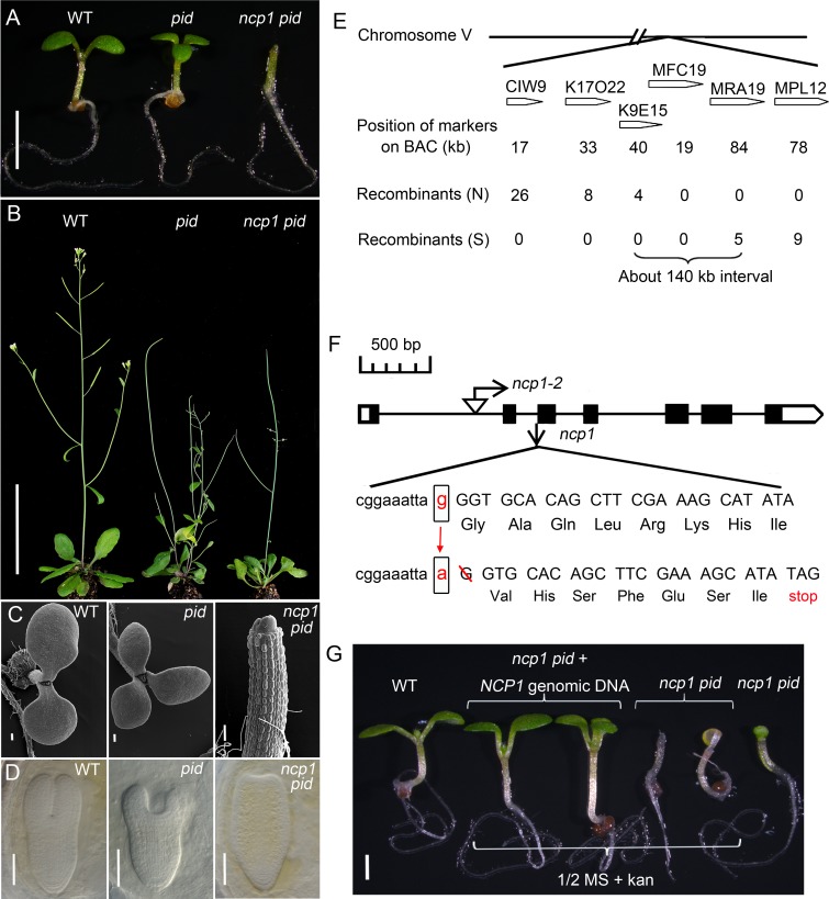
Genetic enhancement has been widely used to identify components in signaling and metabolic pathways. We previously identified npy1 as a genetic enhancer of yuc1 yuc4, which are defective in auxin biosynthesis. NPY1 is a key component of a signaling pathway responsible for auxin-mediated organogenesis [29]. Previous studies have shown that several Arabidopsis auxin mutants/mutant combinations, including npy1, yuc1 yuc4, wag1 wag2, pin1, and wei8 tar2, have no cotyledons when combined with pid, which encodes a protein kinase important for auxin signaling and transport [20,29,30,33]. Therefore, pid provides a sensitized background, and cotyledon formation serves as an easy phenotypic readout for us to genetically identify additional components in auxin-mediated plant development. We conducted a genetic screen for enhancers of pid and isolated a new mutant that lacked cotyledons. We name the mutant as ncp1 (no-cotyledon in pid 1). At seedling stage, ncp1 pid failed to develop cotyledons, but they appeared to have normal hypocotyls and roots (Fig 1A and 1C). The no-cotyledon phenotype of ncp1 pid was highly penetrant: the majority (90%) of the mutants completely lacked both cotyledons, while some plants occasionally developed one cotyledon (Table 1). Interestingly, ncp1 pid plants could develop true leaves, however, they were abnormal in morphology and vascular development (S2 Fig). The ncp1 pid plants were able to transition from vegetative growth to reproductive development, but their inflorescences were all pin-like and failed to produce fertile flowers (Fig 1B). The no-cotyledon phenotype in seedlings of ncp1 pid was caused by defects occurred during embryogenesis. In mature embryos, the cotyledon formation was abolished in ncp1 pid, while two cotyledons in WT and two or three cotyledons developed in pid (Fig 1D).
Fig 1. Identification and molecular cloning of ncp1.
(A) Mutation in NCP1 caused no-cotyledon phenotypes in seedling in pid background. From left to right, WT, pid, and ncp1 pid. (B) Adult plants of WT, pid, and ncp1 pid. (C) Electron micrographs of seedlings of WT (left), pid (middle), and ncp1 pid (right). Note that ncp1 pid failed to develop a cotyledon. (D) Late stage of embryos of WT, pid, and ncp1 pid. (E) Molecular cloning of ncp1. The mutation in ncp1 was mapped to an interval of about 140 kb on Chromosome V. (F) A schematic gene structure of At5g45550 and the location of T-DNA insertion of ncp1-2. Black boxes and lines designate exons and introns. The mutation at the splicing receptor of the second intron in ncp1-1 led to a frame-shift and introduced a premature stop codon in cDNA. (G) Complementation of ncp1-1 with a genomic DNA fragment of At5g45550 gene. From left to right: WT and ncp1 pid with/without the At5g45550 transgene (two seedlings of each). Note that the green transgenic seedlings had two or three cotyledons, while the yellowish non-transgenic seedlings had no cotyledon. The transformed NCP1 gene restored ncp1 pid to pid phenotype. Scale bar, 2 mm (A), 5 cm (B), 100 μm (C), 25 μm (D), 1 mm (G).
Table 1. Genetic analysis of ncp1 pid.
| Mutant genotype analysis | Mutant phenotype analysis | |||||||
|---|---|---|---|---|---|---|---|---|
| No. of seedlings (% of total seedlings mutant seedlings) | No. of seedlings | |||||||
| Parent genotype | Mutant genotype | expected | observed | Seedlings genotyped | with 2 cotyledons | with 3 cotyledons | no cotyledon | with 1 cotyledon |
| pid +/- ncp1-1-/- | pid+/+ ncp1-1-/- | 61 (25) | 75 (30) | 244 | 75 | 0 | 0 | 0 |
| pid +/- ncp1-1-/- | pid +/- ncp1-1-/- | 122 (50) | 105 (43) | 244 | 103 | 2 | 0 | 0 |
| pid +/- ncp1-1-/- | pid -/- ncp1-1-/- | 61 (25) | 64 (26) | 244 | 0 | 0 | 58 | 6 |
The observed no-cotyledon phenotype was dependent on the presence of the pid mutation. We genotyped 48 individual plants that showed the no-cotyledon phenotype and found out that they were all pid homozygous, suggesting that the phenotype was dependent on the presence of the pid mutation. We further analyzed the progenies from a single ncp1+/- pid+/- plant, 22 of 427 seedlings (about 1/20) showed the no-cotyledon phenotype, indicating that the phenotype was caused by two un-linked recessive mutations, i.e. pid and ncp1.
We crossed ncp1+/- pid+/- to Arabidopsis Landsberg ecotype and allowed the F1 plants to self-fertilize to generate a mapping population. In the F2 mapping population, we isolated 1325 seedlings that failed to develop cotyledons from about 26,000 F2 individuals. We found that the no-cotyledon phenotype was linked to two genetic loci: one on the bottom arm of chromosome II and the other on chromosome V. The Chromosome II locus is pid, further supporting that the no-cotyledon phenotype was dependent on pid. We narrowed the mapping interval on Chromosome V down to about 140 kb, between the two genetic markers on K9E15 and MRA19 (Fig 1E). We sequenced all of the open reading frames (ORFs) in the mapping interval and identified a G to A conversion at the splicing junction of the second intron and the third exon of the gene At5g45550. Further analysis of At5g45550 cDNA from the ncp1 mutant plants revealed that the mutation caused a single base-pair shift of the splicing acceptor of the second intron and the deletion of the first G of the third exon. The mutation led to a frame shift after the Lys24, and introduced a premature stop codon (Fig 1F). Therefore, this mutant is likely a null allele.
To further confirm that the identified mutation in At5g45550 was responsible for the observed no-cotyledon phenotype in pid background, we transformed a genomic fragment containing the coding region and its up- and down-stream regulatory sequences of At5g45550 into ncp1-/- pid+/- plants. All of the T1 transgenic plants (341 in total) had two or three cotyledons. We genotyped the T1 plants and found that 86 of them were double mutants, indicating that wild type (WT) copy of At5g45550 complemented the phenotype (Fig 1G).
We also identified a T-DNA insertion allele of ncp1 (GK_719G04) from the NASC stock center, and named it ncp1-2. We generated double mutants ncp1-2 pid and ncp1-2 pid-714 (SAIL_770_E05). Both of the double mutants displayed the same no-cotyledon phenotype as ncp1-1 pid (S3 Fig). Therefore, we conclude that At5g45550 is NCP1 and the identified mutations in At5g45550 are responsible for the no-cotyledon phenotype in pid backgrounds. We used ncp1-1 allele for further detailed analysis and genetic interaction studies in the paper.
NCP1/AtMOB1A plays important roles in Arabidopsis development
NCP1 was identified in the pid mutant background. We segregated out pid and investigated whether ncp1-1 mutation alone caused any developmental defects. At seedling stage, ncp1-1 had shorter root meristems zones when compared to WT. The root phenotypes of ncp1-1 were caused by decreased cell numbers in its root meristem (S4E and S4F Fig). Compared to WT plants, ncp1-1 single mutant plants were slightly taller with shorter siliques and smaller flowers (S4A–S4C Fig). The mutant was much less fertile. Our observed phenotypes of roots, siliques and flowers were consistent with previous findings from the analyses of the T-DNA allele of AtMOB1A, GK_719G04 (ncp1-2) [15]. The shorter root phenotype and the decrease in cell numbers in the root meristems of ncp1 and ncp1 pid may be caused by defects in cell division. To test this hypothesis, we investigated cell division activities in ncp1 and ncp1 pid mutants. CycB1;1:GUS is a widely used marker for the G2/M phase of the cell cycle [34]. The GUS staining domains were dramatically decreased in both ncp1 and ncp1 pid mutants, indicating that cell division activities were decreased in these mutants. These findings could partially account for the observed short root phenotypes of ncp1 and ncp1 pid (S4I and S4J Fig).
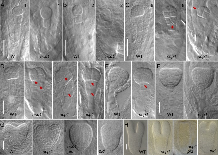
Because the no-cotyledon phenotype of ncp1 pid was caused by defects occurred during embryogenesis, we also analyzed whether disruption of AtMOB1A alone is sufficient to affect embryogenesis. Another indication that AtMOB1A is important for embryogenesis is that about 66.6% of the embryos (n = 785) were aborted in the ncp1-1 siliques (S4D Fig). We carefully analyzed various stages of embryogenesis of the ncp1-1 mutants and discovered that AtMOB1A plays an important role in early embryogenesis. The cell division in some mutant embryos was disturbed as early as 8-cell embryo stage. The upmost suspensor cell divided longitudinally in some ncp1-1 embryos whereas the cell in WT divides horizontally (Fig 2C). At 16-cell stage, both the pro-embryos and suspensor cells were abnormal in ncp1-1 (Fig 2D). At globular stage, the upmost suspensor cell became the hypophysis and remained as a lens-shape cell in WT [35]. But in ncp1-1 mutant, it was no longer lens-shaped and was divided into 2 cells (Fig 2E). The observed defects in the embryogenesis of ncp1-1 mutant would severely affect its embryo development, indicating that NCP1 is important for embryogenesis.
Fig 2. NCP1 is important for embryogenesis.
(A to F) Embryos of WT and ncp1 from 1-cell stage to transition stage. Red arrowheads point to the abnormal cell divisions in pro-embryos and suspensor cells at 8-cell (C) and 16-cell stages in ncp1-1 (D), and in hypophysis at the globular stage in ncp1-1 (E). (G, H) Defects of ncp1-1 pid embryos at heart and torpedo stages. From left to right: WT, ncp1-1, ncp1-1 pid, and pid.
NCP1 encodes a homolog of a key component in the animal Hippo signaling pathway
The predicted NCP1 protein contains 215 amino acid residues. It shares high sequence homology (63% identity) to the Drosophila MOB1 (Mats) (S5 Fig). MOB1 was first identified in yeast as Mps One Binder 1, an essential protein required for the completion of mitosis and maintenance of ploidy [36]. It has been shown that MOB1 is a key component of the Hippo signaling pathway [11]. In the Arabidopsis genome, there are four MOB1-like genes, At5g45550, At4g19045, At5g20430, and At5g20440. They have been renamed as AtMOB1A, AtMOB1B [15], AtMOB1C, and AtMOB1D herein, respectively.
MOB1 is highly conserved in plant species. For example, the MOB1As of Brassica rapa and Arabis alpina are almost identical to AtMOB1A: they differ from AtMOB1A in only one and three amino acid residues out of 215, respectively. Other putative plant MOB1A proteins and AtMOB1A share more than 90% identities (S5 Fig). It is clear from our phylogenetic analysis that animal MOB1 proteins and plant MOB1s belong to different clades (S6 Fig). The MOB1 genes have duplicated in the most recent common ancestor of land plants (Embryophyte) during evolution, and evolved with frequent duplication or deletion in the derived lineages of land plants. Selaginella moellendorffii only has one MOB1 gene whereas Physcomitrella patens has two. Monocots and dicots often have two to four copies of MOB1 genes (S6 Fig).
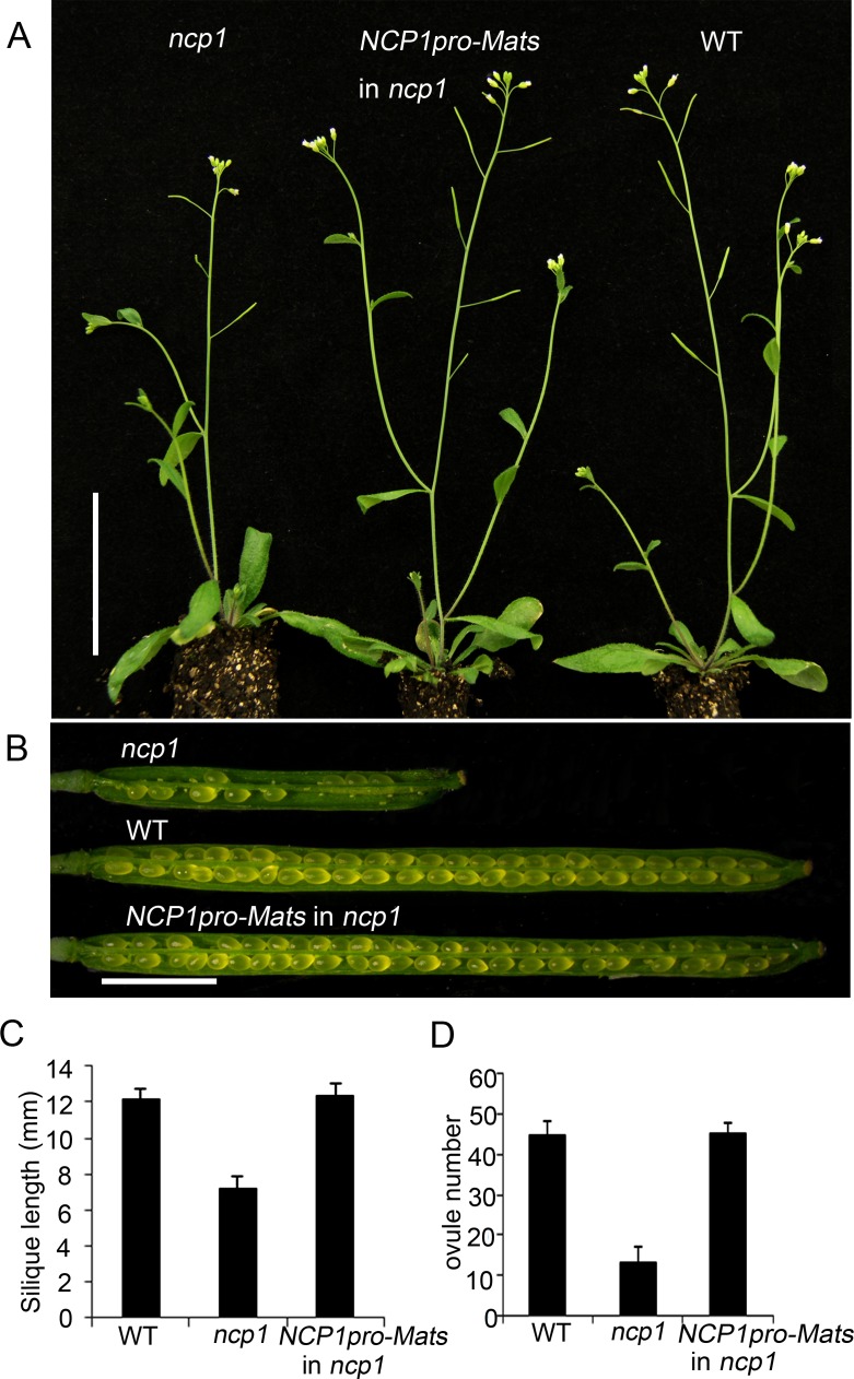
To further demonstrate that NCP1 is functionally related to MOB1 homologs from other organisms, we put the Drosophila MOB1 (Mats) under the control of the NCP1 promoter and transformed the construct into ncp1-1. We confirmed that all of the 88 transgenic ncp1-1 seedlings contained the Mats gene. The adult plants of ncp1-1 mutant showed severe defects in fertility, but the Mats transgenic ncp1-1 mutant plants were able to produce siliques like WT (Fig 3). Our results indicated that the Drosophila Mats gene complemented the defects caused by ncp1-1 mutation, and the function of MOB1/Mats is conserved from plants to Drosophila.
Fig 3. The Drosophila Mats gene can functionally substitute for NCP1.
(A) From left to right: ncp1, ncp1 with Mats under the control of the NCP1 promoter, and WT. (B) Siliques of ncp1, WT, and ncp1 with Mats. (C, D) Quantitative measurement of silique length (C) (n = 20), and ovule number per silique (D) (n = 10). Data are represented as mean ± SEM. Scale bar, 5 cm (A), 2 mm (B).
NCP1/AtMOB1A is expressed during embryogenesis and is localized to several cellular compartments
To investigate the expression pattern of NCP1/AtMOB1A, we generated a construct containing the NCP1 genomic DNA including its regulatory and coding sequences, with the GFP gene inserted immediately before the stop codon. We transformed ncp1 and ncp1 pid+/- mutants with this construct and found that the construct complemented both ncp1 and ncp1 pid, indicating that the NCP1-GFP fusion protein was fully functional. AtMOB1A is uniformly expressed in embryonic and suspensor cells from one-cell to mature embryo stages. The expression patterns of AtMOB1A are consistent with its role in embryo development. AtMOB1A protein is localized to nucleus, cytoplasm and associated to plasma membrane (Fig 4). The observed nuclear localization was consistent with previously findings [16,37].
Fig 4. Expression patterns of AtMOB1A protein during embryogenesis.
(A-H) Embryos from 1-cell stage to mature embryo stage. (A) 1-cell embryo. (B) 2-cell embryo. (C) 4–8 cell embryo. (D) Globular embryo. (E) Transition stage embryo. (F) Heart stage embryo. (G) Torpedo stage embryo. (H) Cotyledon stage embryo. Note that AtMOB1A protein is localized to nucleus, cytoplasm, and associated to plasma membrane. Scale bar, 20 μm.
Synergistic genetic interaction between ncp1/atmob1a and various auxin mutants
The ncp1-1 mutant was isolated as an enhancer of pid, which is a well-known auxin mutant. We further analyzed whether ncp1 could genetically interact with other known auxin mutants. We tested three groups of auxin mutants that are defective in either auxin biosynthesis, or transport, or signaling.
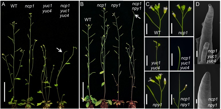
It has been shown that YUC flavin-containing monooxygenases and TAA1/TAR tryptophan amino transferases define a main auxin biosynthetic pathway in Arabidopsis [19,20]. Both YUCs and TAAs play essential roles in all of the major developmental processes including embryogenesis and flower development in Arabidopsis [18,22,23]. When we disrupted NCP1 in yuc1 yuc4 background, the resulting triple mutants developed pin-like inflorescence whereas yuc1 yuc4 never form pins, demonstrating that ncp1 greatly enhanced the phenotypes of auxin biosynthetic mutants (Fig 5A and 5C and 5D).
Fig 5. Genetic interactions between ncp1 and mutants of auxin biosynthesis and polar transport.
(A) ncp1 enhanced yuc1 yuc4 mutants phenotypes. (B) ncp1 enhanced npy1 mutant phenotypes. Arrowheads point to the pin-like inflorescence in ncp1 yuc1 yuc4 (A) and ncp1 npy1 (B). (C) Inflorescences of WT, ncp1, yuc1 yuc4, ncp1 yuc1 yuc4, npy1, ncp1 npy1. (D) SEM micrographs of representative pin-like inflorescences in ncp1 yuc1 yuc4 and ncp1 npy1 mutants. Scale bars, 5 cm (A and B), 5 mm (C), 100 μm (D).
We previously reported that NPY1 is involved in auxin-mediated organogenesis. The pid npy1 double mutants had no cotyledons, and npy1 yuc1 yuc4 triple mutants developed pin-like inflorescences. NPY1 is proposed to play a role in auxin transport and signaling [29,30]. When we introduced ncp1 into npy1 background, the double mutants produced pin-like structures whereas either single mutant did not form any pins (Fig 5B and 5C and 5D).
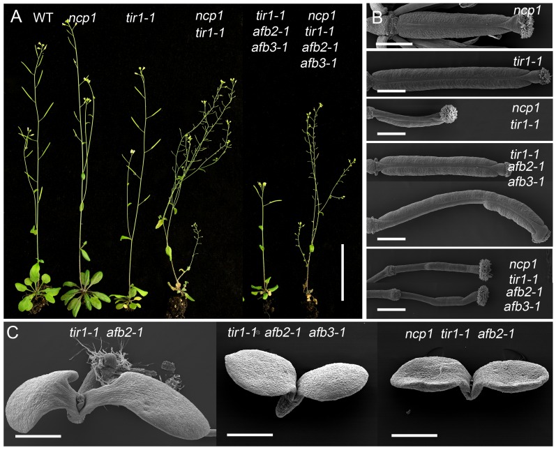
We next tested if ncp1 could synergistically interact with auxin signaling mutants. TIR1/AFBs are the best characterized auxin receptors responsible for regulating expression of auxin inducible genes. We crossed ncp1 to tir1-1 afb2-1 afb3-1 [27] and obtained various combinations of ncp1 and tir1 afb mutants from the F2 populations. The phenotypic analysis was performed in F4 generation. The single mutants of tir1-1, afb2-1, afb3-1, and combinations of their double mutants did not display dramatic developmental defects under normal growth conditions [27]. Interestingly, the ncp1 tir1-1 double mutants showed severe reduction in fertility, which was caused mainly by the defects in gynoecium patterning. The defects were further enhanced in ncp1 tir1-1 afb2-1 afb3-1 and led to complete sterility (Fig 6A and 6B). The adult plants of tir1-1 afb2-1 double mutants showed a reduction in rosette leaf size and inflorescence height, but their seedlings were similar to WT (Fig 6C) [27]. However, the ncp1 tir1-1 afb2-1 triple mutants exhibited strong developmental defects. Six of 34 (18%) triple homozygous seedlings of ncp1 tir1-1 afb2-1 mutants had no roots, whereas the tir1-1 afb2-1 double mutants never displayed such phenotypes. The observed no-root phenotypes closely resembled those of bdl/iaa12 or mp/arf5 mutants. The tir1-1 afb2-1 afb3-1 and tir1-1 afb1-1 afb2-1 afb3-1 mutants also showed the mp-like rootless seedling phenotypes at frequency of 36% and 49%, respectively (Fig 6C) [27]. Our results indicated that ncp1 genetically interacts with auxin signaling pathway.
Fig 6. Genetic interactions between ncp1 and auxin signaling mutants.
(A) From left to right: adult plants of WT, ncp1, tir1-1, ncp1 tir1-1, tir1-1 afb2-1 afb3-1, ncp1 tir1-1 afb2-1 afb3-1. Note the decreased fertility of ncp1 tir1-1 and completely sterile phenotypes of ncp1 tir1-1 afb2-1 afb3-1. (B) From top to bottom: gynoecia of tir1-1, ncp1 tir1-1, tir1-1 afb2-1 afb3-1, ncp1 tir1-1 afb2-1 afb3-1. (C) From left to right: Seedlings of tir1-1 afb2-1, tir1-1 afb2-1 afb3-1, ncp1 tir1-1 afb2-1 afb3-1. Scale bars, 5 cm (A), 500 μm (B and C).
In Arabidopsis, PID has three close homologs: WAG1, WAG2, and PID2, which redundantly control cotyledon development [30]. The ncp1 pid double mutants phenocopied the pid wag1 wag2 pid2 quadruple mutants (S7A and S7B Fig). We further tested if ncp1 could enhance pid wag1 wag2 pid2 phenotypes. The double or higher orders of mutant combinations between ncp1 and wag1 wag2 pid2 did not show obvious phenotypic enhancement, suggesting that PID played a more predominant role in regulating cotyledon development than WAG1, WAG2, and PID2. The ncp1 pid wag1 wag2 pid2 quintuple mutants displayed no-cotyledon phenotype similar to that of pid wag1 wag2 pid2. However, the quintuple mutants showed strong developmental defects in true leaves (S7 Fig). In dark grown seedlings, the initiation of true leaves was delayed in the quintuple mutants, compared to ncp1 pid (S7C Fig). In 14-day-old light grown seedlings, the quintuple mutants developed single or two leaves, and occasionally developed a pin-like true leaf (S7D and S7E Fig). In 36-day-old plants, the quintuple mutants showed two types of phenotypes. The type I plants (44%, n = 61) developed one to three true leaves and a pin-like inflorescence, and were arrested at this developmental stage. The type II plants (56%, n = 61) could produce more than three true leaves, and continued to grow with the phenotypes similar to those of ncp1 pid (S7F Fig).
We showed that NCP1 genetically interacted with PID to control cotyledon development in Arabidopsis. In animals, MOB1 physically interacts with and activates NDR/LATS through recruitment to the plasma membrane [38,39]. Because both PID and NDR/LATS are AGC kinases, we hypothesized that AtMOB1A may use a mechanism analogous to that of animal MOB1. PID may play a role equivalent to that of NDR/LATS. To test this hypothesis, we conducted both pull-down and Co-IP assays to determine whether AtMOB1A physically interacts with PID/WAGs. However, we did not detect direct physical interactions between NCP1/AtMOB1A and PID, or WAG1/2 in our experiments (S8 Fig). These results suggested that there may not be direct interactions between NCP1 and PID/WAGs, or the interactions are transient and difficult to be detected under our assay conditions. There are at least 39 AGCs in Arabidopsis. The observed genetic synergism of AtMOB1A with PID may suggest that AtMOB1A is necessary for the function of other AGCs that have overlapping functions with PID/WAG1/WAG2.
Auxin responses are decreased in ncp1 and ncp1 pid mutants
To assess the role of NCP1 in auxin response, we introduced the auxin reporter DR5-GFP into ncp1, pid, and ncp1 pid mutants background. At heart and torpedo stages of embryogenesis, strong DR5-GFP signals were observed at the cotyledon primordia and hypophysis in WT. In pid, the DR5-GFP signals remained similar to WT. In contrast, the GFP signals were significantly decreased at the cotyledon primordia in ncp1 single and ncp1 pid double mutants. It is worth noting that the auxin responses at hypophysis seemed not changed in ncp1 single and ncp1 pid double mutants (S9A Fig). These observations suggested that NCP1 might be involved in auxin signaling.
It is known that auxin is required for the initiation and growth of lateral root (LR) and root hairs, and exogenous auxin can stimulate these developmental processes [40]. To further investigate the roles of NCP1 in auxin responses, we examined the response of ncp1 mutant to exogenous auxin treatment. Four-day-old seedlings of WT, pid, ncp1, and ncp1 pid germinated on 1/2 strength of Murashige and Skoog medium (MS) plates were transferred and grew on 1/2 MS plates containing 50 nM 2,4-D, a synthetic auxin. It is obvious that the lengths of root hairs and density of LR/LR primordium were dramatically increased in WT, pid and ncp1, however the effects of exogenous auxin on ncp1 pid were much weaker (S9B–S9F Fig). This suggested that ncp1 pid double mutants are partially resistant to auxin in terms of root hair and LR initiation and growth. The pericycle cells in ncp1 and ncp1 pid were similar to WT, suggesting that the LR defects in ncp1 pid was likely due to slow LR primordium growth and the failure to emerge from the epidermis of the primary root. It could also be a defect in pre-branch site formation, which is not morphologically distinct.
ARF7 and ARF19 redundantly control LR development, and they are expressed in lateral and/or primary roots [41,42]. We analyzed the expression of ARF7 and ARF19 in seedlings of ncp1 pid mutants by using ProARF7:GUS and ProARF19:GUS reporter lines [41,42]. The expression levels of ProARF7:GUS and ProARF19:GUS were dramatically decreased in the LR primordium of ncp1, pid, and ncp1 pid mutants, compared to WT. The expression levels of ProARF19:GUS were also reduced in primary roots of ncp1, pid, and ncp1 pid mutants (S10 Fig). These findings suggested that the LR defects in ncp1 pid were partially caused by down-regulation of ARF7 and ARF19.
The expression pattern but not its sub-cellular localization of PIN1-GFP is altered in ncp1 pid double mutants during embryogenesis
PIN1 plays an important role during embryogenesis [28]. It is reported that PIN1-GFP is asymmetrically localized on plasma membrane [43,44]. We introduced the PIN1-GFP marker into ncp1, pid, and ncp1 pid mutants, and carefully checked the subcellular localization of PIN1-GFP from transition to torpedo stage of embryogenesis. No obvious alteration of the subcellular localization of PIN1-GFP was observed in these stages. However, we found that the expression levels of PIN1-GFP were altered in ncp1 mutants compared to WT. At these stages, PIN1-GFP was mainly expressed at the cotyledon primordia and ground tissue, which formed a Y-shape pattern. At transition stage, the expression pattern of PIN1-GFP in ncp1 mutants was similar to that of WT. However, in ncp1 pid double mutants, PIN1-GFP was found to be mainly expressed at the epidermal cell layer of apical part of embryos and ground tissue, which were barely connected by weak PIN1-GFP-expressing cells (S11 Fig). This result suggested that NCP1 plays a role in controlling the expression pattern of PIN1. It was previously reported that the localization pattern of PIN1 appeared normal in roots of Mob1A RNAi seedlings [15]. The discrepancy between our findings and those of the previous study might be because of the tissue specificity.
Discussion
The Hippo signaling pathway has been shown to play a critical role in organ size control and morphogenesis in animals, but it is still an open question whether the Hippo pathway exists in plants. Because MOB1 proteins share high sequence homology in animals and plants, it is tempting to hypothesize that the Hippo pathway may also exist and play a role in plant growth and development. Here we show that AtMOB1A is functionally conserved with the Drosophila protein because atmob1a was fully rescued by its Drosophila counterpart, suggesting that at least part of the Hippo pathway is functional in plants. NCP1/AtMOB1A synergistically interacts with key genes in auxin biosynthesis, transport, and signal transduction pathways to regulate Arabidopsis development. The observed synergistic genetic interactions and the decreased auxin responses in various ncp1 and auxin mutant combinations suggest that there is an intrinsic link between auxin pathway and the hypothesized Hippo pathway in plants. Our finding that the expression levels of ProARF7:GUS and ProARF19:GUS were dramatically decreased in ncp1 pid further supports the notion that AtMOB1A is important for auxin-mediated developmental processes. This work provides a genetic framework for the Hippo pathway in auxin-mediated plant development.
It was reported that about 2% of the progeny of AtMob1A RNAi silenced plants were tetraploid [16], which is a result of cell division defects. Auxin is also known to control plant development by regulating cell division and expansion. Therefore, AtMOB1A may be involved in auxin-controlled cell division. The mutants in animal Hippo pathway display defects in organ overgrowth [1], due to a loss of control of cell proliferation. In ncp1 mutant, the length and the cell number of root meristem were decreased compared to WT (S4 Fig). The different developmental outcomes between animals and plant mob1 mutants suggest that Hippo pathway/MOB1 protein may play different roles in plants and animals regarding cell proliferation. Recently, the Hippo pathway has been shown to control cell fate in animals. For example, the Hippo pathway activity is essential for the maintenance of the differentiated hepatocyte state. Acute inactivation of the Hippo signaling in vivo is sufficient to dedifferentiate adult hepatocytes into cells bearing progenitor characteristics [4]. In Arabidopsis, ncp1 yuc yuc4 and ncp1 npy1 mutants failed to develop flowers (Fig 5). The cotyledons were also eliminated in ncp1 pid, and the hypophysis was lost in ncp1 tir1-1 afb2-1 during embryogenesis (Fig 2 and Fig 6). The observed defects in organ and embryo development in these mutants indicated that the Hippo pathway also plays a critical role in determining cell fate in plants.
It has been shown that the Hippo pathway is highly conserved in mammals and insects. A human MOB1 gene rescued the developmental defects of the Drosophila MOB1 mutant mats [11]. We show that the Drosophila Mats fully rescued developmental defects of the Arabidopsis ncp1 mutant (Fig 3), indicating that at least some of the components of the Hippo pathway are conserved between plants and animals. This functional conservation of MOB1 proteins is consistent with the high similarities of their amino acid sequences (S5 Fig). It has been shown that MOB1 is a phospho-protein in animal systems. Phosphorylation of Thr12 and Thr35 of hMOB1 by MST1 or MST2 is required for the interaction of hMOB1 with NDR/LATS kinases in human [45,46]. Thr12 and Thr35 are absolutely conserved in MOB1s of plants and animals (S5 Fig). Both AtMOB1A and AtMOB1B were identified as phospho-proteins in a proteomic study [47], suggesting AtMOB1A/B is also phosphorylated by some kinase(s). AtMOB1A may also interact with Arabidopsis NDR/LATS kinases. In line with this hypothesis, there are eight NDR-like kinase genes in Arabidopsis [48], and they share high similarities with their human counterparts (S12 Fig).
It is well known that auxin promotes root hair and LR formation [40]. Gain-of-function mutant msg2 of Aux/IAA19 had severely reduced LR and LR formation was not normally induced by exogenous auxin [49]. Root hair and LR formation are also inhibited in arf7 arf19 double mutants [41,42]. pid did not show obvious defects in root development [31]. However, NCP1 and PID synergistically control LR formation and root hair growth in seedlings (S9 Fig). ncp1 pid also displayed strong defects in LR development in response to exogenous auxin treatment (S9 Fig). Expression levels of ProARF7:GUS and ProARF19:GUS were decreased in ncp1 pid. Moreover, ncp1 enhanced tir1-1 afb2-1 mutants’ phenotypes (Fig 6). These findings suggested that NCP1/AtMOB1A plays a positive role in promoting auxin signaling.
In the Hippo pathway, MOB1 binds and activates the AGC kinase NDR/LATS1/2 [38,39]. In Arabidopsis, there are 39 AGC kinases [48]. Some of them have been demonstrated to be involved to auxin pathways, such as PID/WAGs and D6PKs [31,50,51], which phosphorylate PIN1 at different phosphosites with different preference [52]. d6pk0123 quadruple mutants showed somewhat pin-like axillary shoots [51]. pid wag1 wag2 mutants phenocopied ncp1 pid [30]. Because NCP1/AtMOB1A is functionally conserved MOB1 in Arabidopsis, it is possible that PID/WAGs/D6PKs function as a plant counterpart of LATS1/2. It would be interesting to test if PID/WAGs/D6PKs can rescue Drosophila lats mutant phenotypes. AtMOB1A may associate with PID/WAGs/D6PKs and regulates its kinase activity, which subsequently modifies activities of PIN1. In human and Drosophila, MOB1 can activate LATS/NDRs when targeted to the plasma membrane [39,53]. AtMOB1A is localized to nucleus [16,37] and also associated with plasma membrane (Fig 4). PID, WAGs and D6PKs are also associated with plasma membrane [54,55], making it possible for AtMOB1A to activate PID/WAGs/D6PKs. However, we did not detect direct physical interactions between AtMOB1A and PID/WAGs by using pull-down and Co-IP assays. The negative results do not rule out the possibility that AtMOB1A is in a complex with AGC kinases. On the other hand, ncp1 pid wag1 wag2 pid2 showed no-cotyledon phenotypes similar to those of pid wag1 wag2 pid2. But the quintuple mutants displayed enhanced developmental defects in true leaves. These findings support the hypothesis that AtMOB1A may function with PID/WAGs. Alternatively, AtMOB1A and PID/WAGs/D6PKs may regulate transcription levels of auxin related genes. Indeed, we observed the alteration of expression pattern of PIN1-GFP and down-regulation of ARF7:GUS and ARF19:GUS in ncp1 pid double mutants (S10 Fig and S11 Fig). This finding is consistent with the mechanism that the animal Hippo pathway functions through regulating expression of downstream genes via a common growth regulatory effector, the transcriptional co-activator YAP/TAZ [1]. Another possibility is that the Hippo pathway functions in parallel to auxin pathway, yet they crosstalk to control plant development. This would be similar to the crosstalk between Wnt/β-catenin pathway and Hippo pathway to regulate animal development and tumorigenesis. It has been shown that cytoplasmic TAZ of the Hippo pathway can bind to DVL of the Wnt/β-catenin pathway and negatively regulate the Wnt/β-catenin pathway [56].
In conclusion, we demonstrate that AtMOB1A, a key component of the Hippo pathway, plays critical roles in auxin-mediated development in Arabidopsis. AtMOB1A synergistically interacts with auxin biosynthesis, transport, and signaling pathways to regulate Arabidopsis development. MOB1 is a regulator of AGC kinases in animal systems. PID/WAGs, D6PKs are AGC kinases, suggesting that NCP1/AtMOB1A may also regulate kinase activities of PID/WAGs and D6PKs, and possibly other AGC kinases in Arabidopsis. The fact that auxin responses and expression of auxin related genes such as ARF7 and ARF19 were down-regulated in ncp1 pid mutants suggests that NCP1/AtMOB1A may promote auxin signaling. This provides another layer of regulation of plant development by auxin. Further identification of other components of the Hippo pathway in Arabidopsis will help elucidate the mechanisms.
Materials and Methods
Plant materials and growth conditions
Plants were grown under 16-h light/8-h dark cycle at 22℃. The T-DNA insertion lines were obtained from NASC. The mutants used in this work were: pid (SALK_049736), pid-714 (SAIL_770_E05), ncp1-2 (GK_719G04). T-DNA insertion sites were determined by sequencing. Genotyping primers for pid (SALK_049736) and pid-714 (SAIL_770_E05) are: 5’-CCTCAGATTTCGCTTACGCAG-3’, and 5’- GCGAGACGAGTGAATCGTCG-3’, combined with JMLB1 and SAIL-LB1, respectively. For genotyping ncp1-2 (GK_719G04), 5’-ATGGATTCGTGTGGCTTTC-3’, 5’-TGTTTACAGCAAGCCATTC-3’, and PGABI1: 5’-ATATTGACCATCATACTCATTGC-3’ were used. To genotype ncp1-1, 5’-TGACCGTCTTCTTCCTAT-3’ and 5’-TGTTTACAGCAAGCCATTC-3’ were used and the PCR products were digested with MseI. npy1-2, yuc1, yuc4, tir1-1, afb2-1, afb3-1 were previously described [22,27,29]. All T-DNA insertion lines were genotyped as described previously [57–59].
Constructs and transgenic plants
For complementation of ncp1 pid mutants, a genomic DNA fragment containing the coding region as well as up- and down-stream regulatory sequences of At5g45550 was amplified by PCR using the following primers: 5’-CCCCCCGGGGAAACGGTGACCAAAATGCT-3’ and 5’-GCTCTAGAAGACGAGGCTCCAACACG-3’. The PCR product was digested with BamHI and XbaI and subcloned into pPZP211 vector [60] to generate pPZP211-NCP1gDNA. The plasmid was transformed into ncp1 pid+/- mutants via Agrobacterium strain GV3101 using floral dipping method [61]. The transgenic seedlings were selected on 1/2 MS plates containing 50 μg/mL kanamycin.
For expression of the Drosophila Mats under the control of NCP1 promoter, the Mats cDNA was amplified with PCR using the primers: 5’-ACTCCCGGGATGGACTTCTTGTTCGGTTC-3’, and 5’- GCTCTAGACTATATCTGCCGCTCATCCT-3’. The NCP1 promoter was amplified with primers: 5’-ACTGTCGACCTGCCCAATCAGCAAGAA-3’ and 5’-ACTCCCGGGGGCGACAAAAAGCAAGCGAG-3’. The PCR products were digested with SalI, XmaI and XbaI and subcloned into pCambia-1300 to generate pCambia-1300-NCP1p:Mats.
For expression pattern and subcellular analysis of NCP1, the pPZP211-NCP1gDNA construct was modified. The GFP gene was inserted immediate before the stop codon of NCP1 gene with restriction site of ApaI.
Microscopic analysis
SEM samples were prepared as described previously [62], and analyzed using a HITACHI S-4800 FESEM microscope. For whole-mount analysis of vascular structures and embryos, samples were prepared as previously described [63], and photographed under differential interference contrast (DIC) field or dark field on Leica DM 4500 and Leica S8AP0 microscopes. DR5-GFP and PIN1-GFP signals in embryos were viewed on Olympus FV1000MPE following the manufacturer’s instructions.
Phylogenetic analysis
Sequences were aligned using Clustal X version 1.81 [64], then refined manually. Maximum Likelihood method was used to reconstruct the phylogenetic tree using Mega5 [65]. Topological robustness of the phylogenetic tree was assessed by bootstrapping with 1000 replicates [66].
Pull-down and Co-IP assays
For the pull-down assay, cDNA of PID and NCP1 was cloned into pGEX-4T-1 and pET30a vectors to generate the expression constructs. The His-tagged and GST-tagged proteins were expressed in E. coli strain BL21. The subsequent protein purification and pull-down assay with Glutathione Sepharose 4B (GE) or His beads (Bio-Rad Ni-NTA Agarose) were carried out following the manufacturers’ manuals. The bound proteins were eluted and analyzed with anti-GST and anti-HIS antibodies (CWBIO).
To perform Co-IP assay of NCP1 and PID/WAGs, we constructed pEarleyGate104-35S:YFP-NCP1, pSuper1300:PID-Myc, pSuper1300:WAG1-Myc, pSuper1300:WAG2-Myc. YFP-NCP1 and PID-Myc or WAGs-Myc constructs were transformed into tobacco (Nicotiana Benthamiana) by injection. Leaves were grounded into fine powder in liquid nitrogen. Proteins were extracted with the extraction buffer [100 mM HEPES (pH 7.5), 5 mM EDTA, 5 mM EGTA, 10 mM NaF, 5% Glycerol, 10 mM Na3VO4, 10 mM DTT, 1 mM PMSF, 0.1% Triton X-100, 10 μg/mL Aprotinin, 10 μg/mL Leupeptin, 10 μg/mL Antipain]. The protein extracts were spun twice for 30 min at 14,000 g at 4℃. The supernatant was incubated for 3 hr with anti-Myc-tag mAb-agarose (MBL) in IP buffer [20 mM Tris-HCl (pH 7.5), 150 mM NaCl, 1 mM EDTA, 1 mM EGTA, 1 mM Na3VO4, 1 mM NaF, 10 mM glycerophosphate, 0.1% Triton X-100, 1 μg/mL Aprotinin, 1 μg/mL Leupeptin, 1 μg/mL Antipain]. The agarose was washed for three times with 1 ml of PBS. Proteins were then released and collected by boiling in 2×SDS loading buffer for 5 min. IP products were detected by SDS-PAGE and immunoblot analysis using anti-Myc or anti-GFP antibodies (CWBIO). These experiments were repeated at least three times.
Supporting Information
The Hippo pathway is highly conserved between Drosophila and mammals. Shown here is the core part of the pathway in mammals: a Ste20-like Ser/Thr protein kinase Mst1/2, an NDR-family protein kinase Lats1/2, and two kinase regulatory components, Sav and MOB1. Mst1/2 phosphorylates MOB1 and Lats1/2, and activates Lats1/2. MOB1 can bind to Lats1/2 and potentiate its intrinsic kinase activity. The activated Lats1/2 phosphorylates and inactivates the transcriptional co-activator YAP/TAZ. Dephosphorylation of YAP/TAZ promotes its nuclear localization where it interacts with transcription factors and regulates gene expression.
(TIF)
(A) Various morphological phenotypes of true leaves in ncp1 pid mutants. Note the cup-shaped first true leaf in the up-left plant. (B-E) Venation patterns in leaves of WT (B), ncp1 (C), pid (D), and ncp1 pid (E). Note the parallel venation in ncp1 pid (E). Scale bar, 1 mm.
(TIF)
(A) Additional combinations of ncp1 pid double mutants. From left to right: WT, pid, ncp1-2, ncp1-2 pid, pid-714, and ncp1-2 pid-714. Note the no-cotyledon phenotype of ncp1-2 pid and ncp1-2 pid-714 seedlings. (B) Close-up of ncp1-2 pid and ncp1-2 pid-714 seedlings. Note the trichomes on the true leaves of the double mutants. Scale bar, 5 mm (A), 500 μm (B).
(TIF)
(A) The ncp1 plant is slightly taller than the WT plant. (B-E) Flowers of ncp1 are smaller (B, C) and siliques (D) are shorter with some aborted seeds, and root meristems (E) of ncp1 and ncp1 pid are shorter than WT. (F-H) Quantitative measurements of root length (F), root meristem region length (G), and root meristem cell number (H) (n = 20). (I) CycB1;1:GUS expression at 5 days after germination (DAG). (J) Quantification of CycB1;1:GUS spots (n = 10). Data are represented as mean ± SEM. Scale bar, 5 cm (A), 2 mm (D), 100 μm (E).
(TIF)
MOB1 protein sequences of representative plant and animal species are aligned. Dicotyledons: Arabidopsis thaliana, Brassica rapa, Solanum lycopersicum, Cucumis sativus, Populus trichocarpa, Glycine max. Monocotyledons: Oryza sativa, Hordeum Vulgare, Saccharum hybrid. Lycophyte: Selaginella moellendorffii. Moss: Physcomitrella patens. Mammal: Homo sapiens. Insect: Drosophila melanogaster. Percentage of identity and NCBI accession numbers are listed at the end of each sequence. Residues that are identical in all sequences aligned are highlighted in black, and similar residues are in grey. The Thr12 and Thr35 residues are labeled with asterisks.
(TIF)
NCBI accession numbers are listed after the names of species.
(TIF)
(A) Light grown seedlings at 5 DAG. (B) Close-up view of mutant seedlings in (A). (C) SEM micrograph of dark grown seedlings at 3 DAG. Note the top of the seedlings. (D) Seedlings at 14 DAG. (E) Close-up view of true leaf development in ncp1 pid wag1 wag2 pid2 mutants at 14 DAG. (F) Plants at 36 DAG. Note the type I and type II phenotypes of ncp1 pid wag1 wag2 pid2 mutants. Scale bars, 1 mm (A, D), 500 μm (B, E), 100 μm (C), 1 cm (F).
(TIF)
(A, B) Pull-down assay with His (A) and GST tags (B). (C-E) Co-IP assay with YFP and Myc tags.
(TIF)
(A) DR5-GFP auxin response reporter in late heart stage embryos of WT, ncp1, pid and ncp1 pid. Note the arrowheads point to cotyledon primordia, where DR5-GFP signal was reduced in ncp1 and ncp1 pid mutants. (B) Root hair initiation and elongation of WT, ncp1, pid and ncp1 pid seedlings in response to exogenous auxin treatment. 4-day-old seedlings grown on 1/2 MS plate were transferred onto plates containing 0 nM (mock, top panel) or 50 nM 2,4-D (bottom panel) and grew for 4 days. (C) Lateral root initiation of WT, ncp1, pid and ncp1 pid seedlings in response to exogenous auxin treatment. 4-day-old seedlings grown on 1/2 MS plate were transferred onto plates containing 0 nM (mock, top panel) or 50 nM 2,4-D (bottom panel) and grew for 4 days. (D-F) Quantitative measurements of LR density (number of emerged LR per portion of the primary root where LRs are present, D), LR primordium density (number of LR primordium per portion of the primary root where LR primordia are present, E), and LR initiation density (number of non-emerged LR primordia and emerged LRs per portion of the primary root, F) of seedlings in response to exogenous auxin treatment. 4-day-old seedlings grown on 1/2 MS plate were transferred onto plates containing 0 nM (mock) or 50 nM 2,4-D and grew for 4 days. Data are represented as mean ± SEM. Scale bar, 20 μm (A), 500 μm (B), 1 cm (C).
(TIF)
10-d old seedlings were used for GUS staining. (A) ProARF19:GUS, from left to right: seedlings, primary roots, and lateral roots. (B) ProARF7:GUS, from left to right: seedlings and lateral roots. Scale bar, 1 mm (seedlings), 200 μm (primary roots), 50 μm (lateral roots).
(TIF)
Heart (A) and late heart (B) stages of WT, ncp1, pid and ncp1 pid. Scale bar, 20 μm (A and B). Note the arrowheads point to the regions between cotyledon primordium and ground tissue, where PIN1-GFP was expressed in WT embryos but barely in mutants.
(TIF)
NDR/LATS protein sequences of Arabidopsis and human are aligned.
(TIF)
Acknowledgments
We thank Drs. Yunde Zhao, Yongbiao Xue, Kun-Liang Guan, Martin Yanofsky, Maarten Chrispeels, Siva Malka for discussion, critical reading and comments on the manuscript. We thank Dr. Mark Estelle for providing the tir1 afb mutant seeds, Dr. Lei Zhang for the Drosophila Mats cDNA plasmid, Dr. Yuling Jiao for the ProARF7:GUS, and ProARF19:GUS reporter lines, Dr. Yalong Guo and Tingshen Han for helping on phylogenetic analysis, Ms. Jingquan Li and Xiuping Xu for technical assistance on confocal and SEM. We thank two anonymous reviewers for their comments.
Data Availability
All relevant data are within the paper and its Supporting Information files.
Funding Statement
This work was supported by the projects from the National Basic Research Program of China (2014CB943400), National Natural Science Fund of China (31171389; 91217310; 91017008; 31270330) and One-hundred Talent Project of the Chinese Academy of Sciences. The authors gratefully acknowledge the support of K.C. Wong Education Foundation, Hong Kong. The funders had no role in study design, data collection and analysis, decision to publish, or preparation of the manuscript.
References
- 1.Pan D (2010) The hippo signaling pathway in development and cancer. Dev Cell 19: 491–505. 10.1016/j.devcel.2010.09.011 [DOI] [PMC free article] [PubMed] [Google Scholar]
- 2.Mo JS, Park HW, Guan KL (2014) The Hippo signaling pathway in stem cell biology and cancer. Embo Reports 15: 642–656. 10.15252/embr.201438638 [DOI] [PMC free article] [PubMed] [Google Scholar]
- 3.Mahoney JE, Mori M, Szymaniak AD, Varelas X, Cardoso WV (2014) The Hippo Pathway Effector Yap Controls Patterning and Differentiation of Airway Epithelial Progenitors. Developmental Cell 30: 137–150. 10.1016/j.devcel.2014.06.003 [DOI] [PMC free article] [PubMed] [Google Scholar]
- 4.Yimlamai D, Christodoulou C, Galli GG, Yanger K, Pepe-Mooney B, et al. (2014) Hippo Pathway Activity Influences Liver Cell Fate. Cell 157: 1324–1338. 10.1016/j.cell.2014.03.060 [DOI] [PMC free article] [PubMed] [Google Scholar]
- 5.Zhao B, Tumaneng K, Guan KL (2011) The Hippo pathway in organ size control, tissue regeneration and stem cell self-renewal. Nature Cell Biology 13: 877–883. 10.1038/ncb2303 [DOI] [PMC free article] [PubMed] [Google Scholar]
- 6.Wu S, Huang J, Dong J, Pan D (2003) hippo encodes a Ste-20 family protein kinase that restricts cell proliferation and promotes apoptosis in conjunction with salvador and warts. Cell 114: 445–456. [DOI] [PubMed] [Google Scholar]
- 7.Harvey KF, Pfleger CM, Hariharan IK (2003) The Drosophila Mst ortholog, hippo, restricts growth and cell proliferation and promotes apoptosis. Cell 114: 457–467. [DOI] [PubMed] [Google Scholar]
- 8.Justice RW, Zilian O, Woods DF, Noll M, Bryant PJ (1995) The Drosophila tumor suppressor gene warts encodes a homolog of human myotonic dystrophy kinase and is required for the control of cell shape and proliferation. Genes Dev 9: 534–546. [DOI] [PubMed] [Google Scholar]
- 9.Xu T, Wang W, Zhang S, Stewart RA, Yu W (1995) Identifying tumor suppressors in genetic mosaics: the Drosophila lats gene encodes a putative protein kinase. Development 121: 1053–1063. [DOI] [PubMed] [Google Scholar]
- 10.Kango-Singh M, Nolo R, Tao C, Verstreken P, Hiesinger PR, et al. (2002) Shar-pei mediates cell proliferation arrest during imaginal disc growth in Drosophila. Development 129: 5719–5730. [DOI] [PubMed] [Google Scholar]
- 11.Lai ZC, Wei X, Shimizu T, Ramos E, Rohrbaugh M, et al. (2005) Control of cell proliferation and apoptosis by mob as tumor suppressor, mats. Cell 120: 675–685. [DOI] [PubMed] [Google Scholar]
- 12.Huang J, Wu S, Barrera J, Matthews K, Pan D (2005) The Hippo signaling pathway coordinately regulates cell proliferation and apoptosis by inactivating Yorkie, the Drosophila Homolog of YAP. Cell 122: 421–434. [DOI] [PubMed] [Google Scholar]
- 13.Ramos A, Camargo FD (2012) The Hippo signaling pathway and stem cell biology. Trends Cell Biol 22: 339–346. 10.1016/j.tcb.2012.04.006 [DOI] [PMC free article] [PubMed] [Google Scholar]
- 14.Genevet A, Tapon N (2011) The Hippo pathway and apico-basal cell polarity. Biochem J 436: 213–224. 10.1042/BJ20110217 [DOI] [PubMed] [Google Scholar]
- 15.Pinosa F, Begheldo M, Pasternak T, Zermiani M, Paponov IA, et al. (2013) The Arabidopsis thaliana Mob1A gene is required for organ growth and correct tissue patterning of the root tip. Ann Bot 112: 1803–1814. 10.1093/aob/mct235 [DOI] [PMC free article] [PubMed] [Google Scholar]
- 16.Galla G, Zenoni S, Marconi G, Marino G, Botton A, et al. (2011) Sporophytic and gametophytic functions of the cell cycle-associated Mob1 gene in Arabidopsis thaliana L. Gene 484: 1–12. 10.1016/j.gene.2011.05.009 [DOI] [PubMed] [Google Scholar]
- 17.Tao Y, Ferrer JL, Ljung K, Pojer F, Hong F, et al. (2008) Rapid synthesis of auxin via a new tryptophan-dependent pathway is required for shade avoidance in plants. Cell 133: 164–176. 10.1016/j.cell.2008.01.049 [DOI] [PMC free article] [PubMed] [Google Scholar]
- 18.Stepanova AN, Robertson-Hoyt J, Yun J, Benavente LM, Xie DY, et al. (2008) TAA1-mediated auxin biosynthesis is essential for hormone crosstalk and plant development. Cell 133: 177–191. 10.1016/j.cell.2008.01.047 [DOI] [PubMed] [Google Scholar]
- 19.Mashiguchi K, Tanaka K, Sakai T, Sugawara S, Kawaide H, et al. (2011) The main auxin biosynthesis pathway in Arabidopsis. Proc Natl Acad Sci U S A 108: 18512–18517. 10.1073/pnas.1108434108 [DOI] [PMC free article] [PubMed] [Google Scholar]
- 20.Won C, Shen X, Mashiguchi K, Zheng Z, Dai X, et al. (2011) Conversion of tryptophan to indole-3-acetic acid by TRYPTOPHAN AMINOTRANSFERASES OF ARABIDOPSIS and YUCCAs in Arabidopsis. Proc Natl Acad Sci U S A 108: 18518–18523. 10.1073/pnas.1108436108 [DOI] [PMC free article] [PubMed] [Google Scholar]
- 21.Stepanova AN, Yun J, Robles LM, Novak O, He WR, et al. (2011) The Arabidopsis YUCCA1 Flavin Monooxygenase Functions in the Indole-3-Pyruvic Acid Branch of Auxin Biosynthesis. Plant Cell 23: 3961–3973. 10.1105/tpc.111.088047 [DOI] [PMC free article] [PubMed] [Google Scholar]
- 22.Cheng Y, Dai X, Zhao Y (2006) Auxin biosynthesis by the YUCCA flavin monooxygenases controls the formation of floral organs and vascular tissues in Arabidopsis. Genes Dev 20: 1790–1799. [DOI] [PMC free article] [PubMed] [Google Scholar]
- 23.Cheng Y, Dai X, Zhao Y (2007) Auxin synthesized by the YUCCA flavin monooxygenases is essential for embryogenesis and leaf formation in Arabidopsis. Plant Cell 19: 2430–2439. [DOI] [PMC free article] [PubMed] [Google Scholar]
- 24.Vanneste S, Friml J (2009) Auxin: a trigger for change in plant development. Cell 136: 1005–1016. 10.1016/j.cell.2009.03.001 [DOI] [PubMed] [Google Scholar]
- 25.Dharmasiri N, Dharmasiri S, Estelle M (2005) The F-box protein TIR1 is an auxin receptor. Nature 435: 441–445. [DOI] [PubMed] [Google Scholar]
- 26.Kepinski S, Leyser O (2005) The Arabidopsis F-box protein TIR1 is an auxin receptor. Nature 435: 446–451. [DOI] [PubMed] [Google Scholar]
- 27.Dharmasiri N, Dharmasiri S, Weijers D, Lechner E, Yamada M, et al. (2005) Plant development is regulated by a family of auxin receptor F box proteins. Dev Cell 9: 109–119. [DOI] [PubMed] [Google Scholar]
- 28.Friml J, Vieten A, Sauer M, Weijers D, Schwarz H, et al. (2003) Efflux-dependent auxin gradients establish the apical-basal axis of Arabidopsis. Nature 426: 147–153. [DOI] [PubMed] [Google Scholar]
- 29.Cheng Y, Qin G, Dai X, Zhao Y (2007) NPY1, a BTB-NPH3-like protein, plays a critical role in auxin-regulated organogenesis in Arabidopsis. Proc Natl Acad Sci U S A 104: 18825–18829. [DOI] [PMC free article] [PubMed] [Google Scholar]
- 30.Cheng Y, Qin G, Dai X, Zhao Y (2008) NPY genes and AGC kinases define two key steps in auxin-mediated organogenesis in Arabidopsis. Proc Natl Acad Sci U S A 105: 21017–21022. 10.1073/pnas.0809761106 [DOI] [PMC free article] [PubMed] [Google Scholar]
- 31.Christensen SK, Dagenais N, Chory J, Weigel D (2000) Regulation of auxin response by the protein kinase PINOID. Cell 100: 469–478. [DOI] [PubMed] [Google Scholar]
- 32.Benjamins R, Quint A, Weijers D, Hooykaas P, Offringa R (2001) The PINOID protein kinase regulates organ development in Arabidopsis by enhancing polar auxin transport. Development 128: 4057–4067. [DOI] [PubMed] [Google Scholar]
- 33.Furutani M, Vernoux T, Traas J, Kato T, Tasaka M, et al. (2004) PIN-FORMED1 and PINOID regulate boundary formation and cotyledon development in Arabidopsis embryogenesis. Development 131: 5021–5030. [DOI] [PubMed] [Google Scholar]
- 34.Colon-Carmona A, You R, Haimovitch-Gal T, Doerner P (1999) Technical advance: spatio-temporal analysis of mitotic activity with a labile cyclin-GUS fusion protein. Plant J 20: 503–508. [DOI] [PubMed] [Google Scholar]
- 35.Lau S, Slane D, Herud O, Kong J, Jurgens G (2012) Early embryogenesis in flowering plants: setting up the basic body pattern. Annu Rev Plant Biol 63: 483–506. 10.1146/annurev-arplant-042811-105507 [DOI] [PubMed] [Google Scholar]
- 36.Luca FC, Winey M (1998) MOB1, an essential yeast gene required for completion of mitosis and maintenance of ploidy. Mol Biol Cell 9: 29–46. [DOI] [PMC free article] [PubMed] [Google Scholar]
- 37.Van Damme D, Bouget FY, Van Poucke K, Inze D, Geelen D (2004) Molecular dissection of plant cytokinesis and phragmoplast structure: a survey of GFP-tagged proteins. Plant J 40: 386–398. [DOI] [PubMed] [Google Scholar]
- 38.Bichsel SJ, Tamaskovic R, Stegert MR, Hemmings BA (2004) Mechanism of activation of NDR (nuclear Dbf2-related) protein kinase by the hMOB1 protein. J Biol Chem 279: 35228–35235. [DOI] [PubMed] [Google Scholar]
- 39.Hergovich A, Bichsel SJ, Hemmings BA (2005) Human NDR kinases are rapidly activated by MOB proteins through recruitment to the plasma membrane and phosphorylation. Mol Cell Biol 25: 8259–8272. [DOI] [PMC free article] [PubMed] [Google Scholar]
- 40.Overvoorde P, Fukaki H, Beeckman T (2010) Auxin control of root development. Cold Spring Harb Perspect Biol 2: a001537 10.1101/cshperspect.a001537 [DOI] [PMC free article] [PubMed] [Google Scholar]
- 41.Okushima Y, Overvoorde PJ, Arima K, Alonso JM, Chan A, et al. (2005) Functional genomic analysis of the AUXIN RESPONSE FACTOR gene family members in Arabidopsis thaliana: unique and overlapping functions of ARF7 and ARF19. Plant Cell 17: 444–463. [DOI] [PMC free article] [PubMed] [Google Scholar]
- 42.Li J, Dai X, Zhao Y (2006) A role for auxin response factor 19 in auxin and ethylene signaling in Arabidopsis. Plant Physiol 140: 899–908. [DOI] [PMC free article] [PubMed] [Google Scholar]
- 43.Galweiler L, Guan C, Muller A, Wisman E, Mendgen K, et al. (1998) Regulation of polar auxin transport by AtPIN1 in Arabidopsis vascular tissue. Science 282: 2226–2230. [DOI] [PubMed] [Google Scholar]
- 44.Friml J, Yang X, Michniewicz M, Weijers D, Quint A, et al. (2004) A PINOID-dependent binary switch in apical-basal PIN polar targeting directs auxin efflux. Science 306: 862–865. [DOI] [PubMed] [Google Scholar]
- 45.Praskova M, Xia F, Avruch J (2008) MOBKL1A/MOBKL1B phosphorylation by MST1 and MST2 inhibits cell proliferation. Curr Biol 18: 311–321. 10.1016/j.cub.2008.02.006 [DOI] [PMC free article] [PubMed] [Google Scholar]
- 46.Bao Y, Sumita K, Kudo T, Withanage K, Nakagawa K, et al. (2009) Roles of mammalian sterile 20-like kinase 2-dependent phosphorylations of Mps one binder 1B in the activation of nuclear Dbf2-related kinases. Genes Cells 14: 1369–1381. 10.1111/j.1365-2443.2009.01354.x [DOI] [PubMed] [Google Scholar]
- 47.Li H, Wong WS, Zhu L, Guo HW, Ecker J, et al. (2009) Phosphoproteomic analysis of ethylene-regulated protein phosphorylation in etiolated seedlings of Arabidopsis mutant ein2 using two-dimensional separations coupled with a hybrid quadrupole time-of-flight mass spectrometer. Proteomics 9: 1646–1661. 10.1002/pmic.200800420 [DOI] [PubMed] [Google Scholar]
- 48.Bogre L, Okresz L, Henriques R, Anthony RG (2003) Growth signalling pathways in Arabidopsis and the AGC protein kinases. Trends Plant Sci 8: 424–431. [DOI] [PubMed] [Google Scholar]
- 49.Tatematsu K, Kumagai S, Muto H, Sato A, Watahiki MK, et al. (2004) MASSUGU2 encodes Aux/IAA19, an auxin-regulated protein that functions together with the transcriptional activator NPH4/ARF7 to regulate differential growth responses of hypocotyl and formation of lateral roots in Arabidopsis thaliana. Plant Cell 16: 379–393. [DOI] [PMC free article] [PubMed] [Google Scholar]
- 50.Santner AA, Watson JC (2006) The WAG1 and WAG2 protein kinases negatively regulate root waving in Arabidopsis. Plant J 45: 752–764. [DOI] [PubMed] [Google Scholar]
- 51.Zourelidou M, Muller I, Willige BC, Nill C, Jikumaru Y, et al. (2009) The polarly localized D6 PROTEIN KINASE is required for efficient auxin transport in Arabidopsis thaliana. Development 136: 627–636. 10.1242/dev.028365 [DOI] [PubMed] [Google Scholar]
- 52.Zourelidou M, Absmanner B, Weller B, Barbosa IC, Willige BC, et al. (2014) Auxin efflux by PIN-FORMED proteins is activated by two different protein kinases, D6 PROTEIN KINASE and PINOID. Elife 3. [DOI] [PMC free article] [PubMed] [Google Scholar]
- 53.Ho LL, Wei XM, Shimizu T, Lai ZC (2010) Mob as tumor suppressor is activated at the cell membrane to control tissue growth and organ size in Drosophila. Developmental Biology 337: 274–283. 10.1016/j.ydbio.2009.10.042 [DOI] [PubMed] [Google Scholar]
- 54.Barbosa IC, Zourelidou M, Willige BC, Weller B, Schwechheimer C (2014) D6 PROTEIN KINASE activates auxin transport-dependent growth and PIN-FORMED phosphorylation at the plasma membrane. Dev Cell 29: 674–685. 10.1016/j.devcel.2014.05.006 [DOI] [PubMed] [Google Scholar]
- 55.Michniewicz M, Zago MK, Abas L, Weijers D, Schweighofer A, et al. (2007) Antagonistic regulation of PIN phosphorylation by PP2A and PINOID directs auxin flux. Cell 130: 1044–1056. [DOI] [PubMed] [Google Scholar]
- 56.Varelas X, Miller BW, Sopko R, Song S, Gregorieff A, et al. (2010) The Hippo pathway regulates Wnt/beta-catenin signaling. Dev Cell 18: 579–591. 10.1016/j.devcel.2010.03.007 [DOI] [PubMed] [Google Scholar]
- 57.Alonso JM, Stepanova AN, Leisse TJ, Kim CJ, Chen H, et al. (2003) Genome-wide insertional mutagenesis of Arabidopsis thaliana. Science 301: 653–657. [DOI] [PubMed] [Google Scholar]
- 58.Kleinboelting N, Huep G, Kloetgen A, Viehoever P, Weisshaar B (2012) GABI-Kat SimpleSearch: new features of the Arabidopsis thaliana T-DNA mutant database. Nucleic Acids Res 40: D1211–1215. 10.1093/nar/gkr1047 [DOI] [PMC free article] [PubMed] [Google Scholar]
- 59.Sessions A, Burke E, Presting G, Aux G, McElver J, et al. (2002) A high-throughput Arabidopsis reverse genetics system. Plant Cell 14: 2985–2994. [DOI] [PMC free article] [PubMed] [Google Scholar]
- 60.Hajdukiewicz P, Svab Z, Maliga P (1994) The Small, Versatile Ppzp Family of Agrobacterium Binary Vectors for Plant Transformation. Plant Molecular Biology 25: 989–994. [DOI] [PubMed] [Google Scholar]
- 61.Clough SJ, Bent AF (1998) Floral dip: a simplified method for Agrobacterium-mediated transformation of Arabidopsis thaliana. Plant J 16: 735–743. [DOI] [PubMed] [Google Scholar]
- 62.Dinneny JR, Yadegari R, Fischer RL, Yanofsky MF, Weigel D (2004) The role of JAGGED in shaping lateral organs. Development 131: 1101–1110. [DOI] [PubMed] [Google Scholar]
- 63.Berleth T, Jurgens G (1993) The Role of the Monopteros Gene in Organizing the Basal Body Region of the Arabidopsis Embryo. Development 118: 575–587. [Google Scholar]
- 64.Felsenstein J (1985) Confidence-Limits on Phylogenies—an Approach Using the Bootstrap. Evolution 39: 783–791. [DOI] [PubMed] [Google Scholar]
- 65.Tamura K, Peterson D, Peterson N, Stecher G, Nei M, et al. (2011) MEGA5: Molecular Evolutionary Genetics Analysis Using Maximum Likelihood, Evolutionary Distance, and Maximum Parsimony Methods. Molecular Biology and Evolution 28: 2731–2739. 10.1093/molbev/msr121 [DOI] [PMC free article] [PubMed] [Google Scholar]
- 66.Thompson JD, Gibson TJ, Plewniak F, Jeanmougin F, Higgins DG (1997) The CLUSTAL_X windows interface: flexible strategies for multiple sequence alignment aided by quality analysis tools. Nucleic Acids Research 25: 4876–4882. [DOI] [PMC free article] [PubMed] [Google Scholar]
Associated Data
This section collects any data citations, data availability statements, or supplementary materials included in this article.
Supplementary Materials
The Hippo pathway is highly conserved between Drosophila and mammals. Shown here is the core part of the pathway in mammals: a Ste20-like Ser/Thr protein kinase Mst1/2, an NDR-family protein kinase Lats1/2, and two kinase regulatory components, Sav and MOB1. Mst1/2 phosphorylates MOB1 and Lats1/2, and activates Lats1/2. MOB1 can bind to Lats1/2 and potentiate its intrinsic kinase activity. The activated Lats1/2 phosphorylates and inactivates the transcriptional co-activator YAP/TAZ. Dephosphorylation of YAP/TAZ promotes its nuclear localization where it interacts with transcription factors and regulates gene expression.
(TIF)
(A) Various morphological phenotypes of true leaves in ncp1 pid mutants. Note the cup-shaped first true leaf in the up-left plant. (B-E) Venation patterns in leaves of WT (B), ncp1 (C), pid (D), and ncp1 pid (E). Note the parallel venation in ncp1 pid (E). Scale bar, 1 mm.
(TIF)
(A) Additional combinations of ncp1 pid double mutants. From left to right: WT, pid, ncp1-2, ncp1-2 pid, pid-714, and ncp1-2 pid-714. Note the no-cotyledon phenotype of ncp1-2 pid and ncp1-2 pid-714 seedlings. (B) Close-up of ncp1-2 pid and ncp1-2 pid-714 seedlings. Note the trichomes on the true leaves of the double mutants. Scale bar, 5 mm (A), 500 μm (B).
(TIF)
(A) The ncp1 plant is slightly taller than the WT plant. (B-E) Flowers of ncp1 are smaller (B, C) and siliques (D) are shorter with some aborted seeds, and root meristems (E) of ncp1 and ncp1 pid are shorter than WT. (F-H) Quantitative measurements of root length (F), root meristem region length (G), and root meristem cell number (H) (n = 20). (I) CycB1;1:GUS expression at 5 days after germination (DAG). (J) Quantification of CycB1;1:GUS spots (n = 10). Data are represented as mean ± SEM. Scale bar, 5 cm (A), 2 mm (D), 100 μm (E).
(TIF)
MOB1 protein sequences of representative plant and animal species are aligned. Dicotyledons: Arabidopsis thaliana, Brassica rapa, Solanum lycopersicum, Cucumis sativus, Populus trichocarpa, Glycine max. Monocotyledons: Oryza sativa, Hordeum Vulgare, Saccharum hybrid. Lycophyte: Selaginella moellendorffii. Moss: Physcomitrella patens. Mammal: Homo sapiens. Insect: Drosophila melanogaster. Percentage of identity and NCBI accession numbers are listed at the end of each sequence. Residues that are identical in all sequences aligned are highlighted in black, and similar residues are in grey. The Thr12 and Thr35 residues are labeled with asterisks.
(TIF)
NCBI accession numbers are listed after the names of species.
(TIF)
(A) Light grown seedlings at 5 DAG. (B) Close-up view of mutant seedlings in (A). (C) SEM micrograph of dark grown seedlings at 3 DAG. Note the top of the seedlings. (D) Seedlings at 14 DAG. (E) Close-up view of true leaf development in ncp1 pid wag1 wag2 pid2 mutants at 14 DAG. (F) Plants at 36 DAG. Note the type I and type II phenotypes of ncp1 pid wag1 wag2 pid2 mutants. Scale bars, 1 mm (A, D), 500 μm (B, E), 100 μm (C), 1 cm (F).
(TIF)
(A, B) Pull-down assay with His (A) and GST tags (B). (C-E) Co-IP assay with YFP and Myc tags.
(TIF)
(A) DR5-GFP auxin response reporter in late heart stage embryos of WT, ncp1, pid and ncp1 pid. Note the arrowheads point to cotyledon primordia, where DR5-GFP signal was reduced in ncp1 and ncp1 pid mutants. (B) Root hair initiation and elongation of WT, ncp1, pid and ncp1 pid seedlings in response to exogenous auxin treatment. 4-day-old seedlings grown on 1/2 MS plate were transferred onto plates containing 0 nM (mock, top panel) or 50 nM 2,4-D (bottom panel) and grew for 4 days. (C) Lateral root initiation of WT, ncp1, pid and ncp1 pid seedlings in response to exogenous auxin treatment. 4-day-old seedlings grown on 1/2 MS plate were transferred onto plates containing 0 nM (mock, top panel) or 50 nM 2,4-D (bottom panel) and grew for 4 days. (D-F) Quantitative measurements of LR density (number of emerged LR per portion of the primary root where LRs are present, D), LR primordium density (number of LR primordium per portion of the primary root where LR primordia are present, E), and LR initiation density (number of non-emerged LR primordia and emerged LRs per portion of the primary root, F) of seedlings in response to exogenous auxin treatment. 4-day-old seedlings grown on 1/2 MS plate were transferred onto plates containing 0 nM (mock) or 50 nM 2,4-D and grew for 4 days. Data are represented as mean ± SEM. Scale bar, 20 μm (A), 500 μm (B), 1 cm (C).
(TIF)
10-d old seedlings were used for GUS staining. (A) ProARF19:GUS, from left to right: seedlings, primary roots, and lateral roots. (B) ProARF7:GUS, from left to right: seedlings and lateral roots. Scale bar, 1 mm (seedlings), 200 μm (primary roots), 50 μm (lateral roots).
(TIF)
Heart (A) and late heart (B) stages of WT, ncp1, pid and ncp1 pid. Scale bar, 20 μm (A and B). Note the arrowheads point to the regions between cotyledon primordium and ground tissue, where PIN1-GFP was expressed in WT embryos but barely in mutants.
(TIF)
NDR/LATS protein sequences of Arabidopsis and human are aligned.
(TIF)
Data Availability Statement
All relevant data are within the paper and its Supporting Information files.


