Abstract
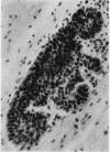
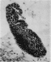
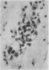
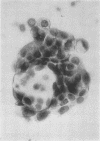
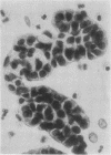
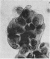
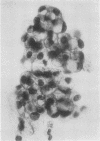
The sputum of an asthmatic often contains large, well-circumscribed clusters of benign columnar epithelial cells which can be misinterpreted as papillary fragments of an adenocarcinoma. Their presence is a manifestation of the excessive shedding of the mucosa of the lower respiratory tract accompanying an attack of bronchial asthma. These clusters are illustrated and compared with clusters of adenocarcinoma cells. To discriminate between the two types of cluster may be difficult and it is therefore important for the cytologist to know when a sputum is from an asthmatic.
Full text
PDF
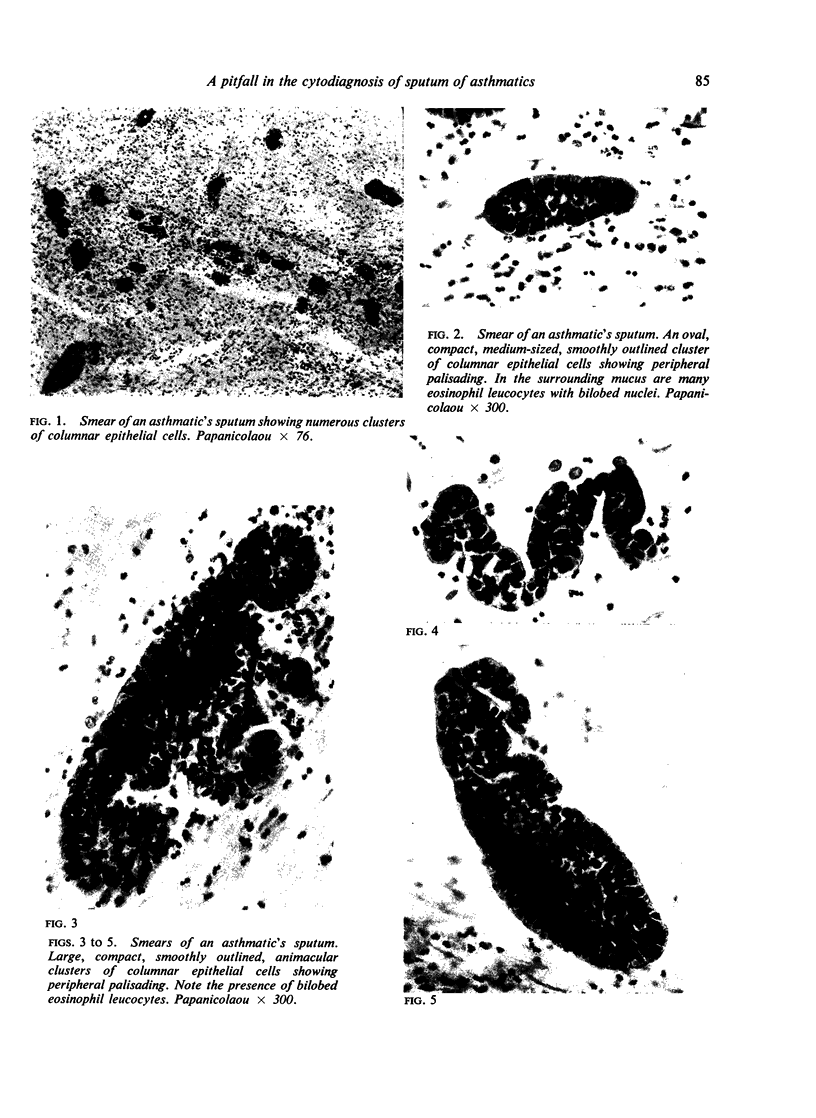
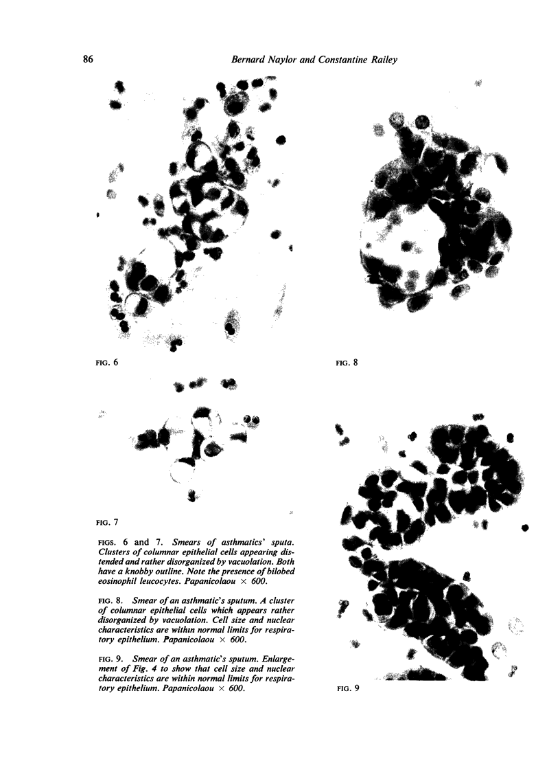
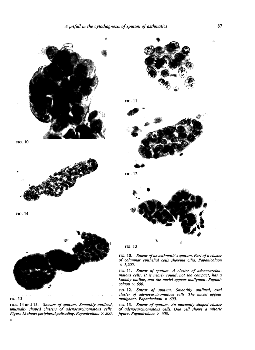
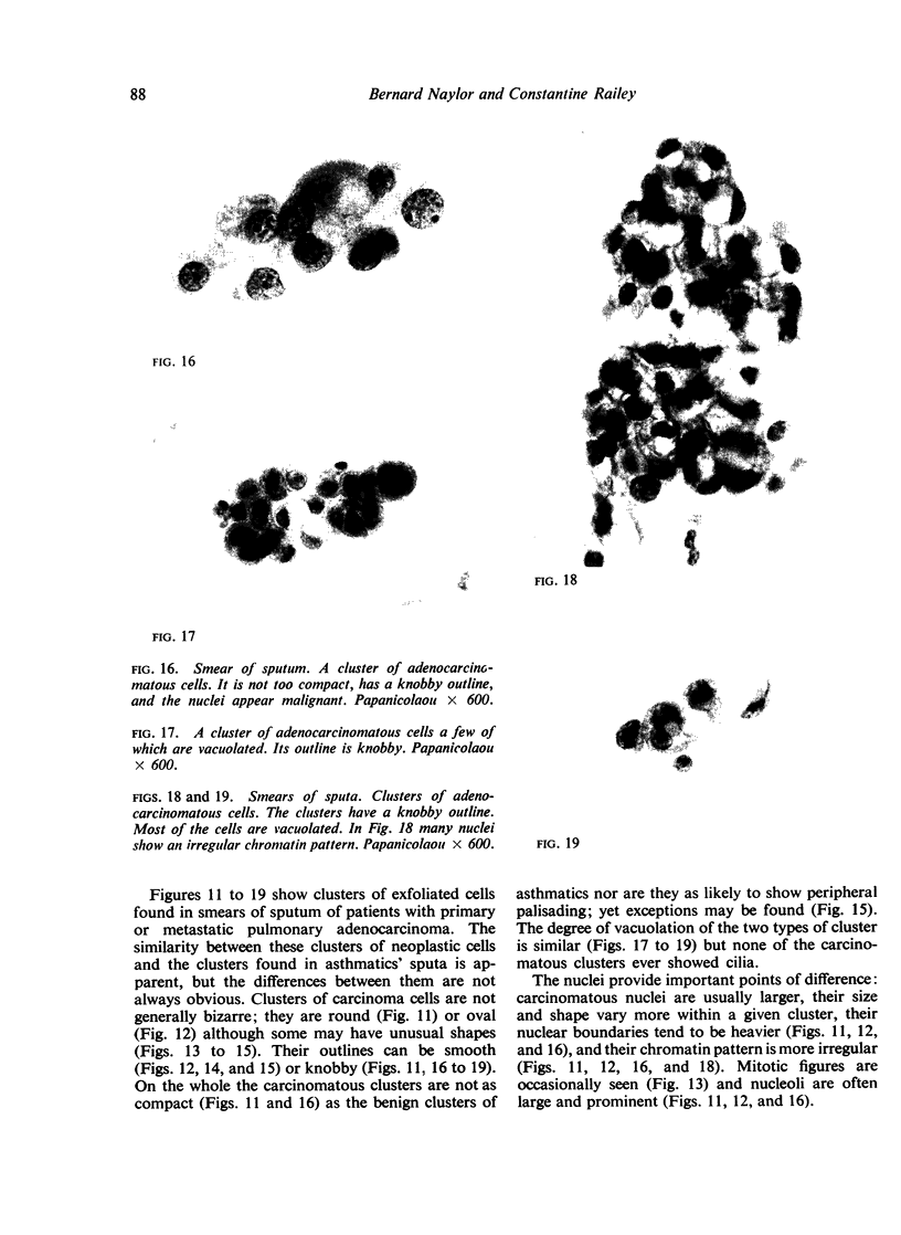
Images in this article
Selected References
These references are in PubMed. This may not be the complete list of references from this article.
- BAMFORTH J. The cytological diagnosis of malignant disease. Practitioner. 1959 Jun;182(1092):702–710. [PubMed] [Google Scholar]
- COHEN R. C., PRENTICE A. I. Metaplastic cells in sputum of patients with pulmonary eosinophilia. Tubercle. 1959 Feb;40(1):44–46. doi: 10.1016/s0041-3879(59)80018-0. [DOI] [PubMed] [Google Scholar]
- FARBER S. M., PHARR S. L., PIERSON B., WOOD D. A. Significant cytologic findings in non-malignant pulmonary disease. Dis Chest. 1957 Jan;31(1):1–13. doi: 10.1378/chest.31.1.1. [DOI] [PubMed] [Google Scholar]
- NAYLOR B. The shedding of the mucosa of the bronchial tree in asthma. Thorax. 1962 Mar;17:69–72. doi: 10.1136/thx.17.1.69. [DOI] [PMC free article] [PubMed] [Google Scholar]
- PHILPS F. R. Vacuolated cells in the sputum simulating adenocarcinoma cells. Br J Cancer. 1956 Mar;10(1):24–25. doi: 10.1038/bjc.1956.3. [DOI] [PMC free article] [PubMed] [Google Scholar]


