Abstract
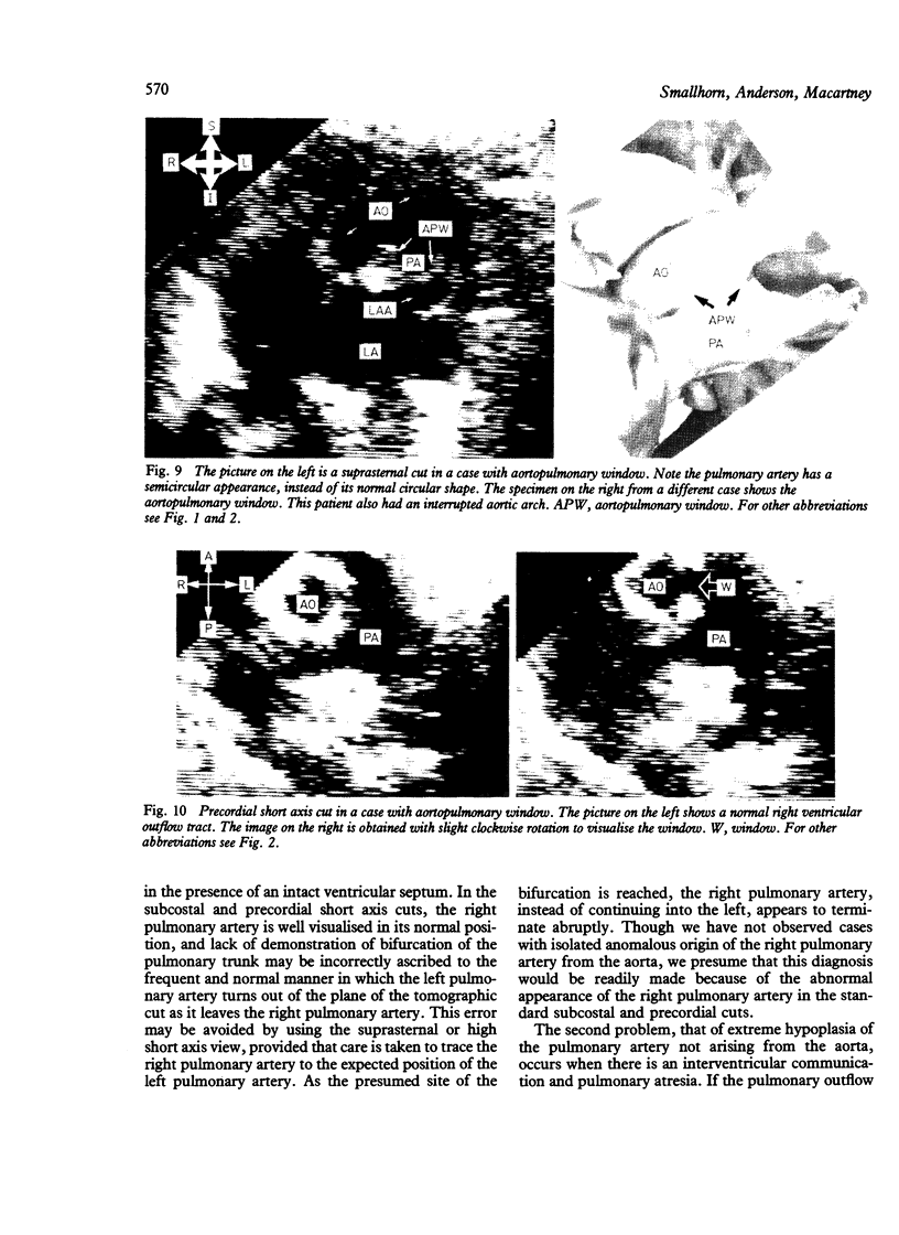
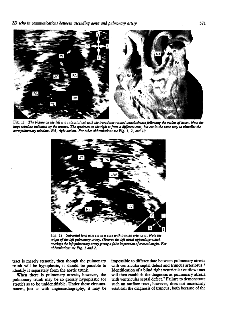
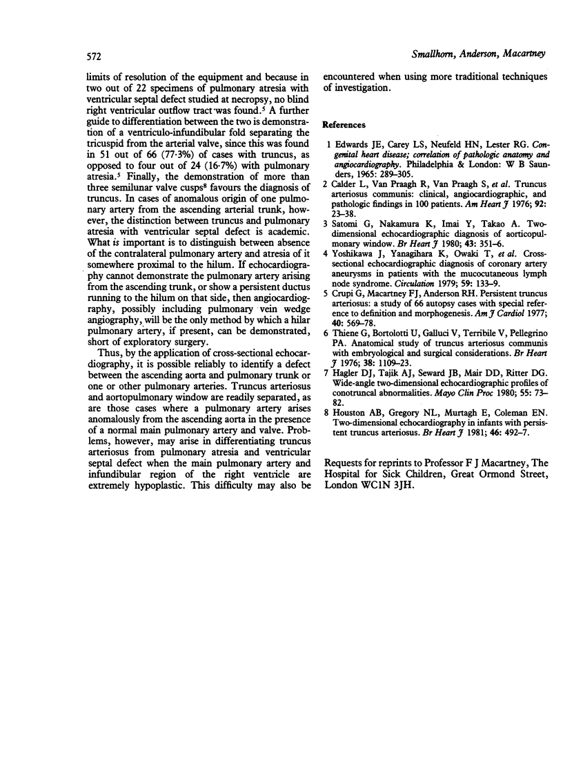
The value of two dimensional echocardiography in identifying communications between the ascending aorta and pulmonary trunk or individual pulmonary arteries was assessed in 24 children, all of whom had either angiocardiographic and surgical or angiocardiographic confirmation alone. Fourteen cases had truncus arteriosus, four aortopulmonary window, four anomalous origin of the left pulmonary artery from the ascending aorta, and two anomalous origin of the right pulmonary artery from the ascending aorta. It was possible to identify reliably each individual abnormality with a combination of suprasternal, precordial, and subcostal cuts. Problems only arose in differentiating truncus arteriosus from pulmonary atresia and ventricular septal defect when the main pulmonary artery and infundibular region of the right ventricle were extremely hypoplastic.
Full text
PDF
Images in this article
Selected References
These references are in PubMed. This may not be the complete list of references from this article.
- Calder L., Van Praagh R., Van Praagh S., Sears W. P., Corwin R., Levy A., Keith J. D., Paul M. H. Truncus arteriosus communis. Clinical, angiocardiographic, and pathologic findings in 100 patients. Am Heart J. 1976 Jul;92(1):23–38. doi: 10.1016/s0002-8703(76)80400-0. [DOI] [PubMed] [Google Scholar]
- Crupi G., Macartney F. J., Anderson R. H. Persistent truncus arteriosus. A study of 66 autopsy cases with special reference to definition and morphogenesis. Am J Cardiol. 1977 Oct;40(4):569–578. doi: 10.1016/0002-9149(77)90073-x. [DOI] [PubMed] [Google Scholar]
- Hagler D. J., Tajik A. J., Seward J. B., Mair D. D., Ritter D. G. Wide-angle two-dimensional echocardiographic profiles of conotruncal abnormalities. Mayo Clin Proc. 1980 Feb;55(2):73–82. [PubMed] [Google Scholar]
- Houston A. B., Gregory N. L., Murtagh E., Coleman E. N. Two-dimensional echocardiography in infants with persistent truncus arteriosus. Br Heart J. 1981 Nov;46(5):492–497. doi: 10.1136/hrt.46.5.492. [DOI] [PMC free article] [PubMed] [Google Scholar]
- Satomi G., Nakamura K., Imai Y., Takao A. Two-dimensional echocardiographic diagnosis of aorticopulmonary window. Br Heart J. 1980 Mar;43(3):351–356. doi: 10.1136/hrt.43.3.351. [DOI] [PMC free article] [PubMed] [Google Scholar]
- Thiene G., Bortolotti U., Gallucci V., Terribile V., Pellegrino P. A. Anatomical study of truncus arteriousus communis with embryological and surgical considerations. Br Heart J. 1976 Nov;38(11):1109–1123. doi: 10.1136/hrt.38.11.1109. [DOI] [PMC free article] [PubMed] [Google Scholar]
- Yoshikawa J., Yanagihara K., Owaki T., Kato H., Takagi Y., Okumachi F., Fukaya T., Tomita Y., Baba K. Cross-sectional echocardiographic diagnosis of coronary artery aneurysms in patients with the mucocutaneous lymph node syndrome. Circulation. 1979 Jan;59(1):133–139. doi: 10.1161/01.cir.59.1.133. [DOI] [PubMed] [Google Scholar]


