Abstract
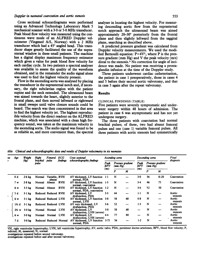
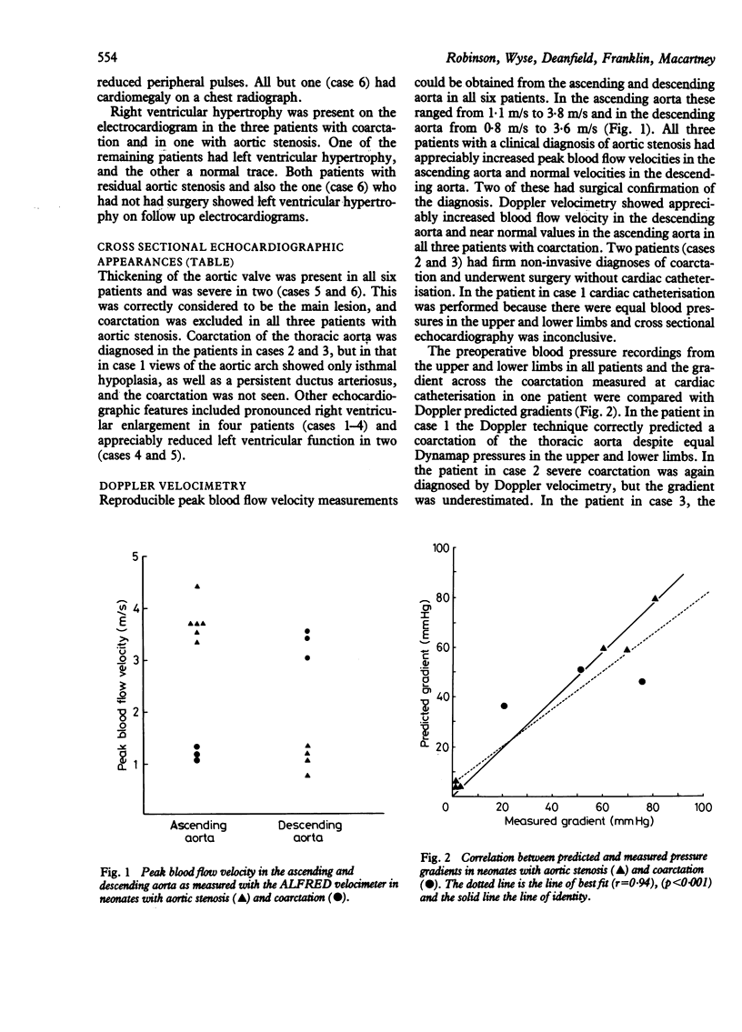
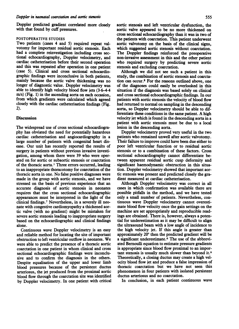
If cross sectional echocardiography in isolation is used to diagnose critical left ventricular outflow obstruction in neonates, false positive and false negative diagnoses may result. Continuous wave Doppler velocimetry was used to measure blood flow velocity in the ascending and descending thoracic aorta in six neonates (aged less than 6 weeks) presenting with reduced or absent peripheral pulses in order to determine the important sites of obstruction. This technique demonstrated abnormal high velocity blood flow jets (three times higher than normal) in the ascending aorta in three patients with normal descending aortic flow velocity, suggesting aortic stenosis. In the other three patients velocity in the ascending aorta was normal but high in the descending aorta, suggesting coarctation. The Doppler diagnosis was confirmed in the five patients who required surgery. Two patients had residual high velocity jets after aortic valvotomy. Both had significant pressure gradients across the aortic valve at cardiac catheterisation with good agreement between actual gradients and those predicted by the Doppler technique. Thus a combined anatomical and physiological approach using cross sectional echocardiography and continuous wave Doppler velocimetry enables accurate non-invasive definition of the site of left ventricular outflow obstruction and may obviate the need for invasive investigation in these sick neonates.
Full text
PDF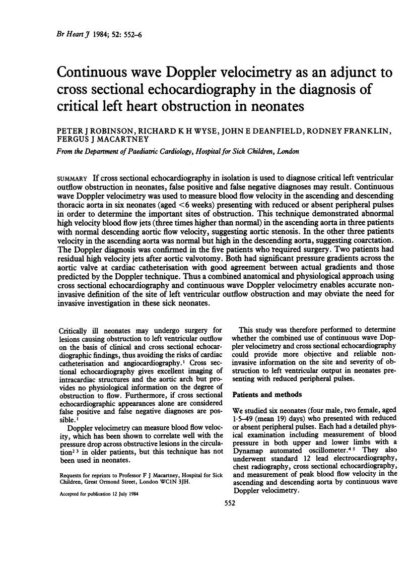
Selected References
These references are in PubMed. This may not be the complete list of references from this article.
- Friesen R. H., Lichtor J. L. Indirect measurement of blood pressure in neonates and infants utilizing an automatic noninvasive oscillometric monitor. Anesth Analg. 1981 Oct;60(10):742–745. [PubMed] [Google Scholar]
- Hatle L., Angelsen B. A., Tromsdal A. Non-invasive assessment of aortic stenosis by Doppler ultrasound. Br Heart J. 1980 Mar;43(3):284–292. doi: 10.1136/hrt.43.3.284. [DOI] [PMC free article] [PubMed] [Google Scholar]
- Hatle L. Noninvasive assessment and differentiation of left ventricular outflow obstruction with Doppler ultrasound. Circulation. 1981 Aug;64(2):381–387. doi: 10.1161/01.cir.64.2.381. [DOI] [PubMed] [Google Scholar]
- Silas J. H., Barker A. T., Ramsay L. E. Clinical evaluation of Dinamap 845 automated blood pressure recorder. Br Heart J. 1980 Feb;43(2):202–205. doi: 10.1136/hrt.43.2.202. [DOI] [PMC free article] [PubMed] [Google Scholar]
- Sink J. D., Smallhorn J. F., Macartney F. J., Taylor J. F., Stark J., de Leval M. R. Management of critical aortic stenosis in infancy. J Thorac Cardiovasc Surg. 1984 Jan;87(1):82–86. [PubMed] [Google Scholar]


