Abstract
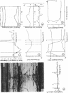
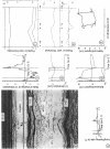
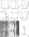
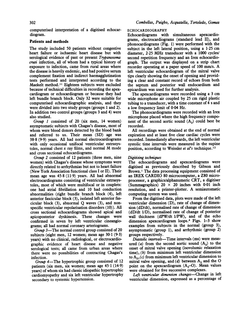
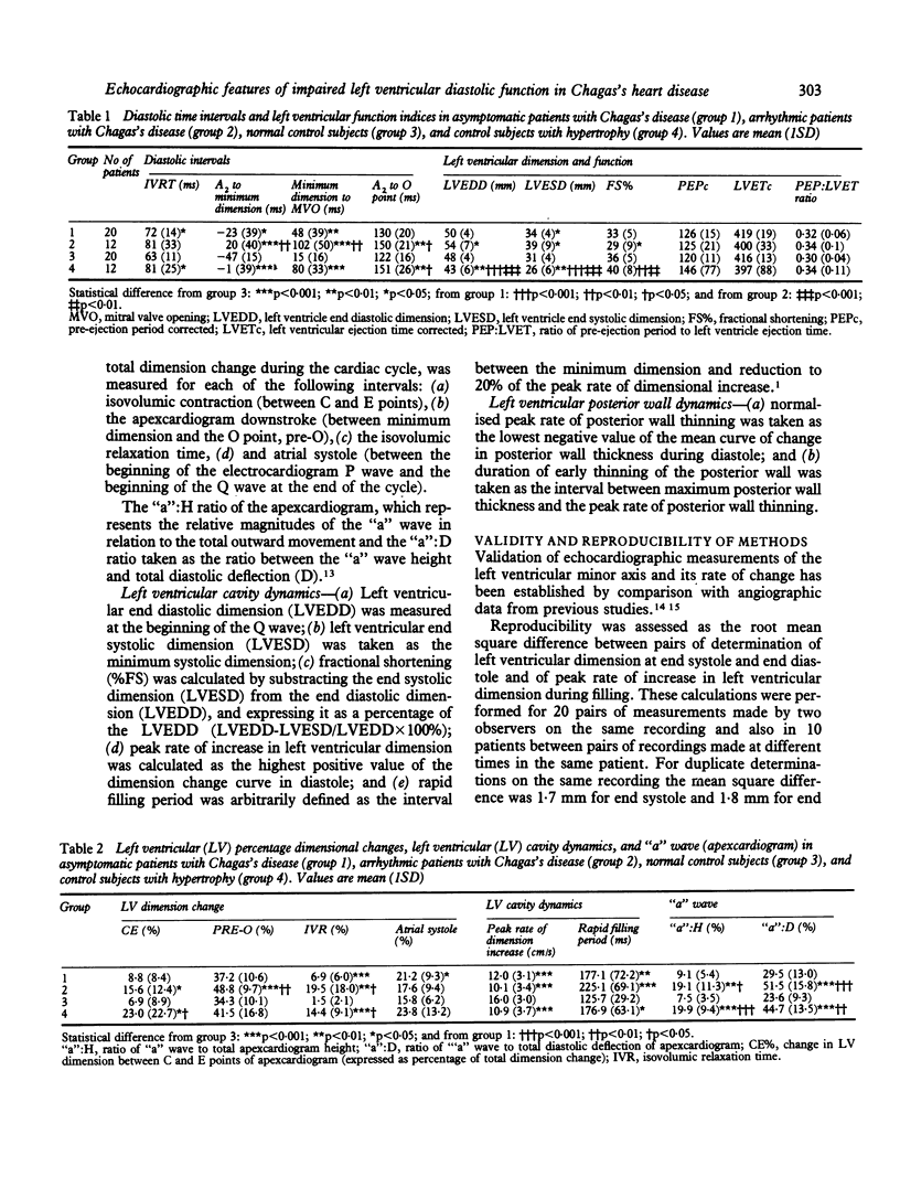
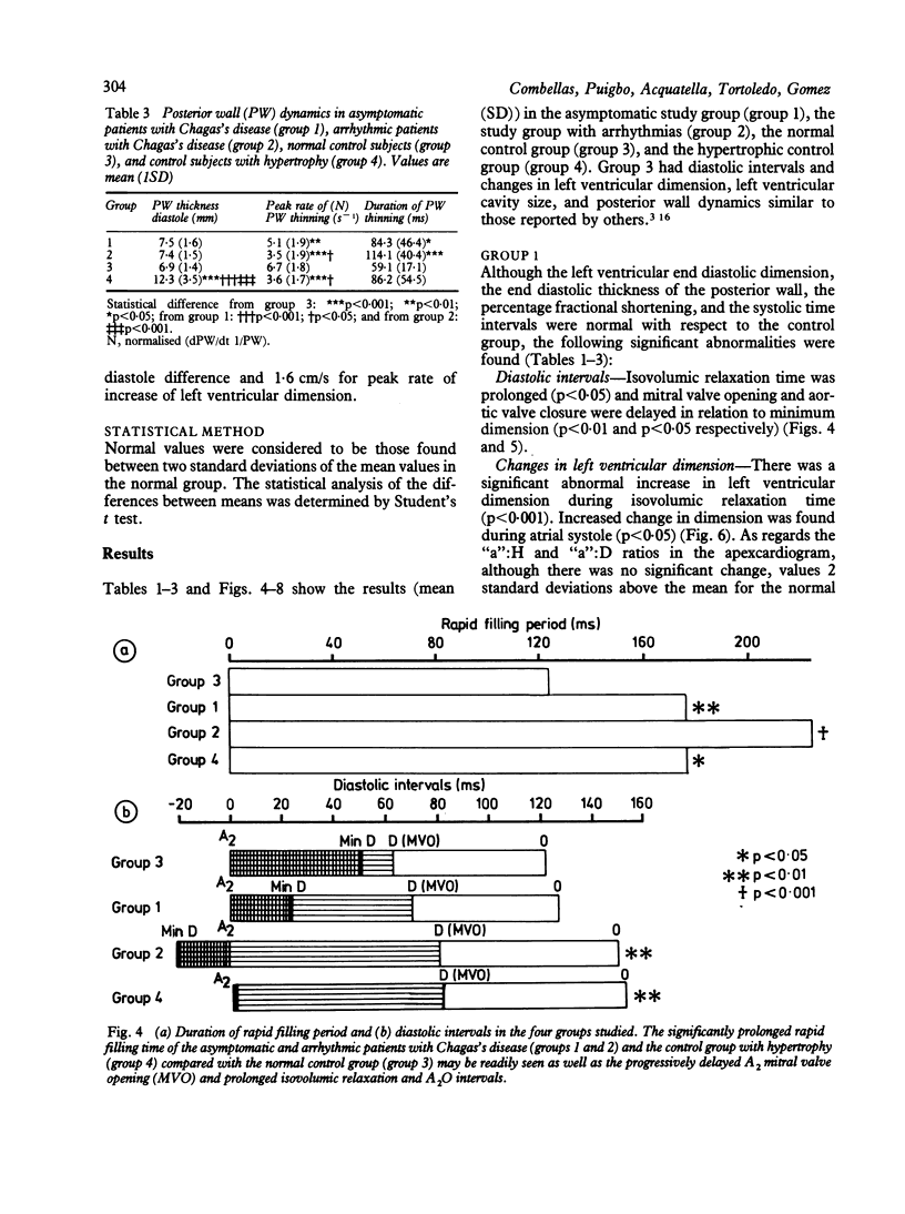
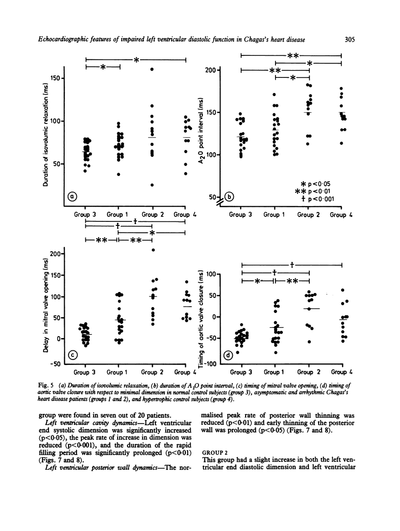
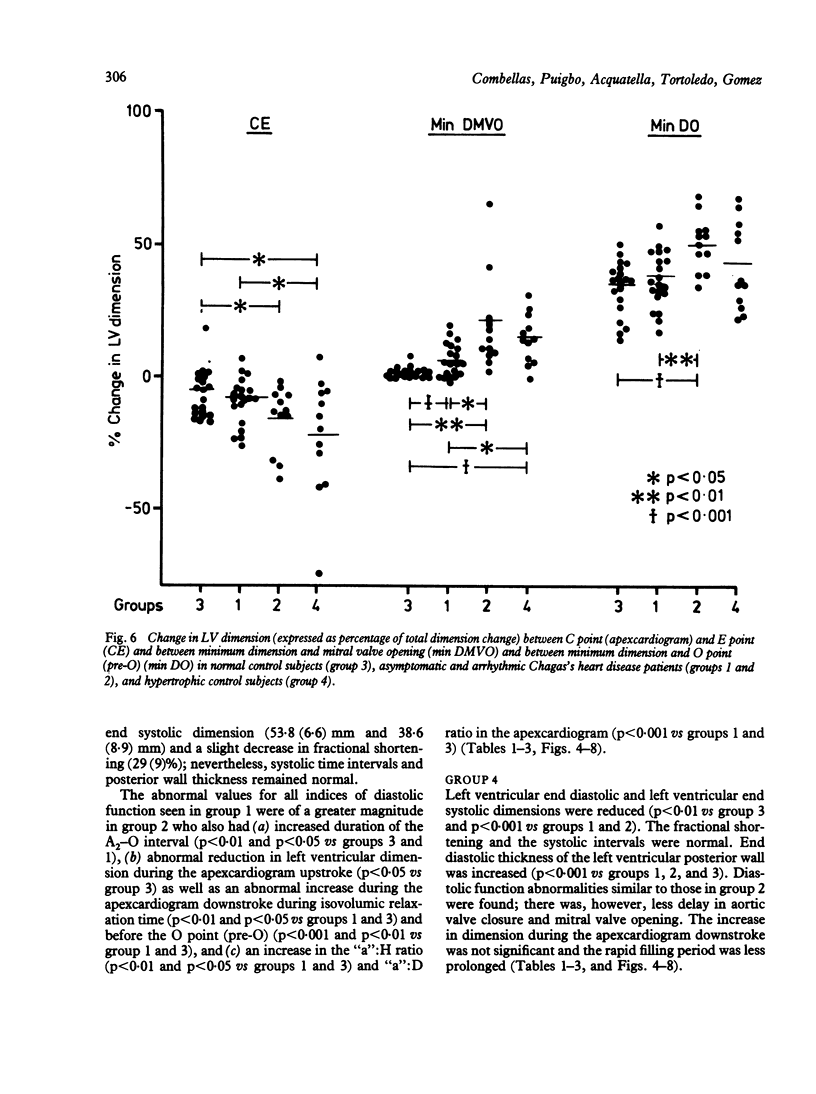
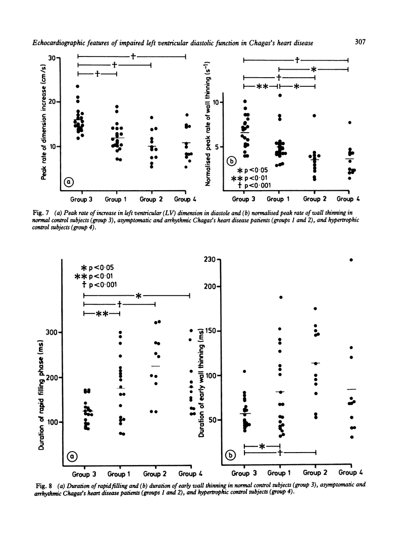
To study left ventricular diastolic function in Chagas's disease, simultaneous echocardiograms, phonocardiograms, and apexcardiograms were recorded in 20 asymptomatic patients with positive Chagas's serology and no signs of heart disease (group 1), 12 with Chagas's heart disease and symptoms of ventricular arrhythmia but no heart failure (group 2), 20 normal subjects (group 3), and 12 patients with left ventricular hypertrophy (group 4). The recordings were digitised to determine left ventricular isovolumic relaxation time and the rate and duration of left ventricular cavity dimension increase and wall thinning. In groups 1 and 2 (a) aortic valve closure (A2) and mitral valve opening were significantly delayed relative to minimum dimension and were associated with prolonged isovolumic relaxation, (b) left ventricular cavity size was abnormally increased during isovolumic relaxation and abnormally reduced during isovolumic contraction, and (c) peak rate of posterior wall thinning and dimension increase were significantly reduced and duration of posterior wall thinning was significantly prolonged; both of these abnormalities occurred at the onset of diastolic filling. These abnormalities were more pronounced in group 2 and were accompanied by an increase in the height of the apexcardiogram "a" wave, an indication of pronounced atrial systole secondary to end diastolic filling impairment due to reduced left ventricular distensibility. Group 4, which had an established pattern of diastolic abnormalities, showed changes similar to those in group 2; however, the delay in aortic valve closure (A2) and in mitral valve opening and the degree of dimension change were greater in the latter group. Thus early isovolumic relaxation and left ventricular abnormalities were pronounced in the patients with Chagas's heart disease and may precede systolic compromise, which may become apparent in later stages of the disease. The digitised method is valuable in the early detection of myocardial damage.
Full text
PDF
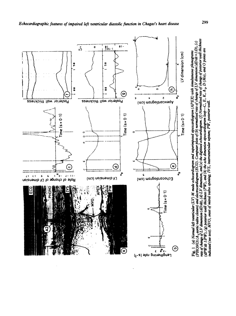
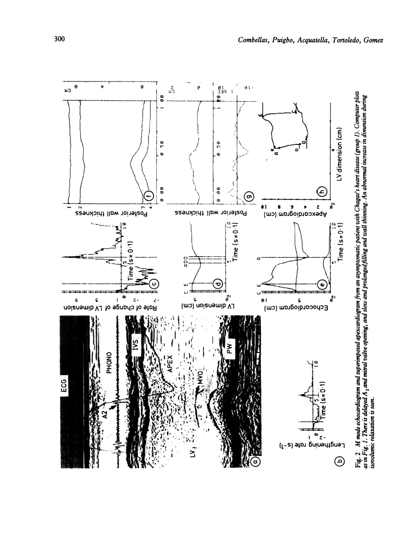
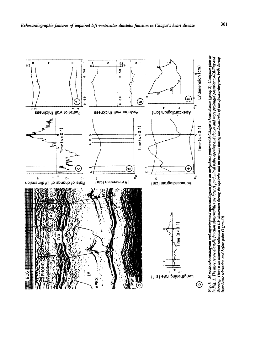
Images in this article
Selected References
These references are in PubMed. This may not be the complete list of references from this article.
- Acquatella H., Schiller N. B., Puigbó J. J., Giordano H., Suárez J. A., Casal H., Arreaza N., Valecillos R., Hirschhaut E. M-mode and two-dimensional echocardiography in chronic Chages' heart disease. A clinical and pathologic study. Circulation. 1980 Oct;62(4):787–799. doi: 10.1161/01.cir.62.4.787. [DOI] [PubMed] [Google Scholar]
- Arreaza N., Puigbó J. J., Acquatella H., Casal H., Giordano H., Valecillos R., Mendoza I., Pérez J. F., Hirschhaut E., Combellas I. Radionuclide evaluation of left-ventricular function in chronic Chagas' cardiomyopathy. J Nucl Med. 1983 Jul;24(7):563–567. [PubMed] [Google Scholar]
- Carrasco H. A., Barboza J. S., Inglessis G., Fuenmayor A., Molina C. Left ventricular cineangiography in Chagas' disease: detection of early myocardial damage. Am Heart J. 1982 Sep;104(3):595–602. doi: 10.1016/0002-8703(82)90232-0. [DOI] [PubMed] [Google Scholar]
- Chen W., Gibson D. Relation of isovolumic relaxation to left ventricular wall movement in man. Br Heart J. 1979 Jul;42(1):51–56. doi: 10.1136/hrt.42.1.51. [DOI] [PMC free article] [PubMed] [Google Scholar]
- Feigenbaum H., Popp R. L., Wolfe S. B., Troy B. L., Pombo J. F., Haine C. L., Dodge H. T. Ultrasound measurements of the left ventricle. A correlative study with angiocardiography. Arch Intern Med. 1972 Mar;129(3):461–467. [PubMed] [Google Scholar]
- Fontes V. F., Sousa J. E., Kormann D. S., Jatene A. D. Avaliaço cineangiocardiográfica da cardiopatie chagásica crónica. Arq Bras Cardiol. 1972 Oct;25(5):375–381. [PubMed] [Google Scholar]
- Gamble W. H., Shaver J. A., Alvares R. F., Salerni R., Reddy P. S. A critical appraisal of diastolic time intervals as a measure of relaxation in left ventricular hypertrophy. Circulation. 1983 Jul;68(1):76–87. doi: 10.1161/01.cir.68.1.76. [DOI] [PubMed] [Google Scholar]
- Gibson D. G., Brown D. J. Measurement of peak rates of left ventricular wall movement in man. Comparison of echocardiography with angiography. Br Heart J. 1975 Jul;37(7):677–683. doi: 10.1136/hrt.37.7.677. [DOI] [PMC free article] [PubMed] [Google Scholar]
- Gibson D. G., Brown D. Measurement of instantaneous left ventricular dimension and filling rate in man, using echocardiography. Br Heart J. 1973 Nov;35(11):1141–1149. doi: 10.1136/hrt.35.11.1141. [DOI] [PMC free article] [PubMed] [Google Scholar]
- Gibson D. G., Traill T. A., Brown D. J. Changes in left ventricular free wall thickness in patients with ischaemic heart disease. Br Heart J. 1977 Dec;39(12):1312–1318. doi: 10.1136/hrt.39.12.1312. [DOI] [PMC free article] [PubMed] [Google Scholar]
- Gibson D. G., Traill T. A., Hall R. J., Brown D. J. Echocardiographic features of secondary left ventricular hypertrophy. Br Heart J. 1979 Jan;41(1):54–59. doi: 10.1136/hrt.41.1.54. [DOI] [PMC free article] [PubMed] [Google Scholar]
- Hanrath P., Mathey D. G., Siegert R., Bleifeld W. Left ventricular relaxation and filling pattern in different forms of left ventricular hypertrophy: an echocardiographic study. Am J Cardiol. 1980 Jan;45(1):15–23. doi: 10.1016/0002-9149(80)90214-3. [DOI] [PubMed] [Google Scholar]
- MAEKELT G. A. [The complement fixation reaction in Chagas' disease]. Z Tropenmed Parasitol. 1960 Aug;11:152–186. [PubMed] [Google Scholar]
- Manolas J., Krayenbuehl H. P., Rutishauser W. Use of apexcardiography to evaluate left ventricular diastolic compliance in human beings. Am J Cardiol. 1979 May;43(5):939–945. doi: 10.1016/0002-9149(79)90356-4. [DOI] [PubMed] [Google Scholar]
- Mattheos M., Shapiro E., Oldershaw P. J., Sacchetti R., Gibson D. G. Non-invasive assessment of changes in left ventricular relaxation by combined phono-, echo-, and mechanocardiography. Br Heart J. 1982 Mar;47(3):253–260. doi: 10.1136/hrt.47.3.253. [DOI] [PMC free article] [PubMed] [Google Scholar]
- Sanderson J. E., Traill T. A., Sutton M. G., Brown D. J., Gibson D. G., Goodwin J. F. Left ventricular relaxation and filling in hypertrophic cardiomyopathy. An echocardiographic study. Br Heart J. 1978 Jun;40(6):596–601. doi: 10.1136/hrt.40.6.596. [DOI] [PMC free article] [PubMed] [Google Scholar]
- Sutton M. G., Tajik A. J., Gibson D. G., Brown D. J., Seward J. B., Guiliani E. R. Echocardiographic assessment of left ventricular filling and septal and posterior wall dynamics in idiopathic hypertrophic subaortic stenosis. Circulation. 1978 Mar;57(3):512–520. doi: 10.1161/01.cir.57.3.512. [DOI] [PubMed] [Google Scholar]
- Traill T. A., Gibson D. G., Brown D. J. Study of left ventricular wall thickness and dimension changes using echocardiography. Br Heart J. 1978 Feb;40(2):162–169. doi: 10.1136/hrt.40.2.162. [DOI] [PMC free article] [PubMed] [Google Scholar]
- Upton M. T., Gibson D. G., Brown D. J. Echocardiographic assessment of abnormal left ventricular relaxation in man. Br Heart J. 1976 Oct;38(10):1001–1009. doi: 10.1136/hrt.38.10.1001. [DOI] [PMC free article] [PubMed] [Google Scholar]
- Upton M. T., Gibson D. G. The study of left ventricular function from digitized echocardiograms. Prog Cardiovasc Dis. 1978 Mar-Apr;20(5):359–384. doi: 10.1016/0033-0620(78)90003-8. [DOI] [PubMed] [Google Scholar]
- Venco A., Gibson D. G., Brown D. J. Relation between apex cardiogram and changes in left ventricular pressure and dimension. Br Heart J. 1977 Feb;39(2):117–125. doi: 10.1136/hrt.39.2.117. [DOI] [PMC free article] [PubMed] [Google Scholar]
- Weissler A. M., Harris W. S., Schoenfeld C. D. Bedside technics for the evaluation of ventricular function in man. Am J Cardiol. 1969 Apr;23(4):577–583. doi: 10.1016/0002-9149(69)90012-5. [DOI] [PubMed] [Google Scholar]


