Abstract
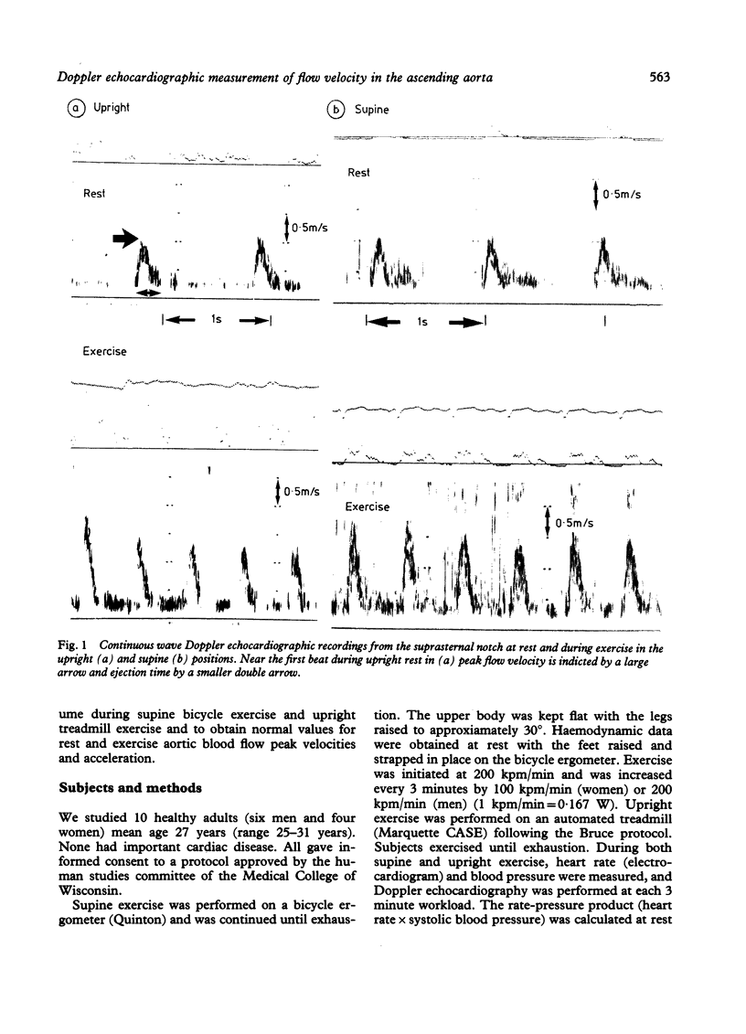
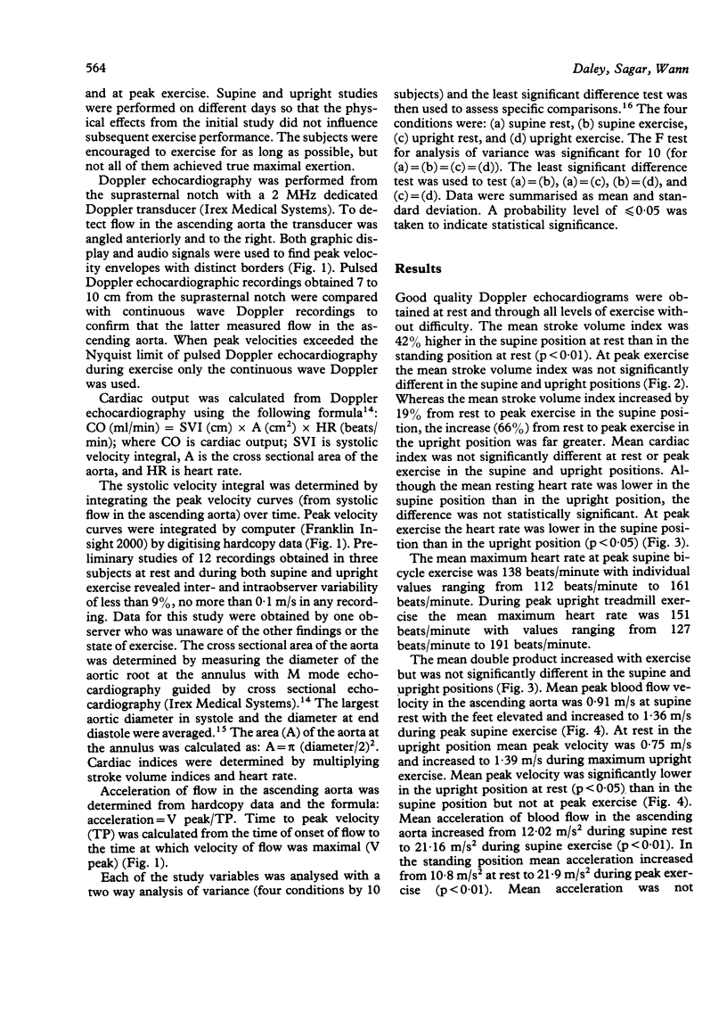
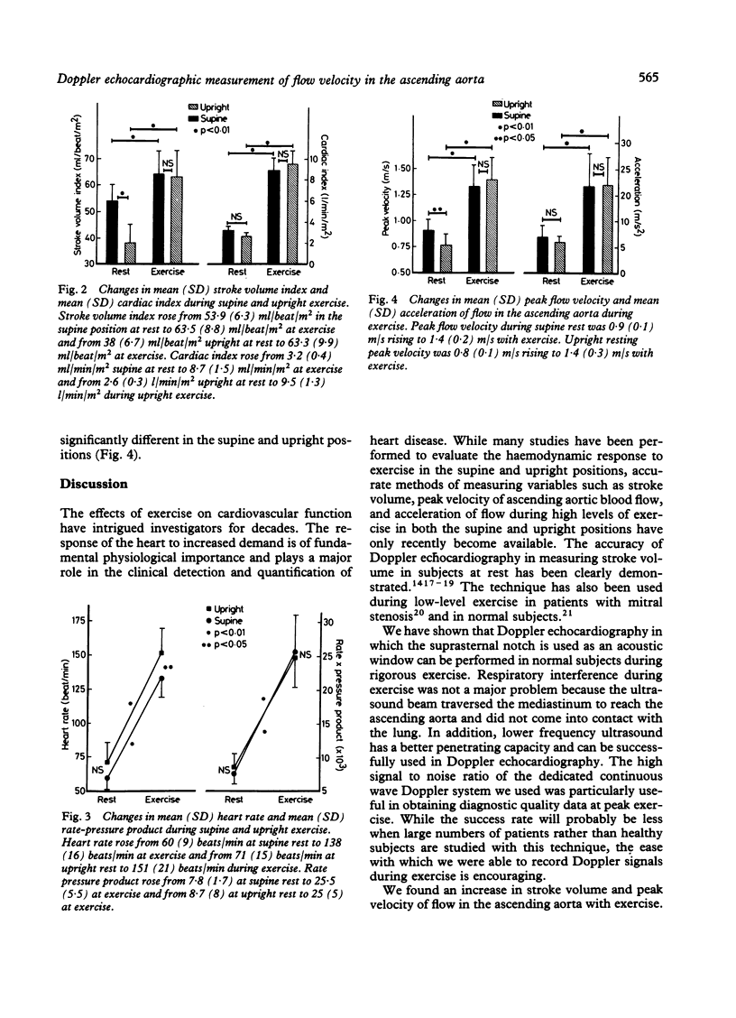
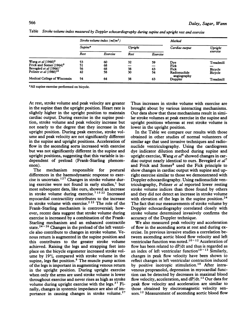
Doppler echocardiography was used to measure stroke volume, peak flow velocity, and acceleration of flow in the ascending aorta in 10 healthy young volunteers during unlimited supine bicycle exercise and upright treadmill exercise. High quality studies were obtained in all subjects through the suprasternal notch acoustic window; there was no appreciable degradation in Doppler signal caused by interference by increased respiration or chest wall motion. Stroke volume index increased from 54 ml/m2 at rest to 63.5 ml/m2 at peak supine exercise and from 38 ml/m2 standing at rest to 63.3 ml/m2 during peak upright exercise. Mean peak flow velocity rose from 0.91 m/s at supine rest to 1.36 m/s during maximum supine exercise. In the upright position mean peak flow velocity increased from 0.75 m/s at rest to 1.39 m/s during maximum exercise. Mean peak velocities were lower in the upright position at rest but were not significantly different at peak exercise. Mean acceleration of flow in the ascending aorta increased from 12.02 m/s2 during supine rest to 21.6 m/s2 during supine exercise and from 10.8 m/s2 at rest on the treadmill to 21.9 m/s2 during peak upright exercise. This study shows that echocardiographic measurement of ascending aortic blood flow by the Doppler technique is feasible even during vigorous exercise; that stroke volume and peak flow velocity at rest are lower in the upright position than in the supine position but equalise at peak exercise; and that acceleration of flow in the ascending aorta is the same in both the supine and upright positions and increases equally at peak exercise in both positions.
Full text
PDF
Images in this article
Selected References
These references are in PubMed. This may not be the complete list of references from this article.
- Bevegård S., Freyschuss U., Strandell T. Circulatory adaptation to arm and leg exercise in supine and sitting position. J Appl Physiol. 1966 Jan;21(1):37–46. doi: 10.1152/jappl.1966.21.1.37. [DOI] [PubMed] [Google Scholar]
- Colocousis J. S., Huntsman L. L., Curreri P. W. Estimation of stroke volume changes by ultrasonic doppler. Circulation. 1977 Dec;56(6):914–917. doi: 10.1161/01.cir.56.6.914. [DOI] [PubMed] [Google Scholar]
- Crawford M. H., White D. H., Amon K. W. Echocardiographic evaluation of left ventricular size and performance during handgrip and supine and upright bicycle exercise. Circulation. 1979 Jun;59(6):1188–1196. doi: 10.1161/01.cir.59.6.1188. [DOI] [PubMed] [Google Scholar]
- FRICK M. H., SOMER T. BASE-LINE EFFECTS ON RESPONSE OF STROKE VOLUME TO LEG EXERCISE IN THE SUPINE POSITION. J Appl Physiol. 1964 Jul;19:639–643. doi: 10.1152/jappl.1964.19.4.639. [DOI] [PubMed] [Google Scholar]
- GREENFIELD J. C., Jr, PATEL D. J. Relation between pressure and diameter in the ascending aorta of man. Circ Res. 1962 May;10:778–781. doi: 10.1161/01.res.10.5.778. [DOI] [PubMed] [Google Scholar]
- Gorlin R., Cohen L. S., Elliott W. C., Klein M. D., Lane F. J. Effect of supine exercise on left ventricular volume and oxygen consumption in man. Circulation. 1965 Sep;32(3):361–371. doi: 10.1161/01.cir.32.3.361. [DOI] [PubMed] [Google Scholar]
- Grimby G., Nilsson N. J., Saltin B. Cardiac output during submaximal and maximal exercise in active middle-aged athletes. J Appl Physiol. 1966 Jul;21(4):1150–1156. doi: 10.1152/jappl.1966.21.4.1150. [DOI] [PubMed] [Google Scholar]
- Horwitz L. D., Atkins J. M., Leshin S. J. Role of the Frank-Starling mechanism in exercise. Circ Res. 1972 Dec;31(6):868–875. doi: 10.1161/01.res.31.6.868. [DOI] [PubMed] [Google Scholar]
- Huntsman L. L., Stewart D. K., Barnes S. R., Franklin S. B., Colocousis J. S., Hessel E. A. Noninvasive Doppler determination of cardiac output in man. Clinical validation. Circulation. 1983 Mar;67(3):593–602. doi: 10.1161/01.cir.67.3.593. [DOI] [PubMed] [Google Scholar]
- Ihlen H., Amlie J. P., Dale J., Forfang K., Nitter-Hauge S., Otterstad J. E., Simonsen S., Myhre E. Determination of cardiac output by Doppler echocardiography. Br Heart J. 1984 Jan;51(1):54–60. doi: 10.1136/hrt.51.1.54. [DOI] [PMC free article] [PubMed] [Google Scholar]
- Klinke W. P., Christie L. G., Nichols W. W., Ray M. E., Curry R. C., Pepine C. J., Conti C. R. Use of catheter-tip velocity--pressure transducer to evaluate left ventricular function in man: effects of intravenous propranolol. Circulation. 1980 May;61(5):946–954. doi: 10.1161/01.cir.61.5.946. [DOI] [PubMed] [Google Scholar]
- Kolettis M., Jenkins B. S., Webb-Peploe M. M. Assessment of left ventricular function by indices derived from aortic flow velocity. Br Heart J. 1976 Jan;38(1):18–31. doi: 10.1136/hrt.38.1.18. [DOI] [PMC free article] [PubMed] [Google Scholar]
- Loeppky J. A., Greene E. R., Hoekenga D. E., Caprihan A., Luft U. C. Beat-by-beat stroke volume assessment by pulsed Doppler in upright and supine exercise. J Appl Physiol Respir Environ Exerc Physiol. 1981 Jun;50(6):1173–1182. doi: 10.1152/jappl.1981.50.6.1173. [DOI] [PubMed] [Google Scholar]
- Magnin P. A., Stewart J. A., Myers S., von Ramm O., Kisslo J. A. Combined doppler and phased-array echocardiographic estimation of cardiac output. Circulation. 1981 Feb;63(2):388–392. doi: 10.1161/01.cir.63.2.388. [DOI] [PubMed] [Google Scholar]
- Nishimura R. A., Callahan M. J., Schaff H. V., Ilstrup D. M., Miller F. A., Tajik A. J. Noninvasive measurement of cardiac output by continuous-wave Doppler echocardiography: initial experience and review of the literature. Mayo Clin Proc. 1984 Jul;59(7):484–489. doi: 10.1016/s0025-6196(12)60438-8. [DOI] [PubMed] [Google Scholar]
- Parker J. O., Case R. B. Normal left ventricular function. Circulation. 1979 Jul;60(1):4–12. doi: 10.1161/01.cir.60.1.4. [DOI] [PubMed] [Google Scholar]
- Poliner L. R., Dehmer G. J., Lewis S. E., Parkey R. W., Blomqvist C. G., Willerson J. T. Left ventricular performance in normal subjects: a comparison of the responses to exercise in the upright and supine positions. Circulation. 1980 Sep;62(3):528–534. doi: 10.1161/01.cir.62.3.528. [DOI] [PubMed] [Google Scholar]
- REEVES J. T., GROVER R. F., BLOUNT S. G., Jr, FILLEY G. F. Cardiac output response to standing and treadmill walking. J Appl Physiol. 1961 Mar;16:283–288. doi: 10.1152/jappl.1961.16.2.283. [DOI] [PubMed] [Google Scholar]
- RUSHMER R. F. Constancy of stroke volume in ventricular responses to exertion. Am J Physiol. 1959 Apr;196(4):745–750. doi: 10.1152/ajplegacy.1959.196.4.745. [DOI] [PubMed] [Google Scholar]
- RUSHMER R. F. INITIAL VENTRICULAR IMPULSE. A POTENTIAL KEY TO CARDIAC EVALUATION. Circulation. 1964 Feb;29:268–283. doi: 10.1161/01.cir.29.2.268. [DOI] [PubMed] [Google Scholar]
- Stenberg J., Astrand P. O., Ekblom B., Royce J., Saltin B. Hemodynamic response to work with different muscle groups, sitting and supine. J Appl Physiol. 1967 Jan;22(1):61–70. doi: 10.1152/jappl.1967.22.1.61. [DOI] [PubMed] [Google Scholar]
- THEILEN E. O., GREGG D. E., ROTTA A. Exercise and cardiac work response at high altitude. Circulation. 1955 Sep;12(3):383–390. doi: 10.1161/01.cir.12.3.383. [DOI] [PubMed] [Google Scholar]
- Vatner S. F., Franklin D., Higgins C. B., Patrick T., Braunwald E. Left ventricular response to severe exertion in untethered dogs. J Clin Invest. 1972 Dec;51(12):3052–3060. doi: 10.1172/JCI107132. [DOI] [PMC free article] [PubMed] [Google Scholar]
- WANG Y., MARSHALL R. J., SHEPHERD J. T. The effect of changes in posture and of graded exercise on stroke volume in man. J Clin Invest. 1960 Jul;39:1051–1061. doi: 10.1172/JCI104120. [DOI] [PMC free article] [PubMed] [Google Scholar]
- WILSON M. F. Left ventricular diameter, posture, and exercise. Circ Res. 1962 Jul;11:90–95. [PubMed] [Google Scholar]


