Abstract
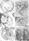
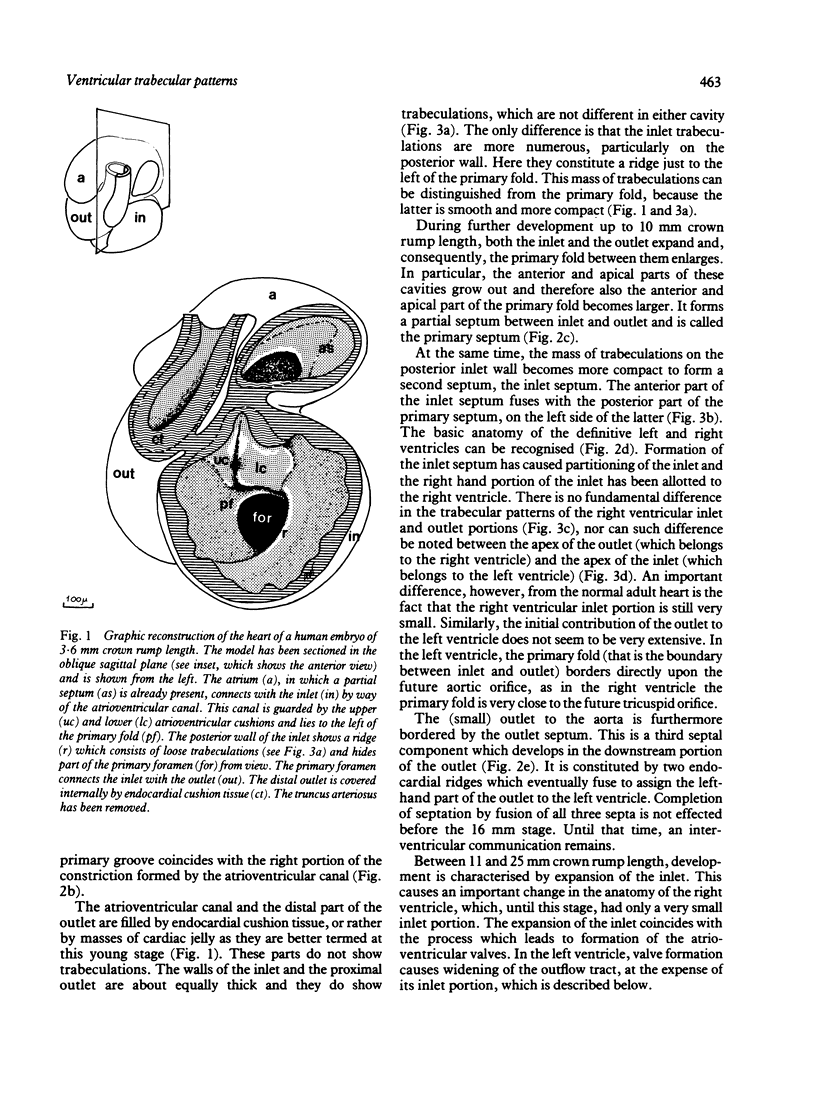
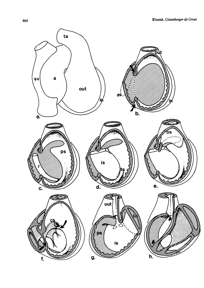
Study of serial sections of human embryos ranging from 3.6 to 25 mm crown rump length shows that the ventricular septum develops from three sources. The primary septum develops between the inlet and outlet which are the two first discernible segments of the ventricular portion of the primary heart tube. Two other septa develop within the inlet and within the outlet, respectively. Before and during septation all ventricular trabeculations are identical. In later stages, the atrioventricular valves and their tension apparatus develop from the inner myocardial layer of the left and right ventricular inlet parts. The outlet trabeculations do not take part in this process. These observations are suggested to explain the typical trabecular patterns of the apices of the mature left and right ventricles, which develop from the inlet and from the outlet, respectively.
Full text
PDF
Images in this article
Selected References
These references are in PubMed. This may not be the complete list of references from this article.
- Anderson R. H., Becker A. E., Van Mierop L. H. What should we call the 'crista'? Br Heart J. 1977 Aug;39(8):856–859. doi: 10.1136/hrt.39.8.856. [DOI] [PMC free article] [PubMed] [Google Scholar]
- Steding G., Seidl W. Contribution to the development of the heart. Part 1: normal development. Thorac Cardiovasc Surg. 1980 Dec;28(6):386–409. doi: 10.1055/s-2007-1022440. [DOI] [PubMed] [Google Scholar]
- Tinkelenberg J. Graphic reconstruction and stereoscopy. J Audiov Media Med. 1980 Apr;3(2):68–71. doi: 10.3109/17453058009154269. [DOI] [PubMed] [Google Scholar]
- Wenink A. C., Gittenberger-de Groot A. C. Straddling mitral and tricuspid valves: morphologic differences and developmental backgrounds. Am J Cardiol. 1982 Jun;49(8):1959–1971. doi: 10.1016/0002-9149(82)90216-8. [DOI] [PubMed] [Google Scholar]
- van Praagh R., Plett J. A., van Praagh S. Single ventricle. Pathology, embryology, terminology and classification. Herz. 1979 Apr;4(2):113–150. [PubMed] [Google Scholar]


