Abstract
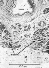
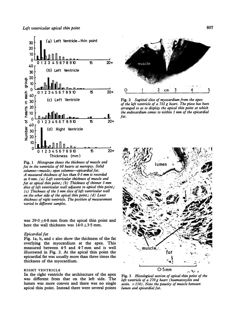
The wall thickness of the apex of the left ventricle has been measured at its thinnest point in 60 adult hearts at necropsy. This measured 2 mm or less in 97% of them and 1 mm or less in 67%.
Full text
PDF
Images in this article
Selected References
These references are in PubMed. This may not be the complete list of references from this article.
- Cook D. J., Bailey I., Strauss H. W., Rouleau J., Wagner H. N., Jr, Pitt B. Thallium-201 for myocardial imaging: appearance of the normal heart. J Nucl Med. 1976 Jul;17(7):583–589. [PubMed] [Google Scholar]
- Fox C. C., Hutchins G. M. The architecture of the human ventricular myocardium. Johns Hopkins Med J. 1972 May;130(5):289–299. [PubMed] [Google Scholar]
- Hunter S. W., Bolduc L., Long V., Quattlebaum F. W. A new myocardial pacemaker lead (sutureless). Chest. 1973 Mar;63(3):430–433. doi: 10.1378/chest.63.3.430. [DOI] [PubMed] [Google Scholar]
- Naclerio E. A., Varriale P. "Screw-in" electrode. New method for permanent ventricular pacing. N Y State J Med. 1974 Dec;74(13):2391–2397. [PubMed] [Google Scholar]
- Vecht R. J., Fontaine C. J., Bradfield J. W. Fatal outcome arising from use of a sutureless "corkscrew" epicardial pacing electrode inserted into apex of left ventricle. Br Heart J. 1976 Dec;38(12):1359–1362. doi: 10.1136/hrt.38.12.1359. [DOI] [PMC free article] [PubMed] [Google Scholar]


