Abstract
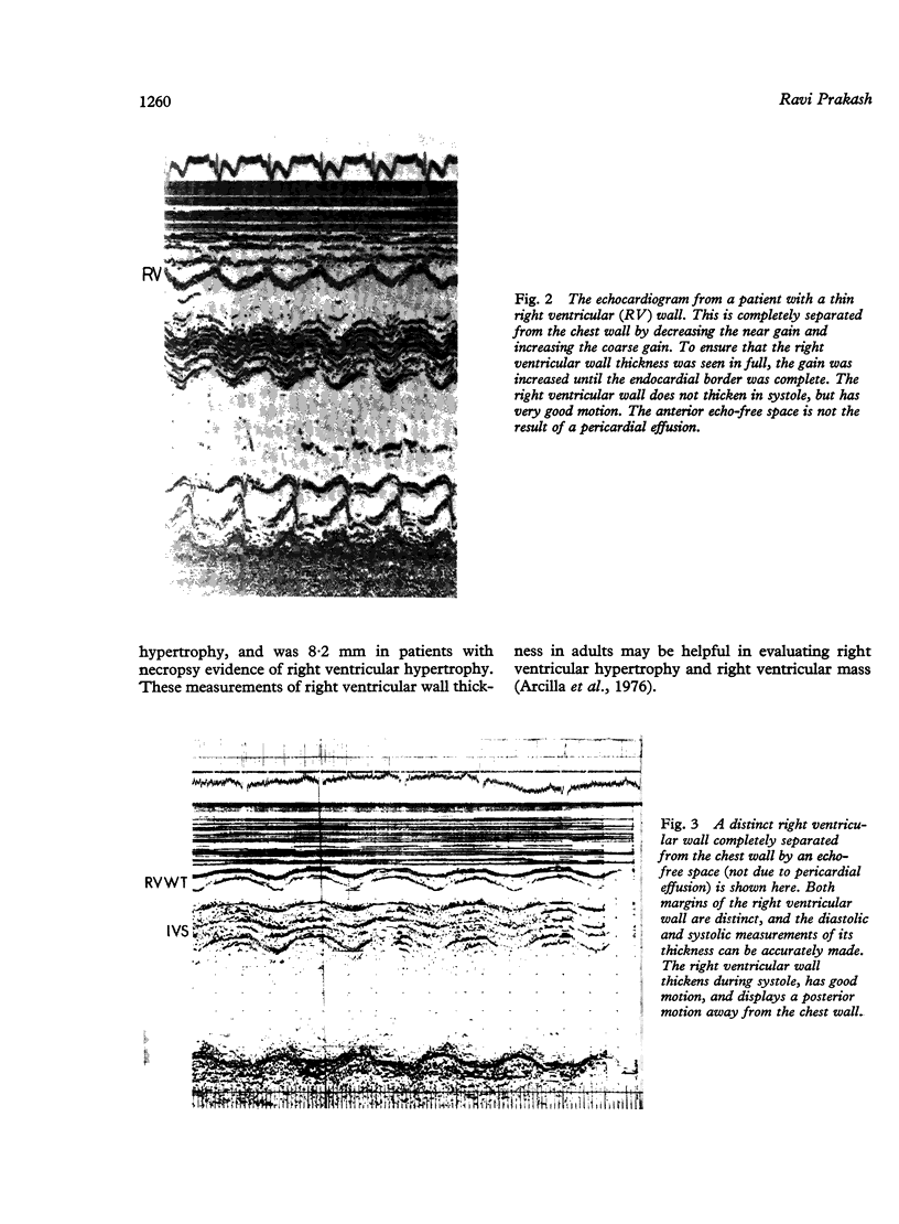
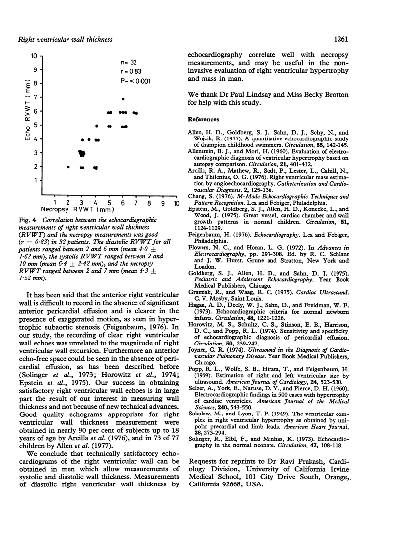
To show that right ventricular wall thickness (RVWT) measurements can be made with precision by echocardiography, we correlated these measurements with those obtained at necropsy in 32 terminal patients. The correlation between the echocardiographic diastolic right ventricular wall thickness (mean 4.0 +/- 1.62 mm) and the necropsy measurement (mean 4.3 +/- 1.52 mm) was good (r = 0.83) in all 32 patients with normal or increased right ventricular wall thickness at necropsy. In 19 patients without necropsy evidence of right ventricular hypertrophy (RVWT less than or equal to 4 mm), the mean diastole and systolic right ventricular wall thickness were 3.0 +/- 0.92 mm and 5.1 +/- 1.64 mm, respectively. In 13 patients with necropsy evidence of right ventricular hypertrophy (RVWT greater than or equal to 5 mm), the mean diastolic and systolic right ventricular wall thicknesses were 5.3 +/- 1.56mm and 8.2 +/- 1.88 mm, respectively. We conclude that technically satisfactory echocardiograms of the right ventricular wall thicknesses. Echocardiography can reliably estimate the diastolic wall thickness and may be helpful in the evaluation of right ventricular hypertrophy.
Full text
PDF
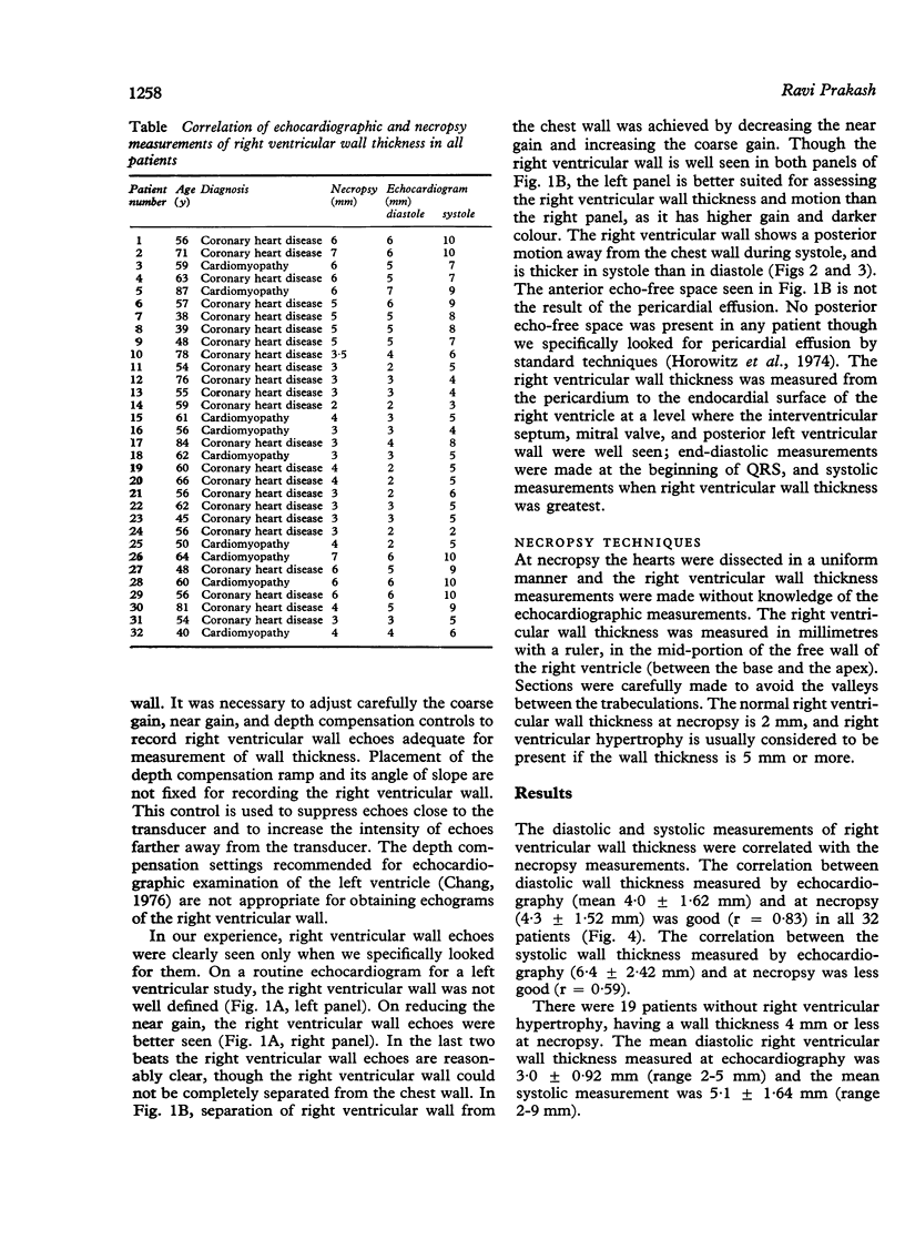
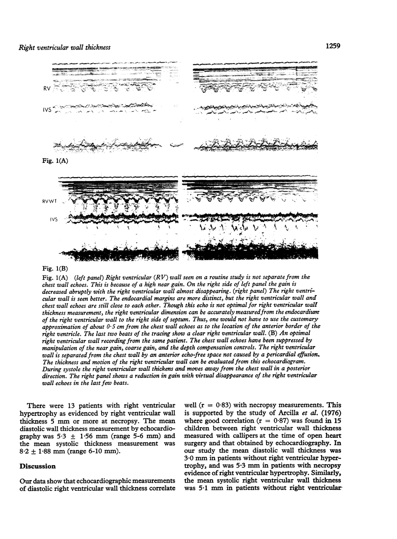
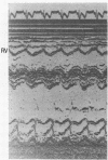
Images in this article
Selected References
These references are in PubMed. This may not be the complete list of references from this article.
- ALLENSTEIN B. J., MORI H. Evaluation of electrocardiographic diagnosis of ventricular hypertrophy based on autopsy comparison. Circulation. 1960 Mar;21:401–412. doi: 10.1161/01.cir.21.3.401. [DOI] [PubMed] [Google Scholar]
- Allen H. D., Goldberg S. J., Sahn D. J., Schy N., Wojcik R. A quantitative echocardiographic study of champion childhood swimmers. Circulation. 1977 Jan;55(1):142–145. doi: 10.1161/01.cir.55.1.142. [DOI] [PubMed] [Google Scholar]
- Arcilla R. A., Mathew R., Sodt P., Lester L., Cahill N., Thilenius O. G. Right ventricular mass estimation by angioechocardiography. Cathet Cardiovasc Diagn. 1976;2(2):125–136. doi: 10.1002/ccd.1810020204. [DOI] [PubMed] [Google Scholar]
- Epstein M. L., Goldberg S. J., Allen H. D., Konecke L., Wood J. Great vessel, cardiac chamber, and wall growth patterns in normal children. Circulation. 1975 Jun;51(6):1124–1129. doi: 10.1161/01.cir.51.6.1124. [DOI] [PubMed] [Google Scholar]
- Hagan A. D., Deely W. J., Sahn D., Friedman W. F. Echocardiographic criteria for normal newborn infants. Circulation. 1973 Dec;48(6):1221–1226. doi: 10.1161/01.cir.48.6.1221. [DOI] [PubMed] [Google Scholar]
- Horowitz M. S., Schultz C. S., Stinson E. B., Harrison D. C., Popp R. L. Sensitivity and specificity of echocardiographic diagnosis of pericardial effusion. Circulation. 1974 Aug;50(2):239–247. doi: 10.1161/01.cir.50.2.239. [DOI] [PubMed] [Google Scholar]
- Popp R. L., Wolfe S. B., Hirata T., Feigenbaum H. Estimation of right and left ventricular size by ultrasound. A study of the echoes from the interventricular septum. Am J Cardiol. 1969 Oct;24(4):523–530. doi: 10.1016/0002-9149(69)90495-0. [DOI] [PubMed] [Google Scholar]
- SELZER A., YORK E., NARUSE D. Y., PIERCE C. H. Electrocardiographic findings in 500 cases with hypertrophy of cardiac ventricles. Am J Med Sci. 1960 Nov;240:543–551. [PubMed] [Google Scholar]
- Solinger R., Elbl F., Minhas K. Echocardiography in the normal neonate. Circulation. 1973 Jan;47(1):108–118. doi: 10.1161/01.cir.47.1.108. [DOI] [PubMed] [Google Scholar]


