Abstract
OBJECTIVE--To identify coronary artery anomalies in patients with tetralogy of Fallot with an aortogram taken with steep caudal and left oblique angulation ("end-on" aortogram). DESIGN--Prospective evaluation of end-on aortogram in the preoperative angiographic assessment of consecutive patients with tetralogy of Fallot. SETTING--Regional paediatric cardiology centre. PATIENTS--34 patients, aged 3 months to 12 years (median age 9 months). METHODS--An aortogram was performed with steep caudal (38 degrees-45 degrees) and left oblique (0 degrees-30 degrees) angulation under general anaesthetic as part of routine preoperative angiographic assessment. RESULTS--The origins and courses of the coronary arteries were visualised in all patients and important coronary artery anomalies were identified in four patients: single left coronary artery; single right coronary artery (two patients); separate high origin of left anterior descending. These anomalous coronary vessels crossed the right ventricular outflow tract. CONCLUSIONS--It is important to identify preoperatively coronary arteries that may interfere with right ventricular outflow tract reconstruction. An aortogram with steep caudal and left oblique angulation is useful in identifying anomalous coronary arteries and more importantly it defines the relation of these vessels to the right ventricular outflow tract.
Full text
PDF
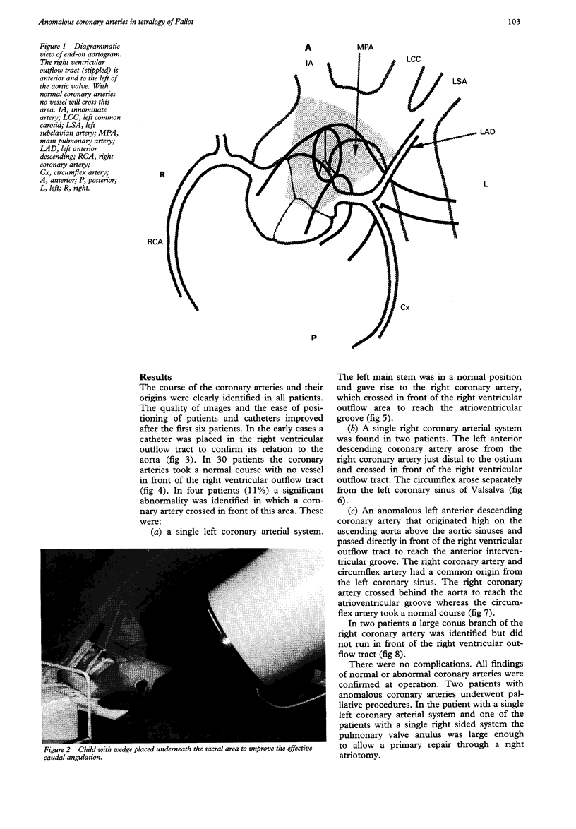
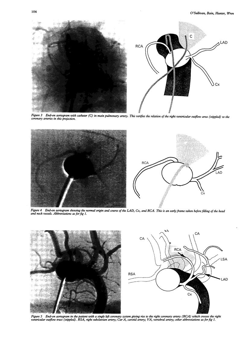
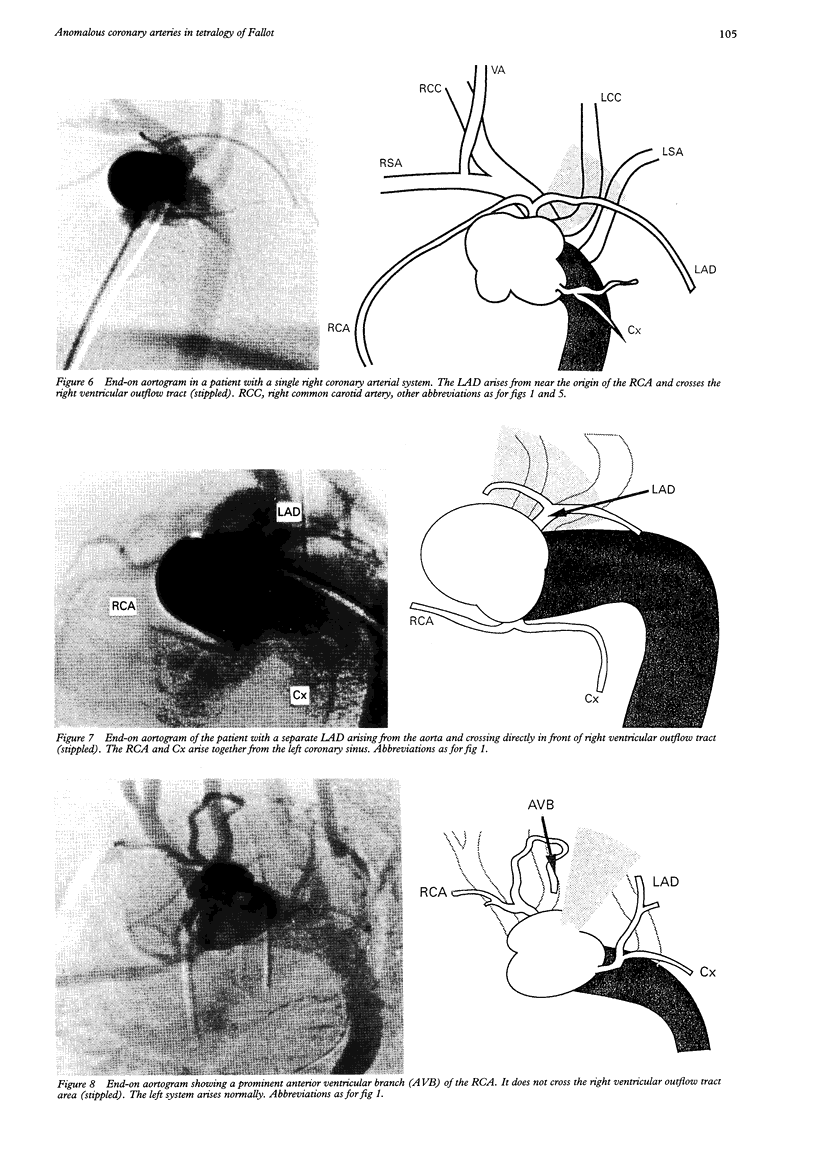
Images in this article
Selected References
These references are in PubMed. This may not be the complete list of references from this article.
- Fellows K. E., Freed M. D., Keane J. F., Praagh R., Bernhard W. F., Castaneda A. C. Results of routine preoperative coronary angiography in tetralogy of Fallot. Circulation. 1975 Mar;51(3):561–566. doi: 10.1161/01.cir.51.3.561. [DOI] [PubMed] [Google Scholar]
- Fellows K. E., Smith J., Keane J. F. Preoperative angiocardiography in infants with tetrad of Fallot. Review of 36 cases. Am J Cardiol. 1981 Jun;47(6):1279–1285. doi: 10.1016/0002-9149(81)90259-9. [DOI] [PubMed] [Google Scholar]
- Jureidini S. B., Appleton R. S., Nouri S. Detection of coronary artery abnormalities in tetralogy of Fallot by two-dimensional echocardiography. J Am Coll Cardiol. 1989 Oct;14(4):960–967. doi: 10.1016/0735-1097(89)90473-7. [DOI] [PubMed] [Google Scholar]
- Mandell V. S., Lock J. E., Mayer J. E., Parness I. A., Kulik T. J. The "laid-back" aortogram: an improved angiographic view for demonstration of coronary arteries in transposition of the great arteries. Am J Cardiol. 1990 Jun 1;65(20):1379–1383. doi: 10.1016/0002-9149(90)91331-y. [DOI] [PubMed] [Google Scholar]
- McManus B. M., Waller B. F., Jones M., Epstein S. E., Roberts W. C. The case for preoperative coronary angiography in patients with tetralogy of Fallot and other complex congenital heart diseases. Am Heart J. 1982 Mar;103(3):451–456. doi: 10.1016/0002-8703(82)90297-6. [DOI] [PubMed] [Google Scholar]


