Abstract
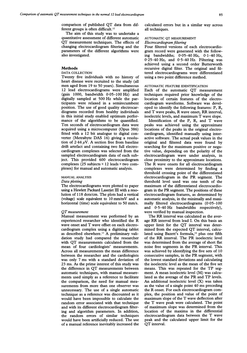
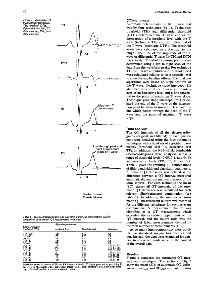
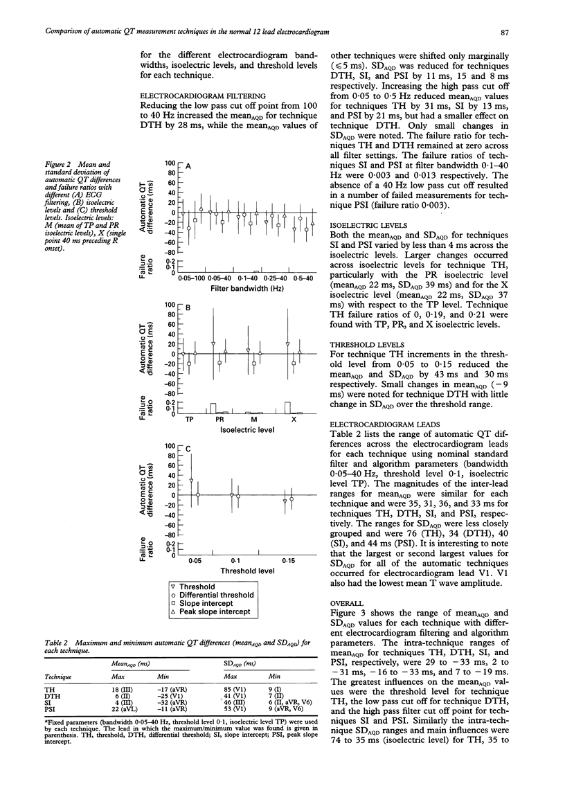
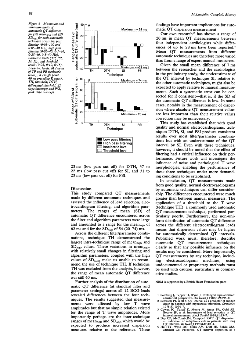
OBJECTIVE--To undertake a quantitative assessment of different automatic QT measurement techniques and investigate the influence of electrocardiogram filtering and algorithm parameters. DESIGN--Four methods for identifying the end of the T wave were compared: (1) threshold crossing of the T wave (TH); (2) threshold crossing of the differential of the T wave (DTH); (3) intercept of an isoelectric level and the maximum T wave slope (SI); and (4) intercept of an isoelectric level and the line passing through the peak and the point of maximum slope of the T wave (PSI). Automatic QT measurements were made by all techniques following different electrocardiogram filtering and, when appropriate, with four different isoelectric levels and with three different threshold levels. SUBJECTS--12 simultaneous standard electrocardiogram leads, containing at least two electrocardiogram complexes, were recorded from 25 healthy volunteers relaxing in a semirecumbent position. MAIN OUTCOME MEASURE--Mean and standard deviation of differences between reference and automatic QT measurements were compared for the four techniques. RESULTS--The mean automatic QT measurements varied by up to 62 ms, which was greater than has been found between manual measurements by experienced clinicians. Technique TH was particularly poor. The other techniques produced consistent results for most electrocardiogram filter, isoelectric level, and threshold level setting; but technique SI underestimated QT relative to the other techniques. CONCLUSION--Different QT measurement techniques produced results which were influenced, to varying degrees, by filtering and technique variables. This is relevant for the inter-comparison of studies using different techniques. Technique TH, a common approach, is not recommended.
Full text
PDF
Selected References
These references are in PubMed. This may not be the complete list of references from this article.
- Ahnve S. Errors in the visual determination of corrected QT (QTc) interval during acute myocardial infarction. J Am Coll Cardiol. 1985 Mar;5(3):699–702. doi: 10.1016/s0735-1097(85)80396-x. [DOI] [PubMed] [Google Scholar]
- Bayés de Luna A., Coumel P., Leclercq J. F. Ambulatory sudden cardiac death: mechanisms of production of fatal arrhythmia on the basis of data from 157 cases. Am Heart J. 1989 Jan;117(1):151–159. doi: 10.1016/0002-8703(89)90670-4. [DOI] [PubMed] [Google Scholar]
- Cowan J. C., Yusoff K., Moore M., Amos P. A., Gold A. E., Bourke J. P., Tansuphaswadikul S., Campbell R. W. Importance of lead selection in QT interval measurement. Am J Cardiol. 1988 Jan 1;61(1):83–87. doi: 10.1016/0002-9149(88)91309-4. [DOI] [PubMed] [Google Scholar]
- Day C. P., McComb J. M., Campbell R. W. QT dispersion: an indication of arrhythmia risk in patients with long QT intervals. Br Heart J. 1990 Jun;63(6):342–344. doi: 10.1136/hrt.63.6.342. [DOI] [PMC free article] [PubMed] [Google Scholar]
- Hii J. T., Wyse D. G., Gillis A. M., Duff H. J., Solylo M. A., Mitchell L. B. Precordial QT interval dispersion as a marker of torsade de pointes. Disparate effects of class Ia antiarrhythmic drugs and amiodarone. Circulation. 1992 Nov;86(5):1376–1382. doi: 10.1161/01.cir.86.5.1376. [DOI] [PubMed] [Google Scholar]
- Laguna P., Thakor N. V., Caminal P., Jané R., Yoon H. R., Bayés de Luna A., Marti V., Guindo J. New algorithm for QT interval analysis in 24-hour Holter ECG: performance and applications. Med Biol Eng Comput. 1990 Jan;28(1):67–73. doi: 10.1007/BF02441680. [DOI] [PubMed] [Google Scholar]
- Merri M., Benhorin J., Alberti M., Locati E., Moss A. J. Electrocardiographic quantitation of ventricular repolarization. Circulation. 1989 Nov;80(5):1301–1308. doi: 10.1161/01.cir.80.5.1301. [DOI] [PubMed] [Google Scholar]
- Mirvis D. M. Spatial variation of QT intervals in normal persons and patients with acute myocardial infarction. J Am Coll Cardiol. 1985 Mar;5(3):625–631. doi: 10.1016/s0735-1097(85)80387-9. [DOI] [PubMed] [Google Scholar]
- Murray A., McLaughlin N. B., Bourke J. P., Doig J. C., Furniss S. S., Campbell R. W. Errors in manual measurement of QT intervals. Br Heart J. 1994 Apr;71(4):386–390. doi: 10.1136/hrt.71.4.386. [DOI] [PMC free article] [PubMed] [Google Scholar]
- O'Donnell J., Lovelace D. E., Knoebel S. B., McHenry P. L. Behavior of the terminal T wave during exercise in normal subjects, patients with symptomatic coronary artery disease and apparently healthy subjects with abnormal ST segment depression. J Am Coll Cardiol. 1985 Jan;5(1):78–84. doi: 10.1016/s0735-1097(85)80087-5. [DOI] [PubMed] [Google Scholar]
- Schwartz P. J., Wolf S. QT interval prolongation as predictor of sudden death in patients with myocardial infarction. Circulation. 1978 Jun;57(6):1074–1077. doi: 10.1161/01.cir.57.6.1074. [DOI] [PubMed] [Google Scholar]
- Somberg J., Tepper D., Wynn J. Prolonged repolarization: a historical perspective. Am Heart J. 1985 Feb;109(2):395–398. doi: 10.1016/0002-8703(85)90625-8. [DOI] [PubMed] [Google Scholar]


