Abstract
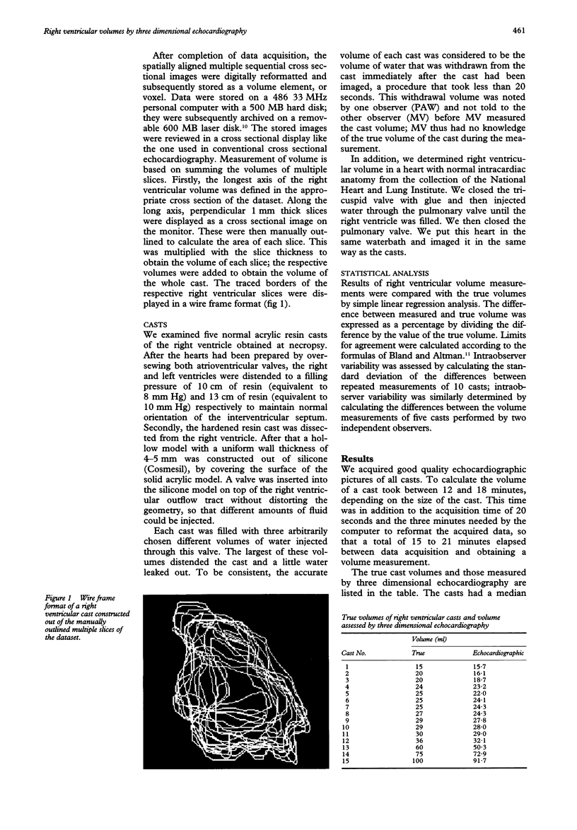
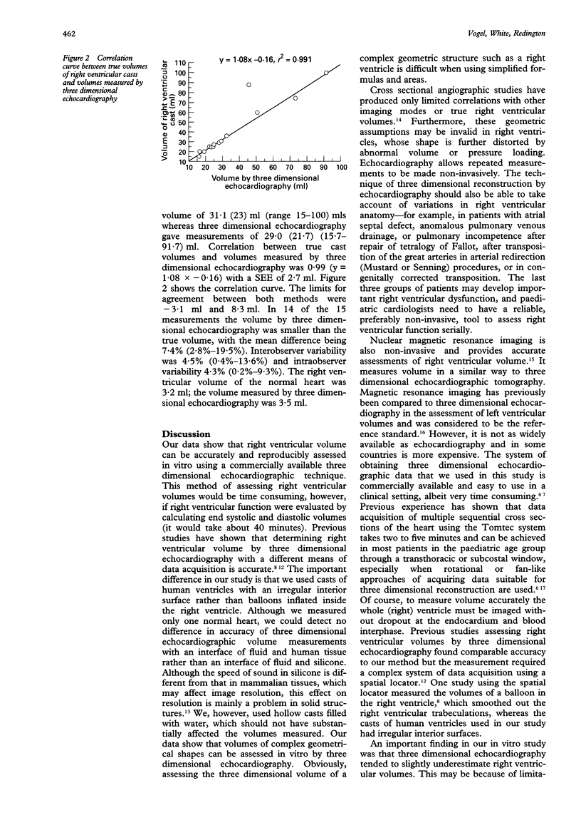
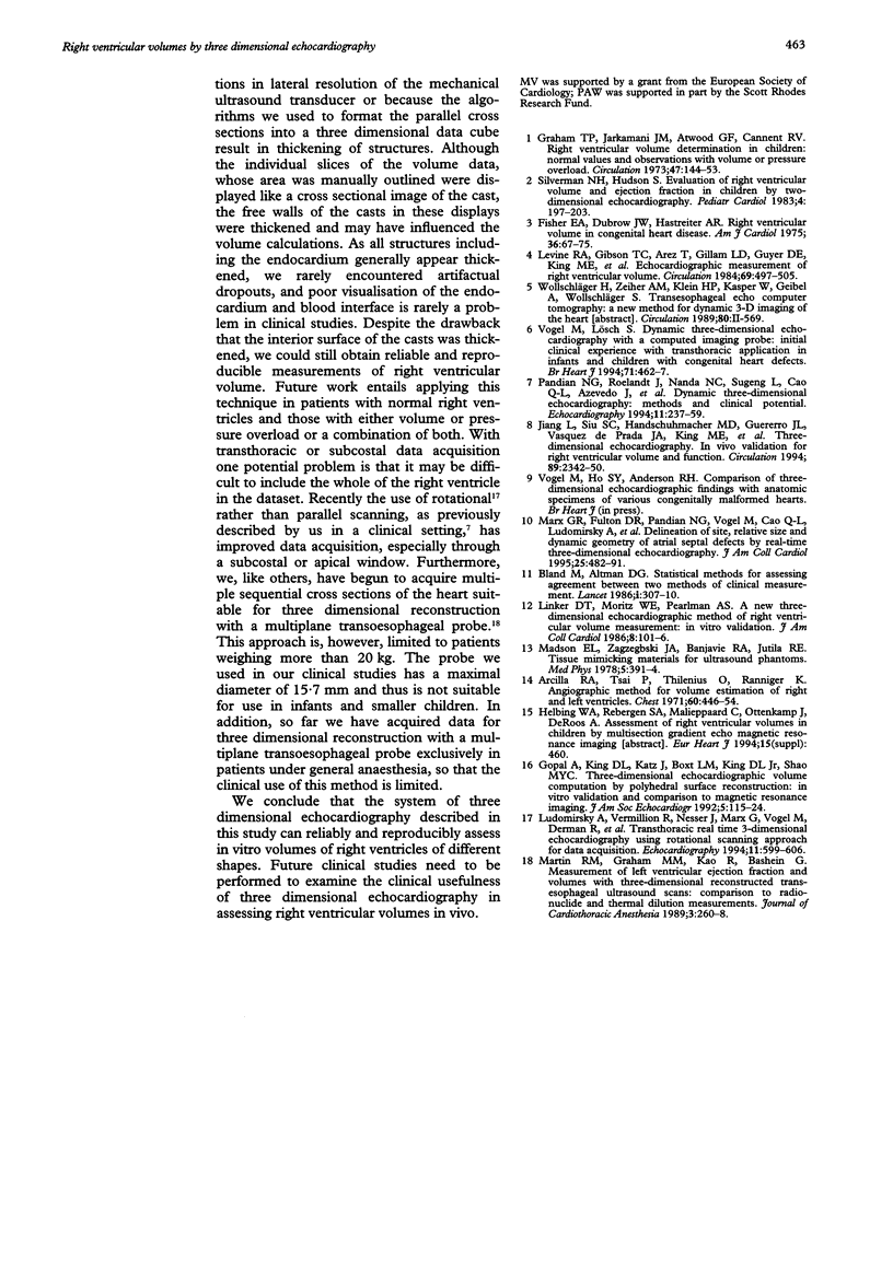
OBJECTIVE--Evaluation of ability of three dimensional echocardiography to accurately assess right ventricular volumes in vitro. METHODS--Silicone casts of normal human right ventricles were examined. Each was filled with three different volumes of water to yield 15 different measurements. The casts were examined in a waterbath with three dimensional echocardiography using a 7.5 MHz ultrasound probe mounted in a scan frame. It was steered by a stepper motor, which moved the probe in steps of 0.25 mm over a distance of 5.9 cm inside the frame, acquiring an image at each step. 236 parallel slices of the cast were thus obtained, forming the three dimensional dataset. The longest axis of the right ventricular volume was defined and the area of perpendicular 1 mm thick slices was outlined manually to calculate the area of each slice. This was multiplied by the slice thickness to obtain the volume of each slice; the respective volumes were added to obtain the volume of the whole cast. RESULTS--The casts had a median volume of 31.1 (23) ml (range 15-100); three dimensional echocardiography gave a median volume of 29.0 (21.7) ml (15.7-91.7). Interobserver variability was 4.5% (0.4%-13.6%) and intraobserver variability 4.3% (0.2%-9.3%). Correlation between real cast volumes and volumes measured by three dimensional echocardiography was 0.99 (y = 1.08 x -0.16) with an SEE of 2.7 ml. Limits for agreement between methods ranged from -3.1 ml to 8.3 ml. In 14 of the 15 measurements, volume by three dimensional echocardiography was smaller than real volume, with the mean difference being 7.4% (2.8%-19.5%). This may be due to the thickening of surfaces of structures when imaged by ultrasonography. CONCLUSION--Right ventricular volumes can accurately be determined by three dimensional echocardiography.
Full text
PDF


Images in this article
Selected References
These references are in PubMed. This may not be the complete list of references from this article.
- Arcilla R. A., Tsai P., Thilenius O., Ranniger K. Angiographic method for volume estimation of right and left ventricles. Chest. 1971 Nov;60(5):446–454. doi: 10.1378/chest.60.5.446. [DOI] [PubMed] [Google Scholar]
- Benchimol A., Desser K. B., Hastreiter A. R. Right ventricular volume in congenital heart disease. Am J Cardiol. 1975 Jul;36(1):67–75. [PubMed] [Google Scholar]
- Bland J. M., Altman D. G. Statistical methods for assessing agreement between two methods of clinical measurement. Lancet. 1986 Feb 8;1(8476):307–310. [PubMed] [Google Scholar]
- Gopal A. S., King D. L., Katz J., Boxt L. M., King D. L., Jr, Shao M. Y. Three-dimensional echocardiographic volume computation by polyhedral surface reconstruction: in vitro validation and comparison to magnetic resonance imaging. J Am Soc Echocardiogr. 1992 Mar-Apr;5(2):115–124. doi: 10.1016/s0894-7317(14)80541-5. [DOI] [PubMed] [Google Scholar]
- Graham T. P., Jr, Jarmakani J. M., Atwood G. F., Canent R. V., Jr Right ventricular volume determinations in children. Normal values and observations with volume or pressure overload. Circulation. 1973 Jan;47(1):144–153. doi: 10.1161/01.cir.47.1.144. [DOI] [PubMed] [Google Scholar]
- Jiang L., Siu S. C., Handschumacher M. D., Luis Guererro J., Vazquez de Prada J. A., King M. E., Picard M. H., Weyman A. E., Levine R. A. Three-dimensional echocardiography. In vivo validation for right ventricular volume and function. Circulation. 1994 May;89(5):2342–2350. doi: 10.1161/01.cir.89.5.2342. [DOI] [PubMed] [Google Scholar]
- Levine R. A., Gibson T. C., Aretz T., Gillam L. D., Guyer D. E., King M. E., Weyman A. E. Echocardiographic measurement of right ventricular volume. Circulation. 1984 Mar;69(3):497–505. doi: 10.1161/01.cir.69.3.497. [DOI] [PubMed] [Google Scholar]
- Linker D. T., Moritz W. E., Pearlman A. S. A new three-dimensional echocardiographic method of right ventricular volume measurement: in vitro validation. J Am Coll Cardiol. 1986 Jul;8(1):101–106. doi: 10.1016/s0735-1097(86)80098-5. [DOI] [PubMed] [Google Scholar]
- Ludomirsky A., Vermilion R., Nesser J., Marx G., Vogel M., Derman R., Pandian N. Transthoracic real-time three-dimensional echocardiography using the rotational scanning approach for data acquisition. Echocardiography. 1994 Nov;11(6):599–606. doi: 10.1111/j.1540-8175.1994.tb01104.x. [DOI] [PubMed] [Google Scholar]
- Madsen E. L., Zagzebski J. A., Banjavie R. A., Jutila R. E. Tissue mimicking materials for ultrasound phantoms. Med Phys. 1978 Sep-Oct;5(5):391–394. doi: 10.1118/1.594483. [DOI] [PubMed] [Google Scholar]
- Martin R. W., Graham M. M., Kao R., Bashein G. Measurement of left ventricular ejection fraction and volumes with three-dimensional reconstructed transesophageal ultrasound scans: comparison to radionuclide and thermal dilution measurements. J Cardiothorac Anesth. 1989 Jun;3(3):260–268. doi: 10.1016/0888-6296(89)90105-1. [DOI] [PubMed] [Google Scholar]
- Marx G. R., Fulton D. R., Pandian N. G., Vogel M., Cao Q. L., Ludomirsky A., Delabays A., Sugeng L., Klas B. Delineation of site, relative size and dynamic geometry of atrial septal defects by real-time three-dimensional echocardiography. J Am Coll Cardiol. 1995 Feb;25(2):482–490. doi: 10.1016/0735-1097(94)00372-w. [DOI] [PubMed] [Google Scholar]
- Silverman N. H., Hudson S. Evaluation of right ventricular volume and ejection fraction in children by two-dimensional echocardiography. Pediatr Cardiol. 1983 Jul-Sep;4(3):197–203. doi: 10.1007/BF02242255. [DOI] [PubMed] [Google Scholar]
- Vogel M., Lösch S. Dynamic three-dimensional echocardiography with a computed tomography imaging probe: initial clinical experience with transthoracic application in infants and children with congenital heart defects. Br Heart J. 1994 May;71(5):462–467. doi: 10.1136/hrt.71.5.462. [DOI] [PMC free article] [PubMed] [Google Scholar]


