Abstract
OBJECTIVES: The presence of angina pectoris and myocardial scarring in patients with hypertrophic cardiomyopathy (HCM) suggests that myocardial ischemia is a factor in the pathophysiology of the disease. The clinical evaluation of ischaemia is problematic in HCM as baseline electrocardiographic abnormalities are frequent and thallium-201 perfusion abnormalities correlate poorly with anginal symptoms. Coronary sinus pH measurement using a catheter mounted pH electrode is a validated sensitive technique for the detection of myocardial ischaemia. METHODS AND RESULTS: 11 patients with HCM and chest pain (eight men; mean (SD) (range) age 36 (11) (19-53) years) and six controls (two men; mean (SD) (range) age 49 (11) (31-62) years) with atypical pain and normal coronary angiograms were studied. Eight patients with HCM had baseline ST segment depression of > or = 1 mm and four had reversible perfusion defects during stress 201TI scintigraphy. A catheter mounted hydrogen ion sensitive electrode was introduced into the coronary sinus and pH monitored continuously during dipyridamole infusion (0.56 mg/kg over four min). The maximal change in coronary sinus pH during dipyridamole stress was greater in patients with HCM than in controls (0.082 (0.083) (0 to -0.275) v 0.005 (0.006) (0 to -0.012), P = 0.02). In six patients (four men; mean (SD) (range) age 29 (9) (19-40 years) the development of chest pain was associated with a gradual decline in coronary sinus pH (mean 0.123 (0.089)), peaking at 442 (106) s. There were no relations among left ventricular dimensions, maximal wall thickness, and maximum pH change. In patients with HCM there was a correlation between maximum pH change and maximum heart rate during dipyridamole infusion (r = 0.70, P = 0.02). CONCLUSION: This study provides further evidence that chest pain in patients with HCM is caused by myocardial ischaemia. The role of myocardial ischaemia in the pathophysiology of the disease remains to be determined but coronary sinus pH monitoring provides a method for quantifying and prospectively assessing its effects on clinical presentation and prognosis.
Full text
PDF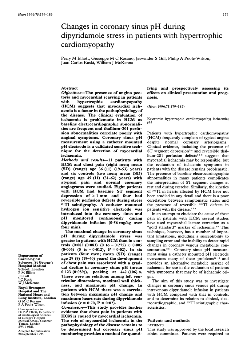
Selected References
These references are in PubMed. This may not be the complete list of references from this article.
- Cannon R. O., 3rd, Dilsizian V., O'Gara P. T., Udelson J. E., Schenke W. H., Quyyumi A., Fananapazir L., Bonow R. O. Myocardial metabolic, hemodynamic, and electrocardiographic significance of reversible thallium-201 abnormalities in hypertrophic cardiomyopathy. Circulation. 1991 May;83(5):1660–1667. doi: 10.1161/01.cir.83.5.1660. [DOI] [PubMed] [Google Scholar]
- Cannon R. O., 3rd, Rosing D. R., Maron B. J., Leon M. B., Bonow R. O., Watson R. M., Epstein S. E. Myocardial ischemia in patients with hypertrophic cardiomyopathy: contribution of inadequate vasodilator reserve and elevated left ventricular filling pressures. Circulation. 1985 Feb;71(2):234–243. doi: 10.1161/01.cir.71.2.234. [DOI] [PubMed] [Google Scholar]
- Cobbe S. M., Poole-Wilson P. A. Catheter tip pH electrodes for continuous intravascular recording. J Med Eng Technol. 1980 May;4(3):122–124. doi: 10.3109/03091908009161104. [DOI] [PubMed] [Google Scholar]
- Cobbe S. M., Poole-Wilson P. A. Continuous coronary sinus and arterial pH monitoring during pacing-induced ischaemia in coronary artery disease. Br Heart J. 1982 Apr;47(4):369–374. doi: 10.1136/hrt.47.4.369. [DOI] [PMC free article] [PubMed] [Google Scholar]
- Cobbe S. M., Poole-Wilson P. A. Continuous coronary sinus and arterial pH monitoring during pacing-induced ischaemia in coronary artery disease. Br Heart J. 1982 Apr;47(4):369–374. doi: 10.1136/hrt.47.4.369. [DOI] [PMC free article] [PubMed] [Google Scholar]
- Crake T., Crean P. A., Shapiro L. M., Rickards A. F., Poole-Wilson P. A. Coronary sinus pH during percutaneous transluminal coronary angioplasty: early development of acidosis during myocardial ischaemia in man. Br Heart J. 1987 Aug;58(2):110–115. doi: 10.1136/hrt.58.2.110. [DOI] [PMC free article] [PubMed] [Google Scholar]
- Frank S., Braunwald E. Idiopathic hypertrophic subaortic stenosis. Clinical analysis of 126 patients with emphasis on the natural history. Circulation. 1968 May;37(5):759–788. doi: 10.1161/01.cir.37.5.759. [DOI] [PubMed] [Google Scholar]
- Frenneaux M. P., Counihan P. J., Caforio A. L., Chikamori T., McKenna W. J. Abnormal blood pressure response during exercise in hypertrophic cardiomyopathy. Circulation. 1990 Dec;82(6):1995–2002. doi: 10.1161/01.cir.82.6.1995. [DOI] [PubMed] [Google Scholar]
- Gertz E. W., Wisneski J. A., Neese R., Bristow J. D., Searle G. L., Hanlon J. T. Myocardial lactate metabolism: evidence of lactate release during net chemical extraction in man. Circulation. 1981 Jun;63(6):1273–1279. doi: 10.1161/01.cir.63.6.1273. [DOI] [PubMed] [Google Scholar]
- Gertz E. W., Wisneski J. A., Neese R., Houser A., Korte R., Bristow J. D. Myocardial lactate extraction: multi-determined metabolic function. Circulation. 1980 Feb;61(2):256–261. doi: 10.1161/01.cir.61.2.256. [DOI] [PubMed] [Google Scholar]
- Gould K. L. Noninvasive assessment of coronary stenoses by myocardial perfusion imaging during pharmacologic coronary vasodilatation. I. Physiologic basis and experimental validation. Am J Cardiol. 1978 Feb;41(2):267–278. doi: 10.1016/0002-9149(78)90165-0. [DOI] [PubMed] [Google Scholar]
- Maron B. J., Gottdiener J. S., Epstein S. E. Patterns and significance of distribution of left ventricular hypertrophy in hypertrophic cardiomyopathy. A wide angle, two dimensional echocardiographic study of 125 patients. Am J Cardiol. 1981 Sep;48(3):418–428. doi: 10.1016/0002-9149(81)90068-0. [DOI] [PubMed] [Google Scholar]
- Maron B. J., Wolfson J. K., Epstein S. E., Roberts W. C. Intramural ("small vessel") coronary artery disease in hypertrophic cardiomyopathy. J Am Coll Cardiol. 1986 Sep;8(3):545–557. doi: 10.1016/s0735-1097(86)80181-4. [DOI] [PubMed] [Google Scholar]
- McKenna W., Deanfield J., Faruqui A., England D., Oakley C., Goodwin J. Prognosis in hypertrophic cardiomyopathy: role of age and clinical, electrocardiographic and hemodynamic features. Am J Cardiol. 1981 Mar;47(3):532–538. doi: 10.1016/0002-9149(81)90535-x. [DOI] [PubMed] [Google Scholar]
- O'Gara P. T., Bonow R. O., Maron B. J., Damske B. A., Van Lingen A., Bacharach S. L., Larson S. M., Epstein S. E. Myocardial perfusion abnormalities in patients with hypertrophic cardiomyopathy: assessment with thallium-201 emission computed tomography. Circulation. 1987 Dec;76(6):1214–1223. doi: 10.1161/01.cir.76.6.1214. [DOI] [PubMed] [Google Scholar]
- O'Gorman D. J., Sheridan D. J. Abnormalities of the coronary circulation associated with left ventricular hypertrophy. Clin Sci (Lond) 1991 Dec;81(6):703–713. doi: 10.1042/cs0810703. [DOI] [PubMed] [Google Scholar]
- Pasternac A., Noble J., Streulens Y., Elie R., Henschke C., Bourassa M. G. Pathophysiology of chest pain in patients with cardiomyopathies and normal coronary arteries. Circulation. 1982 Apr;65(4):778–789. doi: 10.1161/01.cir.65.4.778. [DOI] [PubMed] [Google Scholar]
- Pitcher D., Wainwright R., Maisey M., Curry P., Sowton E. Assessment of chest pain in hypertrophic cardiomyopathy using exercise thallium-201 myocardial scintigraphy. Br Heart J. 1980 Dec;44(6):650–656. doi: 10.1136/hrt.44.6.650. [DOI] [PMC free article] [PubMed] [Google Scholar]
- Romhilt D. W., Bove K. E., Norris R. J., Conyers E., Conradi S., Rowlands D. T., Scott R. C. A critical appraisal of the electrocardiographic criteria for the diagnosis of left ventricular hypertrophy. Circulation. 1969 Aug;40(2):185–195. doi: 10.1161/01.cir.40.2.185. [DOI] [PubMed] [Google Scholar]
- Rubin K. A., Morrison J., Padnick M. B., Binder A. J., Chiaramida S., Margouleff D., Padmanabhan V. T., Gulotta S. J. Idiopathic hypertrophic subaortic stenosis: evaluation of anginal symptoms with thallium-201 myocardial imaging. Am J Cardiol. 1979 Nov;44(6):1040–1045. doi: 10.1016/0002-9149(79)90166-8. [DOI] [PubMed] [Google Scholar]
- Scheler S., Motz W., Strauer B. E. Transient myocardial ischaemia in hypertensives: missing link with left ventricular hypertrophy. Eur Heart J. 1992 Sep;13 (Suppl 500):62–65. doi: 10.1093/eurheartj/13.suppl_d.62. [DOI] [PubMed] [Google Scholar]
- Shapiro L. M., McKenna W. J. Distribution of left ventricular hypertrophy in hypertrophic cardiomyopathy: a two-dimensional echocardiographic study. J Am Coll Cardiol. 1983 Sep;2(3):437–444. doi: 10.1016/s0735-1097(83)80269-1. [DOI] [PubMed] [Google Scholar]
- Stafford W. J., Trohman R. G., Bilsker M., Zaman L., Castellanos A., Myerburg R. J. Cardiac arrest in an adolescent with atrial fibrillation and hypertrophic cardiomyopathy. J Am Coll Cardiol. 1986 Mar;7(3):701–704. doi: 10.1016/s0735-1097(86)80484-3. [DOI] [PubMed] [Google Scholar]
- Takata J., Counihan P. J., Gane J. N., Doi Y., Chikamori T., Ozawa T., McKenna W. J. Regional thallium-201 washout and myocardial hypertrophy in hypertrophic cardiomyopathy and its relation to exertional chest pain. Am J Cardiol. 1993 Jul 15;72(2):211–217. doi: 10.1016/0002-9149(93)90162-6. [DOI] [PubMed] [Google Scholar]
- Tanaka M., Fujiwara H., Onodera T., Wu D. J., Matsuda M., Hamashima Y., Kawai C. Quantitative analysis of narrowings of intramyocardial small arteries in normal hearts, hypertensive hearts, and hearts with hypertrophic cardiomyopathy. Circulation. 1987 Jun;75(6):1130–1139. doi: 10.1161/01.cir.75.6.1130. [DOI] [PubMed] [Google Scholar]
- von Dohlen T. W., Prisant L. M., Frank M. J. Significance of positive or negative thallium-201 scintigraphy in hypertrophic cardiomyopathy. Am J Cardiol. 1989 Sep 1;64(8):498–503. doi: 10.1016/0002-9149(89)90428-1. [DOI] [PubMed] [Google Scholar]


