Abstract
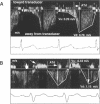
OBJECTIVE: To analyse profiles of coronary artery flow velocity at rest in patients with aortic stenosis and to determine whether changes of the coronary artery flow velocities are related to symptoms in patients with aortic stenosis. DESIGN: A prospective study investigating the significance of aortic valve area, pressure gradient across the aortic valve, systolic left ventricular wall stress index, ejection fraction, and left ventricular mass index in the coronary flow velocity profile of aortic stenosis; and comparing flow velocity profiles between symptomatic and asymptomatic patients with aortic stenosis using transoesophageal Doppler echocardiography to obtain coronary artery flow velocities of the left anterior descending coronary artery. SETTING: Tertiary referral cardiac centre. PATIENTS: Fifty eight patients with aortic stenosis and 15 controls with normal coronary arteries. RESULTS: Adequate recordings of the profile of coronary artery flow velocities were obtained in 46 patients (79%). Left ventricular wall stress was the only significant haemodynamic variable for determining peak systolic velocity (r = -0.83, F = 88.5, P < 0.001). The pressure gradient across the aortic valve was the only contributor for explaining peak diastolic velocity (r = 0.56, F = 20.9, P < 0.001). Controls and asymptomatic patients with aortic stenosis (n = 12) did not differ for peak systolic velocity [32.8 (SEM 9.7) v 27.0 (8.7) cm/s, NS] and peak diastolic velocity [58.3 (18.7) v 61.9 (13.5) cm/s, NS]. In contrast, patients with angina (n = 12) or syncope (n = 8) had lower peak systolic velocities and higher peak diastolic velocities than asymptomatic patients (P < 0.01). Peak systolic and diastolic velocities were -7.7 (22.5) cm/s and 81.7 (17.6) cm/s for patients with angina, and -19.5 (22.3) cm/s and 94.0 (20.9) cm/s for patients with syncope. Asymptomatic patients and patients with dyspnoea (n = 14) did not differ. CONCLUSIONS: Increased pressure gradient across the aortic valve and enhanced systolic wall stress result in characteristic changes of the profile of coronary flow velocities in patients with aortic stenosis. Decreased or reversed systolic flow velocities are compensated by enhanced diastolic flow velocities, particularly in patients with angina and syncope. This characteristic pattern of the profile of coronary artery flow velocities in patients with angina or syncope may be useful for differentiating those patients from asymptomatic patients.
Full text
PDF
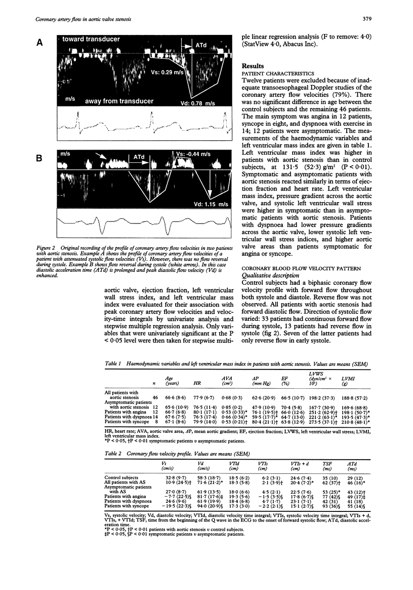
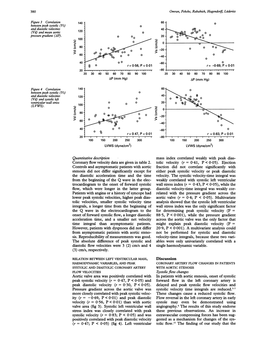
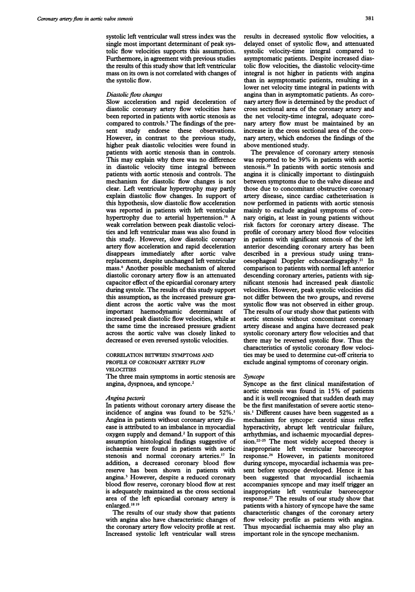
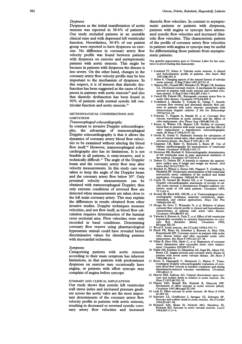
Images in this article
Selected References
These references are in PubMed. This may not be the complete list of references from this article.
- Baltazar R. F., Go E. H., Benesh S., Mower M. M. Case report: myocardial ischemia: an overlooked substrate in syncope of aortic stenosis. Am J Med Sci. 1992 Feb;303(2):105–108. doi: 10.1097/00000441-199202000-00008. [DOI] [PubMed] [Google Scholar]
- CHAPMAN C. B., BAKER O., REYNOLDS J., BONTE F. J. Use of biplane cinefluorography for measurement of ventricular volume. Circulation. 1958 Dec;18(6):1105–1117. doi: 10.1161/01.cir.18.6.1105. [DOI] [PubMed] [Google Scholar]
- Carroll R. J., Falsetti H. L. Retrograde coronary artery flow in aortic valve disease. Circulation. 1976 Sep;54(3):494–499. doi: 10.1161/01.cir.54.3.494. [DOI] [PubMed] [Google Scholar]
- Currie P. J., Seward J. B., Reeder G. S., Vlietstra R. E., Bresnahan D. R., Bresnahan J. F., Smith H. C., Hagler D. J., Tajik A. J. Continuous-wave Doppler echocardiographic assessment of severity of calcific aortic stenosis: a simultaneous Doppler-catheter correlative study in 100 adult patients. Circulation. 1985 Jun;71(6):1162–1169. doi: 10.1161/01.cir.71.6.1162. [DOI] [PubMed] [Google Scholar]
- Devereux R. B., Reichek N. Echocardiographic determination of left ventricular mass in man. Anatomic validation of the method. Circulation. 1977 Apr;55(4):613–618. doi: 10.1161/01.cir.55.4.613. [DOI] [PubMed] [Google Scholar]
- Dineen E., Brent B. N. Aortic valve stenosis: comparison of patients with to those without chronic congestive heart failure. Am J Cardiol. 1986 Feb 15;57(6):419–422. doi: 10.1016/0002-9149(86)90764-2. [DOI] [PubMed] [Google Scholar]
- Fujiwara T., Nogami A., Masaki H., Yamane H., Matsuoka S., Yoshida H., Fukuda H., Katsumura T., Kajiya F. Coronary flow velocity waveforms in aortic stenosis and the effects of valve replacement. Ann Thorac Surg. 1989 Oct;48(4):518–522. doi: 10.1016/s0003-4975(10)66853-1. [DOI] [PubMed] [Google Scholar]
- Hakki A. H., Kimbiris D., Iskandrian A. S., Segal B. L., Mintz G. S., Bemis C. E. Angina pectoris and coronary artery disease in patients with severe aortic valvular disease. Am Heart J. 1980 Oct;100(4):441–449. doi: 10.1016/0002-8703(80)90655-9. [DOI] [PubMed] [Google Scholar]
- Hongo M., Goto T., Watanabe N., Nakatsuka T., Tanaka M., Kinoshita O., Yamada H., Okubo S., Sekiguchi M. Relation of phasic coronary flow velocity profile to clinical and hemodynamic characteristics of patients with aortic valve disease. Circulation. 1993 Sep;88(3):953–960. doi: 10.1161/01.cir.88.3.953. [DOI] [PubMed] [Google Scholar]
- Iliceto S., Marangelli V., Memmola C., Rizzon P. Transesophageal Doppler echocardiography evaluation of coronary blood flow velocity in baseline conditions and during dipyridamole-induced coronary vasodilation. Circulation. 1991 Jan;83(1):61–69. doi: 10.1161/01.cir.83.1.61. [DOI] [PubMed] [Google Scholar]
- Kenny A., Wisbey C. R., Shapiro L. M. Profiles of coronary blood flow velocity in patients with aortic stenosis and the effect of valve replacement: a transthoracic echocardiographic study. Br Heart J. 1994 Jan;71(1):57–62. doi: 10.1136/hrt.71.1.57. [DOI] [PMC free article] [PubMed] [Google Scholar]
- LEAK D. Effort syncope in aortic stenosis. Br Heart J. 1959 Apr;21(2):289–292. doi: 10.1136/hrt.21.2.289. [DOI] [PMC free article] [PubMed] [Google Scholar]
- Lombard J. T., Selzer A. Valvular aortic stenosis. A clinical and hemodynamic profile of patients. Ann Intern Med. 1987 Feb;106(2):292–298. doi: 10.7326/0003-4819-106-2-292. [DOI] [PubMed] [Google Scholar]
- Marcus M. L., Doty D. B., Hiratzka L. F., Wright C. B., Eastham C. L. Decreased coronary reserve: a mechanism for angina pectoris in patients with aortic stenosis and normal coronary arteries. N Engl J Med. 1982 Nov 25;307(22):1362–1366. doi: 10.1056/NEJM198211253072202. [DOI] [PubMed] [Google Scholar]
- Reichek N., Wilson J., St John Sutton M., Plappert T. A., Goldberg S., Hirshfeld J. W. Noninvasive determination of left ventricular end-systolic stress: validation of the method and initial application. Circulation. 1982 Jan;65(1):99–108. doi: 10.1161/01.cir.65.1.99. [DOI] [PubMed] [Google Scholar]
- Selzer A. Changing aspects of the natural history of valvular aortic stenosis. N Engl J Med. 1987 Jul 9;317(2):91–98. doi: 10.1056/NEJM198707093170206. [DOI] [PubMed] [Google Scholar]
- Seward J. B., Khandheria B. K., Oh J. K., Abel M. D., Hughes R. W., Jr, Edwards W. D., Nichols B. A., Freeman W. K., Tajik A. J. Transesophageal echocardiography: technique, anatomic correlations, implementation, and clinical applications. Mayo Clin Proc. 1988 Jul;63(7):649–680. doi: 10.1016/s0025-6196(12)65529-3. [DOI] [PubMed] [Google Scholar]
- Villari B., Hess O. M., Meier C., Pucillo A., Gaglione A., Turina M., Krayenbuehl H. P. Regression of coronary artery dimensions after successful aortic valve replacement. Circulation. 1992 Mar;85(3):972–978. doi: 10.1161/01.cir.85.3.972. [DOI] [PubMed] [Google Scholar]
- WOOD P. Aortic stenosis. Am J Cardiol. 1958 May;1(5):553–571. doi: 10.1016/0002-9149(58)90138-3. [DOI] [PubMed] [Google Scholar]
- Yoshikawa J., Akasaka T., Yoshida K., Takagi T. Systolic coronary flow reversal and abnormal diastolic flow patterns in patients with aortic stenosis: assessment with an intracoronary Doppler catheter. J Am Soc Echocardiogr. 1993 Sep-Oct;6(5):516–524. doi: 10.1016/s0894-7317(14)80471-9. [DOI] [PubMed] [Google Scholar]


