Abstract
Objective:
To quantitatively and qualitatively compare MRI of the temporomandibular joint (TMJ) using a standard TMJ surface coil and a head coil at 3.0 T.
Methods:
22 asymptomatic volunteers were MR imaged using a 2-channel surface coil (standard TMJ coil) and a 32-channel head coil at 3.0 T (Philips Ingenia; Philips Healthcare, Netherlands). Imaging protocol consisted of an oblique sagittal proton density weighted turbo spin echo sequence (repetition time/echo time, 2700/26 ms). For quantitative assessment, a spherical phantom was imaged using the same sequence including a noise scan and a B1+ scan. Signal-to-noise ratio (SNR) maps and B1+ maps were calculated on a voxelwise basis. For qualitative evaluation, all volunteers underwent MRI of both TMJs with the jaw in the closed position. Two independent blinded readers assessed accuracy of TMJ anatomical representation and overall image quality on a 5-point scale. Quantitative and qualitative measurements were compared between coils using t-tests and Wilcoxon signed-rank test, respectively.
Results:
Quantitative analysis showed similar B1+ and significantly higher SNR for the head coil than the TMJ surface coil. Qualitative analysis showed significantly better visibility and delineation of clinically relevant anatomical structures of the TMJ, including the articular disc, bilaminar zone and lateral pterygoid muscle. Furthermore, better overall image quality was observed for the head coil than for the TMJ surface coil.
Conclusions:
A 32-channel head coil is preferable to a standard 2-channel TMJ surface coil when imaging the TMJ at 3.0 T, because it yields higher SNR, thus increasing accuracy of the anatomical representation of the TMJ.
Keywords: magnetic resonance imaging, joints, temporomandibular joint, temporomandibular joint disc, signal-to-noise ratio
Introduction
Temporomandibular disorders (TMDs) is a collective term for various pathologies of the temporomandibular joint (TMJ) and the masticatory muscles.1 TMDs are characterized by common features, including pain, TMJ sounds and restricted mandibular movement.2 With an estimated prevalence of 12% in the adult population, they represent the most common cause of orofacial pain of non-dental origin.3,4 Furthermore, TMDs are associated with a wide variety of frequent conditions, such as tension and migraine headache, depression, fatigue, sleep apnoea, obesity and Type 2 diabetes mellitus.5 In the USA, TMDs cause an estimated 17,800,000 lost workdays for every 100,000,000 working adults per year.3 Therefore, TMDs have not only a great impact on the quality of life of patients but also cause great socioeconomic costs.6,7 Besides identification of risk factors and early detection of pathological processes underlying TMDs, the diagnostic process can be further optimized.8,9
MRI is nowadays considered the state-of-the-art method for the evaluation of TMJ structures, namely the articular disc, retrodiscal tissue, cartilage and bone.10,11 Most commonly, MRI of the TMJ is performed at 1.5 T using dedicated surface coils.12 Theory suggests that TMJ imaging at higher field strength yields higher spatial resolution at comparable scan duration given that signal intensity and signal-to-noise ratio (SNR) increase with magnetic field strength.13 Indeed, recent studies using dedicated surface coils at 3.0 T have demonstrated that TMJ imaging benefits from higher field strength, particularly due to improved depiction of clinically relevant structures and better overall image quality.12,14–17
In spite of technological advances, recent TMJ imaging studies still employ dedicated surface coils, such as the commercially available SENSE FlexM™ (Philips Healthcare, Best, Netherlands) or custom-made bilateral phased-array coils, respectively.14,16 However, several limiting factors restrict their use. These include the availability at non-specialized imaging sites, need for proper positioning, field of view (FoV) and SNR. Modern head coils, increasingly available on 3.0-T systems, can be set up easily with increased SNR. Although they have primarily been designed for high-resolution brain imaging, they also cover the TMJ and surrounding structures. Therefore, head coils potentially improve TMJ imaging at 3.0 T. Yet, a direct comparison between head and TMJ surface coils at 3.0 T is still unavailable.
The aim of the current study was the quantitative and qualitative evaluation of commercially available dedicated 2-channel TMJ surface coils vs a standard 32-channel head coil for imaging of the TMJ at 3.0 T.
Methods and materials
This prospective MRI study in asymptomatic volunteers was approved by the local institutional review board. It included imaging of a phantom and a cohort of asymptomatic healthy volunteers. All participants gave written informed consent.
Subjects
This study included phantom measurements for SNR and a volunteer study. Therefore, 22 asymptomatic volunteers (8 females; mean age 23.9 years; range, 19–26 years; and 14 males; mean age 26.7 years; range, 20–35 years) were subsequently included. Exclusion criteria were pregnancy, metallic implants (including retainers), claustrophobia and TMD history.
MRI
MRI was performed on a 3.0-T Philips Ingenia system (Philips Healthcare) using a high-resolution 32-channel head coil (SENSE Head coil 32 elements, Philips Healthcare) and a 2-channel TMJ surface coil (SENSE FlexS, Philips Healthcare). Proton density weighted turbo spin echo (PDw-TSE) sequences in oblique sagittal planes were acquired according to a previously published high-resolution protocol for imaging the TMJ at 3.0 T that yielded good and robust image quality.17 The sequence parameters were identical for both coils [repetition time (TR) = 2700 ms, echo time (TE) = 26 ms, FoV = 150 × 150 mm2, pixel size = 0.5 × 0.5 mm2, reconstructed pixel size = 0.25 × 0.25 mm2, slice thickness = 2 mm, number of slices = 2 × 12 resulting in a total of 24 slices, number of signal averages = 1 and total acquisition time = 3 hours 52 min].
Phantom imaging
For the phantom study, a “Braino” phantom (GE Medical Systems, Milwaukee, WI) was used.17 It contained 12.5 mM of N-acetyl-l-aspartic acid, 10 mM of creatine hydrate, 3 mM of choline chloride, 7.5 mM of myo-inositol, 12.5 mM of l-glutamic acid, 5 mM of dl-lactic acid, sodium azide (0.1%), 50 mM of potassium phosphate monobasic, 56 mM of sodium hydroxide and 1 ml l−1 of Gd-DPTA (Magnevist®; Bayer Schering Pharma AG, Berlin, Germany). Image acquisition was accompanied by an identical scan without radiofrequency (RF) excitation to assess signal noise and allow SNR calculation on a voxelwise basis. To ensure that potential differences in SNR were not due to different distributions of B1+ fields, B1 mapping was performed as described previously using a gradient-echo-based sequence consisted of a constant flip angle and two interleaved, different repetition times resulting in 10 slices at a spatial resolution of 3.0 × 3.0 × 8.0 mm³ (actual flip-angle method).18,19 Subsequently, the pixel value in the obtained B1 image was scaled as a percentage of the nominal transmit RF amplitude, whereas a value of 100% indicated that all RF pulses had precisely their nominal flip angles owing to the adjustment of the RF power optimization preparation phase.
Volunteer imaging
MR images of both TMJs were taken with the mouth closed. The scans with the two different coil systems were performed successively which means that all volunteers were imaged twice, using the head coil and TMJ surface coil. To avoid potential bias due to positioning and scanner drift, half of the participants were first imaged using the TMJ surface coil and the other half vice versa. Care was taken to ensure the correct and identical positioning of the participants and coils throughout the study. The FoV was always positioned perpendicular to the condylar transverse axis. Both TMJs were imaged simultaneously.
Image analysis
Quantitative SNR measurements
For SNR analysis, data were examined on a voxelwise basis, which allowed the calculation of SNR maps within the whole FoV. Therefore, phantom image data and corresponding noise data of every coil channel were post-processed individually using dedicated software routines (MATLAB®, MathWorks®, Natick, MA). In particular, the SNR calculation followed the procedure as reported extensively by Nordmeyer-Massner et al13 and applied recently for evaluation of the SNR in studies investigating MRI of the TMJ:17,19
(ρ, exact magnitude of the available transverse magnetization at the voxel position; σ, standard deviation of the corresponding noise components; d, complex values that a given pixel exhibits in each single-channel data; H, the complex conjugate transpose; ψ, noise covariance matrix computed from the noise scan).20,21 The combination of these data finally results in voxel-based SNR maps. In addition, the phantom was segmented for each acquired slice manually by one coauthor (blinded for review), yielding one SNR value for each slice for each coil setup. For B1+ analysis, the corresponding B1+ values along a horizontal line profile were extracted using MIPAV (Center for Information Technology, National Institutes of Health, Bethesda, MD; http://mipav.cit.nih.gov) for both B1+ maps following the procedure reported in a previous study.19
Qualitative image evaluation
The in vivo data were independently assessed by two fellowship-trained radiologists (blinded for review). All images were anonymized (subject's initials blinded) and evaluated in random order using a dedicated workstation (Impax® 6.0.; Agfa-Gevaert Group, Mortsel, Belgium) by the observers who were blinded to the coil used. They rated the overall image quality and the depiction of clinically relevant structures, including the articular disc, bilaminar zone, mandibular fossa, mandibular condyle and lateral pterygoid muscle. In particular, the articular disc was evaluated with respect to anatomical subregions (anterior band, intermediate zone, posterior band). The accuracy of representation of anatomical structures was rated from 1 to 5 according to a previously published grading system (1: excellent visibility; 2: good visibility; 3: moderate visibility; 4: poor visibility; and 5: not visible) (complete lack of diagnostic information).14
Statistical analysis
The Shapiro–Wilk test was performed to assess the normal distribution of the SNR (significance level α = 0.05). Paired-sample t-tests were used to evaluate for statistically significant SNR differences between the 32-channel head coil and the 2-channel SENSE FlexS coil. Bonferroni correction was used to account for multiple testing.
To determine the interreader agreement in the qualitative MR image analysis, Kappa statistics was used. Kappa values of 0.41–0.60 were considered as moderate agreement, values of 0.61–0.80 were considered as substantial agreement, values of 0.81–0.99 were considered as almost perfect agreement, and values of 1.00 were considered as perfect agreement.22 To investigate statistically significant between-group differences between coils with respect to the visibility and delineation of clinically relevant anatomical structures, respectively, Wilcoxon signed-rank tests were performed [p < 0.05, corrected for multiple comparisons (n = 13)]. SPSS® software v. 22.0 (IBM Corp., New York, NY; formerly SPSS Inc., Chicago, IL) was used for all statistical analyses.
Results
Quantitative data from phantom imaging
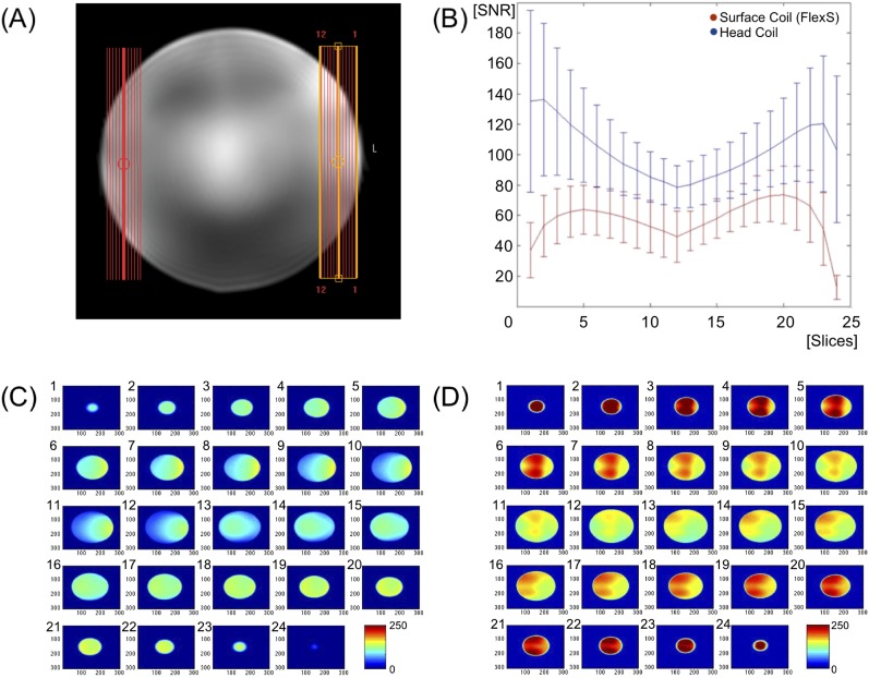
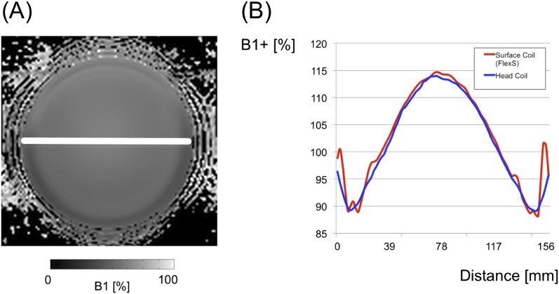
The SNR analysis revealed normal distribution for both coils (FlexS, p = 0.478; HeadCoil, p = 0.582). Furthermore, the head coil yielded increased SNR compared with the TMJ surface coil (mean ± standard deviation (SD), FlexS, 57.18 ± 12.08; HeadCoil, 102.97 ± 17.03; Figure 1). In addition, B1+ distribution was similar along the plotted profile for both coils (mean ± SD; FlexS, 102.67 ± 8.56; HeadCoil, 101.73 ± 8.61; for detailed presentation of the B1+ distribution, see Figure 2).
Figure 1.
Signal-to-noise ratio analysis. (a) The field of view imaged in a spherical phantom in sagittal orientation. In particular, 2 stacks consisting of 12 slices each were imaged resulting in 24 slices. Slices are numbered consecutively from left to right. (b) A diagram displaying the signal-to-noise ratio (SNR) for both coils. The x-axis represents the slice number, the y-axis represents the SNR. Head coil values are depicted in blue and temporomandibular joint (TMJ) surface coil values in red. For each slice, means and standard deviations are reported for both coils. (c, d) The voxelwise distribution of SNR for each slice, respectively, where (c) represents the TMJ surface coil, whereas (d) represents the head coil. In all images, the SNR values are color coded from 0 (blue) to 150 (red). For colour image see online.
Figure 2.
B1+ analysis. (a) A representative B1+ map for the spherical phantom acquired at 3.0 T using a 32-channel head coil. The B1+ image is scaled as a percentage of nominal transmit radiofrequency amplitude. B1+ values are greyscale coded from 0 (black) to 100 (white). The white line starts at the right margin of the phantom (0 mm) and ends at the left margin of the phantom (156 mm) and indicates the profile from which the B1+ values have been obtained for further analysis. (b) The extracted B1+ values for the surface coil (red) and the head coil (blue). Note that B1+ distribution along the profile line was similar for both coils. For colour image see online.
Qualitative data from volunteer imaging
Bilateral TMJ images of all 22 volunteers were evaluated. Interrater reliability was “almost perfect” for the respective anatomical structures (kappa 0.812–0.963). Table 1 shows means and SDs for each anatomical structure, calculated separately for each observer and coil as well as Cohen's kappa values.
Table 1.
Depiction of different anatomical structures of the temporomandibular joint at 3.0 T for a 2-channel surface coil (SENSE FlexS, Philips Healthcare, Best, Netherlands) and a 32-channel head coil (SENSE head coil, Philips Healthcare)
| Anatomical structure | Reader 1 |
Reader 2 |
Interrater reliability |
|||
|---|---|---|---|---|---|---|
| Mean | SD | Mean | SD | Kappa | p-value | |
| 2-channel surface coil (FlexS) | ||||||
| Disc | ||||||
| Visibility anterior band | 2.114 | 0.655 | 2.136 | 0.668 | 0.961 | <0.001 |
| Visibility intermediate zone | 2.159 | 0.745 | 2.227 | 0.743 | 0.892 | <0.001 |
| Visibility posterior band | 2.205 | 0.765 | 2.227 | 0.774 | 0.894 | <0.001 |
| Delineation | 2.273 | 0.694 | 2.318 | 0.674 | 0.923 | <0.001 |
| Bilaminar zone | ||||||
| Visibility | 2.395 | 0.821 | 2.419 | 0.823 | 0.895 | <0.001 |
| Delineation | 2.419 | 0.982 | 2.442 | 0.959 | 0.903 | <0.001 |
| Mandibular fossa | ||||||
| Visibility | 2.045 | 0.608 | 2.068 | 0.587 | 0.868 | <0.001 |
| Delineation | 2.068 | 0.587 | 2.068 | 0.545 | 0.812 | <0.001 |
| Mandibular condyle | ||||||
| Visibility | 1.955 | 0.608 | 1.932 | 0.587 | 0.868 | <0.001 |
| Delineation | 2.114 | 0.618 | 2.045 | 0.569 | 0.868 | <0.001 |
| Lateral pterygoid muscle | ||||||
| Visibility | 2.023 | 0.698 | 2.000 | 0.647 | 0.881 | <0.001 |
| Delineation | 2.023 | 0.590 | 2.068 | 0.625 | 0.914 | <0.001 |
| Overall image quality | 2.091 | 0.603 | 2.114 | 0.618 | 0.872 | <0.001 |
| 32-channel head coil (HeadCoil) | ||||||
| Disc | ||||||
| Visibility anterior band | 1.682 | 0.561 | 1.659 | 0.568 | 0.868 | <0.001 |
| Visibility intermediate zone | 1.682 | 0.708 | 1.705 | 0.701 | 0.888 | <0.001 |
| Visibility posterior band | 1.705 | 0.701 | 1.682 | 0.674 | 0.887 | <0.001 |
| Delineation | 1.750 | 0.615 | 1.727 | 0.585 | 0.958 | <0.001 |
| Bilaminar zone | ||||||
| Visibility | 2.045 | 1.275 | 2.023 | 1.267 | 0.963 | <0.001 |
| Delineation | 2.341 | 1.328 | 2.386 | 1.316 | 0.934 | <0.001 |
| Mandibular fossa | ||||||
| Visibility | 1.727 | 0.660 | 1.773 | 0.677 | 0.923 | <0.001 |
| Delineation | 1.886 | 0.689 | 1.909 | 0.709 | 0.881 | <0.001 |
| Mandibular condyle | ||||||
| Visibility | 1.636 | 0.650 | 1.614 | 0.618 | 0.960 | <0.001 |
| Delineation | 1.841 | 0.608 | 1.864 | 0.632 | 0.875 | <0.001 |
| Lateral pterygoid muscle | ||||||
| Visibility | 1.659 | 0.568 | 1.682 | 0.518 | 0.865 | <0.001 |
| Delineation | 1.705 | 0.632 | 1.727 | 0.624 | 0.880 | <0.001 |
| Overall image quality | 1.591 | 0.542 | 1.636 | 0.574 | 0.913 | <0.001 |
SD, standard deviation.
For each observer and coil, the mean and SD are given for the visibility and delineation of each anatomical structure. Grading was based on a grading system previously published and ranged from 1 (perfect) to 5 (non-diagnostic).14
For interrater reliability, kappa values and the corresponding p-values are reported.
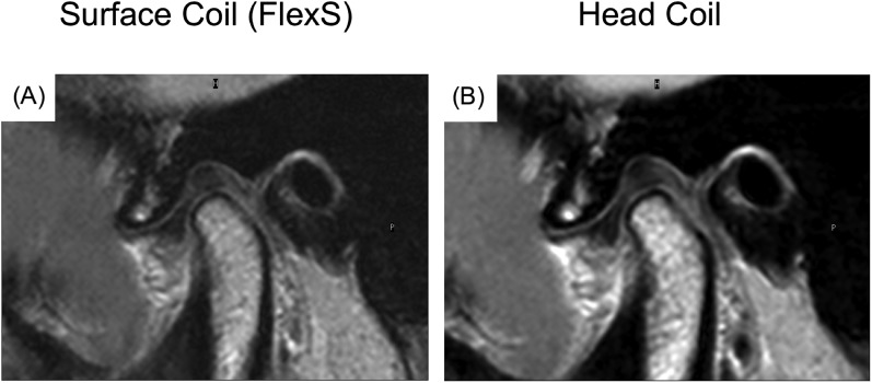
For both observers, qualitative analysis demonstrated significantly better overall image quality using the head coil than the TMJ surface coil (p < 0.05, corrected for multiple comparisons). Furthermore, the head coil yielded significantly better accuracy of representation of clinically relevant structures, including the anterior and posterior band as well as the intermediate zone of the articular disc, bilaminar zone and lateral pterygoid muscle (Wilcoxon signed-rank tests, p < 0.05, corrected for multiple comparisons). Table 2 provides a detailed presentation of between-group differences regarding accuracy of representation of all anatomical structures (Figure 3).
Table 2.
Between-coil differences of visibility and delineation of clinically relevant temporomandibular joint (TMJ) structures
| Anatomic Structure | Reader 1 |
Reader 2 |
||
|---|---|---|---|---|
| z-value | p-value | z-value | p-value | |
| 2-channel surface coil (FlexS) compared to 32-channel head coil (HeadCoil) | ||||
| Disc | ||||
| Visibility anterior band | −3.288 | 0.001 | −3.740 | <0.001 |
| Visibility intermediate zone | −3.091 | 0.002 | −4.146 | <0.001 |
| Visibility posterior band | −3.314 | 0.001 | −3.679 | <0.001 |
| Delineation | −3.651 | <0.001 | −4.461 | <0.001 |
| Bilaminar zone | ||||
| Visibility | −3.869 | <0.001 | −4.874 | <0.001 |
| Delineation | −1.429 | 0.153 | −1.377 | 0.168 |
| Mandibular fossa | ||||
| Visibility | −2.645 | 0.008 | −2.852 | 0.004 |
| Delineation | −1.617 | 0.106 | −1.025 | 0.305 |
| Mandibular condyle | ||||
| Visibility | −2.403 | 0.016 | −2.532 | 0.011 |
| Delineation | −2.425 | 0.015 | −2.098 | 0.036 |
| Lateral pterygoid muscle | ||||
| Visibility | −3.234 | 0.001 | −3.274 | 0.001 |
| Delineation | −3.130 | 0.002 | −3.396 | 0.001 |
| Overall image quality | −3.740 | <0.001 | −3.778 | <0.001 |
To evaluate potential differences between the 2-channel TMJ surface coil (SENSE FlexS, Philips Healthcare, Best, Netherlands) and the 32-channel head coil (SENSE head coil, Philips Healthcare) with respect to the visibility and delineation of clinically relevant TMJ structures, Wilcoxon signed-rank tests were performed for each structure, respectively.
z-values and the corresponding p-values are reported separately for each observer. Italics indicate that tests remained significant after correction for multiple comparisons (n = 13).
Figure 3.
Qualitative analysis. Proton density weighted oblique sagittal images in closed-mouth position at 3.0 T. (a, b) The temporomandibular joint (TMJ) of an asymptomatic volunteer assessed using the 2-channel TMJ surface coil (a; SENSE FlexS; Philips Healthcare, Best, Netherlands) and the 32-channel head coil (b; SENSE head coil; Philips Healthcare), respectively. The overall image quality and accuracy of representation of TMJ structures were consistently rated better on images obtained with the head coil. In particular, overall image quality was rated a score of 2 (good) for (a) and a score of 1 (excellent) for (b); for detailed presentation of parameters regarding visibility and delineation of all anatomical structures of the TMJ, see Table 2.
Discussion
In this study, the 32-channel head coil provided higher SNR and more detailed depiction of key anatomical TMJ structures than a 2-channel TMJ surface coil. The results indicate that the 32-channel head coil might be preferred over the surface coil for TMJ imaging.
Only few studies have so far investigated technical aspects of TMJ imaging. One investigation evaluated MR image quality of different protocols at 1.5 and 3.0 T.14 Similarly to our study, the accuracy of representation of anatomical structures was rated qualitatively. However, the quantitative analysis only included the assessment of contrast-to-noise ratio as a surrogate for SNR based on a region-of-interest (ROI) approach. ROIs were placed manually within the articular disc and retrodiscal tissue. Subsequently, SDs of the ROI signal intensities were extracted and used to calculate the contrast-to-noise ratio. Although the authors concluded that the increased image quality and higher contrast-to-noise ratio were associated with higher SNR owing to an increase of the magnetic field strength according to the Boltzmann equation, SNR itself was not directly measured.13 In another study, various MR sequences for TMJ imaging at 3.0 T were compared and SNR was also assessed using a ROI-based approach.23 Then, the SNR was computed by comparing the signal intensity in the articular disc to the signal intensity in the image background.23
To the best of our knowledge, no study has directly compared different coils for TMJ imaging in a 3.0-T scanner. In contrast to the aforementioned studies, we also applied a more elaborate approach to calculate SNR voxelwise over all slices by additionally measuring it in a phantom. This provided information enabling a detailed evaluation of the spatial distribution of the SNR with respect to the ROI and surrounding areas (Figure 1).
In contrast to commonly used simple ROI-based SNR estimations, our method yielded SNR maps for the entire FoV and correctly accounted for possible noise correlations among individual coil channels.17,23 In addition, noise estimation was more robust because of the high number of pixels used compared with a ROI within an anatomic image. Applying this method, we found that SNR was significantly higher for the 32-channel head coil than the 2-channel TMJ surface coil. Moreover, the measured SNR values for both coils were higher than in comparable studies.23 This observation might be explained by the very high sensitivity of the algorithm used to calculate SNR and the difference between sequences and coil setups relative to the other studies. It is to note that the spatial heterogeneities detected with the aforementioned voxelwise approach seem to be higher when using the 32-channel head coil than the surface coil, as indicated by a higher SD of the SNR, which might be caused by different B1+ distributions for each coil setup. However, B1+ analysis yielded similar flip angle distributions along the phantom, suggesting that B1+ inhomogeneities were similar for both coils and thus did not explain differences in SNR. Hence, with respect to SNR, our detailed analysis demonstrates the superiority of 32-channel head coils compared with 2-channel TMJ surface coils.
In the present study, interrater agreement was “almost perfect” for all anatomical structures and in general higher than in comparable studies.16 This might be explained by the standardized assessment, the extensive experience of TMJ imaging at the study site and the more detailed depiction of all clinically relevant TMJ structures in oblique sagittal orientation.
Compared with the 2-channel TMJ surface coil, the 32-channel head coil yielded a more accurate representation of TMJ structures, including the articular disc, the bilaminar zone and the lateral pterygoid muscle. Overall image quality was also higher for the head coil than the TMJ surface coil. Statistical differences remained significant after correction for multiple comparisons. These findings are perfectly in line with the observation in our phantom study where the head coil provided higher SNRs than the TMJ surface coil (see above). It is to note that no intrareader analysis was performed in the current study. However, the high interreader agreement as well as the low SDs for all scores and both readers suggest a strong consistency and reproducibility of the presented data. Furthermore, qualitative evaluation considerably depends on the chosen MR sequence parameters. In the current study, we adopted a proposed high-resolution MRI protocol particularly designed for imaging the TMJ at 3.0 T.17 This approach ensured a standardized comparison between the image quality obtained with both coils and allowed for an adequate comparability between the current and previously published studies.
Taken together, results of the current study indicate that imaging of the TMJ using a 32-channel head coil yields not only higher SNR but also a superior visualization of relevant structures than a dedicated 2-channel TMJ surface coil.
We acknowledge several study limitations. (1) Sample size. The current study included only 22 asymptomatic volunteers, resulting in 44 TMJ images. However, the subject number is in line with previous reports demonstrating that the investigation of even smaller groups of asymptomatic volunteers yields robust and significant results.14,17 (2) Asymptomatic pathologies. All 22 volunteers reported to be free of TMD symptoms. Nevertheless, during imaging, in 9 of 44 TMJs, disc displacements were observed using both coils. Disc displacement does not affect image quality and thus did not interfere with our TMJ image evaluation. However, future studies in different patient cohorts are needed to evaluate if the better SNR and image quality from the 32-channel head coils will translate into a better patient management and outcome for different pathologies underlying TMDs. (3) Scan planning. In clinical routine, imaging is performed in both closed- and open-mouth position for optimal detection of potential jaw motion–related pathologies. It is to note that imaging was performed only in the sagittal orientation and closed-mouth position in the present study, whereas clinical imaging is often additionally performed in the coronal plane. For the purpose of this study, we consider the evaluation of performed sequences as sufficient to infer on differences in imaging quality between different coil types. (4) SNR analysis. In the current study, SNR analysis was performed in a phantom only. In general, performing the SNR analysis in the assessed volunteers might allow for a reliable evaluation of the variation of the SNR-gain for different tissue types of the TMJ. However, since B1+ distributions, the field strength as well as the scan parameters were identical for both coils, assessing the potential impact of the coils on measured SNR in a phantom can be assumed to represent an adequate surrogate for potential differences in vivo. Nevertheless, further studies investigating the effect of different tissue types regarding B1+ and SNR in the context of different coil setups are needed to address this question. (5) Lack of a gold standard. Surgical and/or pathological reinvestigations of the imaged structures would be necessary to uncover the quality of correlation between imaging results and anatomical structures. Recently, it was shown that MRI of cadaver TMJs at 3.0 T provides an excellent anatomical characterization, thus suggesting that also in vivo MRI at this field strength might reveal detailed anatomical structures.23
Clinical implications
The present study indicates that TMJ imaging at 3.0 T using a 32-channel head coil bears a great potential for clinical routine use. First, it was previously shown that the diagnosis of anterior disc displacement benefits from imaging at 3.0 T compared with 1.5 T, presumably due to higher SNR.16 Therefore, it can be expected that SNR gain resulting from head coil use might contribute to a further improvement of the diagnostic accuracy of TMDs.9 However, it has to be taken into careful consideration that it is unclear as to what extent the reported difference in image quality between the assessed coils might translate into diagnostic accuracy, in particular when considering the heterogeneity of pathologies underlying TMDs ranging from subtle soft-tissue lesions to distinct dislocations of the temporomandibular disc. The current study assessed asymptomatic volunteers only and therefore does not necessarily provide enough insight to infer on how the reported differences in coil performance might influence clinical performance for different pathological conditions underlying TMDs. Extensive studies are needed to systematically evaluate the extent of potential benefits of head coils for patients with different pathologies of the TMJ with respect to the sensitivity and specificity of the diagnostic process as well as its possible impact on therapeutical decision-making. Second, surface coil positioning is a tricky and time-consuming task requiring experience, whereas patient placement in head coils is easy to standardize and more time efficient. Finally, commercially available 32-channel head coils are broadly available at most MR sites, facilitating MRI of the TMJ also outside of highly specialized centres.
In conclusion, a 32-channel head coil is preferable to a standard 2-channel TMJ surface coil when imaging the TMJ at 3.0 T, because it yields higher SNR, thus increasing accuracy of TMJ anatomical representation.
References
- 1.McNeill C. Management of temporomandibular disorders: concepts and controversies. J Prosthet Dent 1997; 77: 510–22. doi: http://dx.doi.org/10.1016/S0022-3913(97)70145-8 [DOI] [PubMed] [Google Scholar]
- 2.Weinberg LA. The etiology, diagnosis, and treatment of TMJ dysfunction-pain syndrome. Part I: Etiology. J Prosthet Dent 1979; 42: 654–64. doi: http://dx.doi.org/10.1016/0022-3913(79)90197-5 [DOI] [PubMed] [Google Scholar]
- 3.Maixner W, Diatchenko L, Dubner R, Fillingim RB, Greenspan JD, Knott C, et al. Orofacial pain prospective evaluation and risk assessment study–the OPPERA study. J Pain 2011; 12: T4–11.e1–2. doi: http://dx.doi.org/10.1016/j.jpain.2011.08.002 [DOI] [PMC free article] [PubMed] [Google Scholar]
- 4.Slade GD, Fillingim RB, Sanders AE, Bair E, Greenspan JD, Ohrbach R, et al. Summary of findings from the OPPERA prospective cohort study of incidence of first-onset temporomandibular disorder: implications and future directions. J Pain 2013; 14 (Suppl. 12): T116–24. doi: http://dx.doi.org/10.1016/j.jpain.2013.09.010 [DOI] [PMC free article] [PubMed] [Google Scholar]
- 5.Hoffmann RG, Kotchen JM, Kotchen TA, Cowley T, Dasgupta M, Cowley AWJ. Temporomandibular disorders and associated clinical comorbidities. Clin J Pain 2011; 27: 268–74. doi: http://dx.doi.org/10.1097/AJP.0b013e31820215f5 [DOI] [PubMed] [Google Scholar]
- 6.De Rossi SS. Orofacial pain: a primer. Dent Clin North Am 2013; 57: 383–92. doi: http://dx.doi.org/10.1016/j.cden.2013.04.001 [DOI] [PubMed] [Google Scholar]
- 7.White BA, Williams LA, Leben JR. Health care utilization and cost among health maintenance organization members with temporomandibular disorders. J Orofac Pain 2001; 15: 158–69. [PubMed] [Google Scholar]
- 8.Manfredini D, Guarda-Nardini L, Winocur E, Piccotti F, Ahlberg J, Lobbezoo F. Research diagnostic criteria for temporomandibular disorders: a systematic review of axis I epidemiologic findings. Oral Surg Oral Med Oral Pathol Oral Radiol Endod 2011; 112: 453–62. doi: http://dx.doi.org/10.1016/j.tripleo.2011.04.021 [DOI] [PubMed] [Google Scholar]
- 9.Schiffman E, Ohrbach R, Truelove E, Look J, Anderson G, Goulet JP, et al. ; International RDC/TMD Consortium Network, International association for Dental Research; Orofacial Pain Special Interest Group, International Association for the Study of Pain. Diagnostic criteria for temporomandibular disorders (DC/TMD) for cinical and research applications: recommendations of the International RDC/TMD Consortium Network* and Orofacial Pain Special Interest Groupdagger. J Oral Facial Pain Headache 2014; 28: 6–27. doi: http://dx.doi.org/10.11607/jop.1151 [DOI] [PMC free article] [PubMed] [Google Scholar]
- 10.Oğütcen-Toller M, Taskaya-Yilmaz N, Yilmaz F. The evaluation of temporomandibular joint disc position in TMJ disorders using MRI. Int J Oral Maxillofac Surg 2002; 31: 603–7. [DOI] [PubMed] [Google Scholar]
- 11.Aiken A, Bouloux G, Hudgins P. MR imaging of the temporomandibular joint. Magn Reson Imaging Clin N Am 2012; 20: 397–412. doi: http://dx.doi.org/10.1016/j.mric.2012.05.002 [DOI] [PubMed] [Google Scholar]
- 12.Iwasaki H, Kubo H, Harada M, Nishitani H. Temporomandibular joint and 3.0 T pseudodynamic magnetic resonance imaging. Part 1: evaluation of condylar and disc dysfunction. Dentomaxillofac Radiol 2010; 39: 475–85. doi: http://dx.doi.org/10.1259/dmfr/29741224 [DOI] [PMC free article] [PubMed] [Google Scholar]
- 13.Nordmeyer-Massner JA, Wyss M, Andreisek G, Pruessmann KP, Hodler J. In vitro and in vivo comparison of wrist MR imaging at 3.0 and 7.0 tesla using a gradient echo sequence and identical eight-channel coil array designs. J Magn Reson Imaging 2011; 33: 661–7. doi: http://dx.doi.org/10.1002/jmri.22419 [DOI] [PubMed] [Google Scholar]
- 14.Stehling C, Vieth V, Bachmann R, Nassenstein I, Kugel H, Kooijman H, et al. High-resolution magnetic resonance imaging of the temporomandibular joint: image quality at 1.5 and 3.0 Tesla in volunteers. Invest Radiol 2007; 42: 428–34. doi: http://dx.doi.org/10.1097/01.rli.0000262081.23997.6b [DOI] [PubMed] [Google Scholar]
- 15.Iwasaki H, Kubo H, Harada M, Nishitani H, Ohashi Y. Temporomandibular joint and 3.0 T pseudodynamic magnetic resonance imaging. Part 2: evaluation of articular disc obscurity. Dentomaxillofac Radiol 2010; 39: 486–93. doi: http://dx.doi.org/10.1259/dmfr/92017549 [DOI] [PMC free article] [PubMed] [Google Scholar]
- 16.Schmid-Schwap M, Drahanowsky W, Bristela M, Kundi M, Piehslinger E, Robinson S. Diagnosis of temporomandibular dysfunction syndrome–image quality at 1.5 and 3.0 Tesla magnetic resonance imaging. Eur Radiol 2009; 19: 1239–45. doi: http://dx.doi.org/10.1007/s00330-008-1264-7 [DOI] [PubMed] [Google Scholar]
- 17.Manoliu A, Spinner G, Wyss M, Erni S, Ettlin DA, Nanz D, et al. Quantitative and qualitative comparison of MR imaging of the temporomandibular joint at 1.5 and 3.0 Tesla using an optimized high-resolution protocol. Dentomaxillofac Radiol 2015; 45: 20150240. doi: http://dx.doi.org/10.1259/dmfr.20150240 [DOI] [PMC free article] [PubMed] [Google Scholar]
- 18.Yarnykh VL. Actual flip-angle imaging in the pulsed steady state: a method for rapid three-dimensional mapping of the transmitted radiofrequency field. Magn Reson Med 2007; 57: 192–200. doi: http://dx.doi.org/10.1002/mrm.21120 [DOI] [PubMed] [Google Scholar]
- 19.Manoliu A, Spinner G, Wyss M, Ettlin DA, Nanz D, Kuhn FP, et al. Magnetic resonance imaging of the temporomandibular joint at 7.0 T using high-permittivity dielectric pads: a feasibility study. Invest Radiol 2015; 50: 843–9. doi: http://dx.doi.org10.1097/RLI.0000000000000196 [DOI] [PubMed] [Google Scholar]
- 20.Roemer PB, Edelstein WA, Hayes CE, Souza SP, Mueller OM. The NMR phased array. Magn Reson Med 1990; 16: 192–225. doi: http://dx.doi.org/10.1002/mrm.1910160203 [DOI] [PubMed] [Google Scholar]
- 21.Pruessmann KP, Weiger M, Scheidegger MB, Boesiger P. SENSE: sensitivity encoding for fast MRI. Magn Reson Med 1999; 42: 952–62. doi: http://dx.doi.org/10.1002/(SICI)1522-2594(199911)42:5<952::AID-MRM16>3.0.CO;2-S [PubMed] [Google Scholar]
- 22.Landis JR, Koch GG. The measurement of observer agreement for categorical data. Biometrics 1977; 33: 159–74. doi: http://dx.doi.org/10.2307/2529310 [PubMed] [Google Scholar]
- 23.Sanal HT, Bae WC, Pauli C, Du J, Statum S, Znamirowski R, et al. Magnetic resonance imaging of the temporomandibular joint disc: feasibility of novel quantitative magnetic resonance evaluation using histologic and biomechanical reference standards. J Orofac Pain 2011; 25: 345–53. [PMC free article] [PubMed] [Google Scholar]


