Abstract
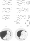
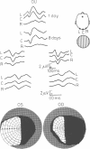
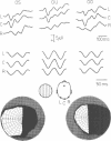
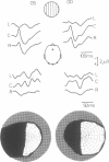
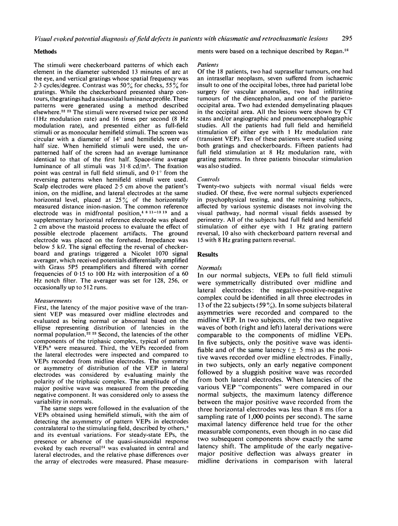
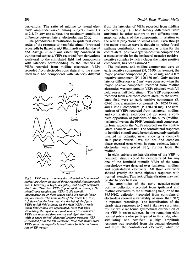
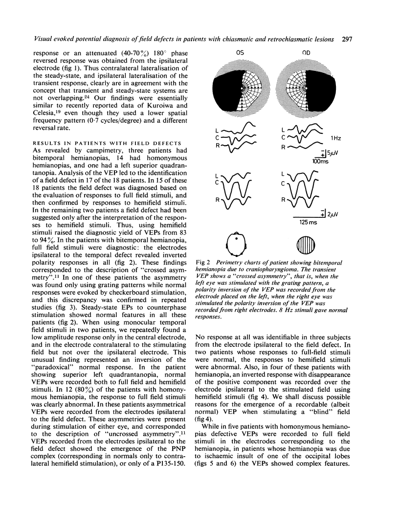
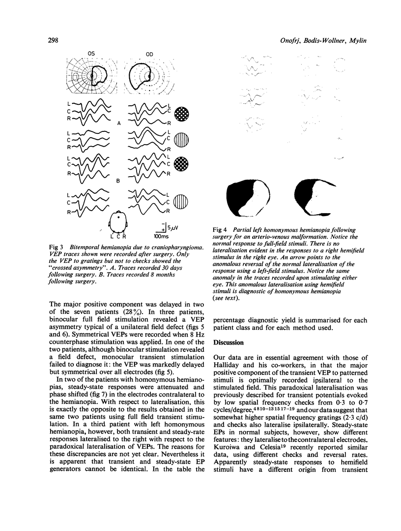
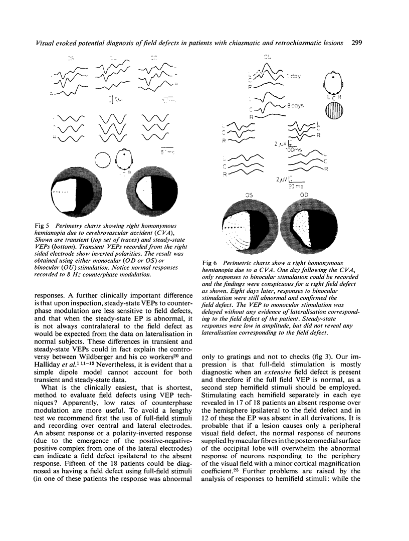
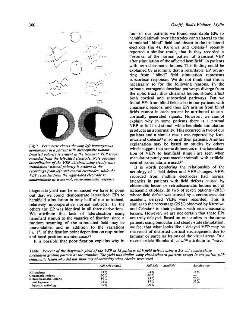
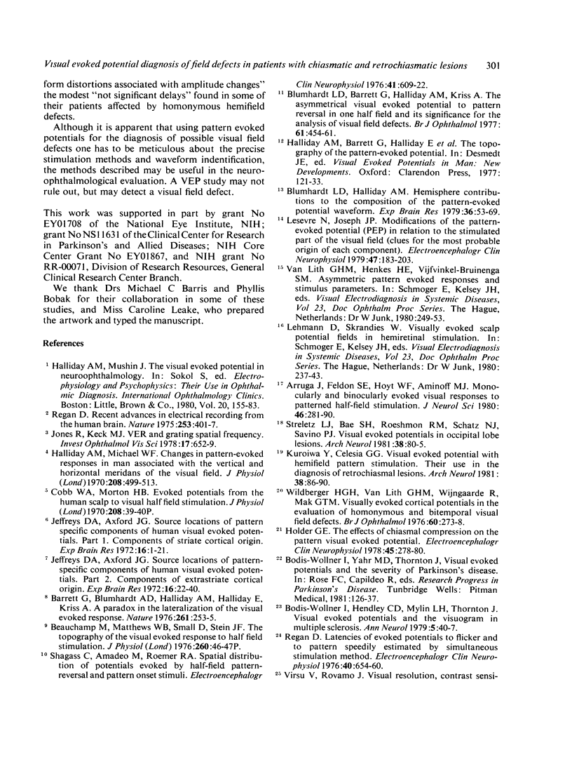
Visual evoked potentials were studied in 18 patients with visual field defects assessed by perimetry. Stimuli were reversing checkerboard and grating patterns of 13', full field and hemifield. Two reversal rates were studied: 1 Hz (transient VEPs) and 8 Hz (steady-state VEPs). Using monocular hemifield stimulation the "paradoxical lateralisation" of transient but not of steady-state VEPs was confirmed. Diagnostic yield was 90% using transient, but less than 20% using steady-state stimuli.
Full text
PDF

Images in this article
Selected References
These references are in PubMed. This may not be the complete list of references from this article.
- Arruga J., Feldon S. E., Hoyt W. F., Aminoff M. J. Monocularly and binocularly evoked visual responses to patterned half-field stimulation. J Neurol Sci. 1980 Jun;46(3):281–290. doi: 10.1016/0022-510x(80)90052-0. [DOI] [PubMed] [Google Scholar]
- Barett G., Blumhardt L., Halliday A. M., Halliday E., Kriss A. A paradox in the lateralisation of the visual evoked response. Nature. 1976 May 20;261(5557):253–255. doi: 10.1038/261253a0. [DOI] [PubMed] [Google Scholar]
- Blumenhardt L. D., Halliday A. M. Hemisphere contributions to the composition of the pattern-evoked potential waveform. Exp Brain Res. 1979 Jun 1;36(1):53–69. doi: 10.1007/BF00238467. [DOI] [PubMed] [Google Scholar]
- Blumhardt L. D., Barrett G., Halliday A. M., Kriss A. The effect of experimental 'scotomata' on the ipsilateral and contralateral responses to pattern-reversal in one half-field. Electroencephalogr Clin Neurophysiol. 1978 Sep;45(3):376–392. doi: 10.1016/0013-4694(78)90189-x. [DOI] [PubMed] [Google Scholar]
- Blumhardt L. D., Barrett G., Halliday A. M. The asymmetrical visual evoked potential to pattern reversal in one half field and its significance for the analysis of visual field defects. Br J Ophthalmol. 1977 Jul;61(7):454–461. doi: 10.1136/bjo.61.7.454. [DOI] [PMC free article] [PubMed] [Google Scholar]
- Bodis-Wollner I., Hendley C. D., Mylin L. H., Thornton J. Visual evoked potentials and the visuogram in multiple sclerosis. Ann Neurol. 1979 Jan;5(1):40–47. doi: 10.1002/ana.410050107. [DOI] [PubMed] [Google Scholar]
- Halliday A. M., Michael W. F. Changes in pattern-evoked responses in man associated with the vertical and horizontal meridians of the visual field. J Physiol. 1970 Jun;208(2):499–513. doi: 10.1113/jphysiol.1970.sp009134. [DOI] [PMC free article] [PubMed] [Google Scholar]
- Holder G. E. The effects of chiasmal compression on the pattern visual evoked potential. Electroencephalogr Clin Neurophysiol. 1978 Aug;45(2):278–280. doi: 10.1016/0013-4694(78)90011-1. [DOI] [PubMed] [Google Scholar]
- Jeffreys D. A., Axford J. G. Source locations of pattern-specific components of human visual evoked potentials. I. Component of striate cortical origin. Exp Brain Res. 1972;16(1):1–21. doi: 10.1007/BF00233371. [DOI] [PubMed] [Google Scholar]
- Jeffreys D. A., Axford J. G. Source locations of pattern-specific components of human visual evoked potentials. II. Component of extrastriate cortical origin. Exp Brain Res. 1972;16(1):22–40. doi: 10.1007/BF00233372. [DOI] [PubMed] [Google Scholar]
- Jones R., Keck M. J. Visual evoked response as a function of grating spatial frequency. Invest Ophthalmol Vis Sci. 1978 Jul;17(7):652–659. [PubMed] [Google Scholar]
- Kuroiwa Y., Celesia G. G. Visual evoked potentials with hemifield pattern stimulation. Their use in the diagnosis of retrochiasmatic lesions. Arch Neurol. 1981 Feb;38(2):86–90. doi: 10.1001/archneur.1981.00510020044005. [DOI] [PubMed] [Google Scholar]
- Lesevre N., Joseph J. P. Modifications of the pattern-evoked potential (PEP) in relation to the stimulated part of the visual field (clues for the most probable origin of each component). Electroencephalogr Clin Neurophysiol. 1979 Aug;47(2):183–203. doi: 10.1016/0013-4694(79)90220-7. [DOI] [PubMed] [Google Scholar]
- Regan D. Latencies of evoked potentials to flicker and to pattern speedily estimated by simultaneous method. Electroencephalogr Clin Neurophysiol. 1976 Jun;40(6):654–660. doi: 10.1016/0013-4694(76)90140-1. [DOI] [PubMed] [Google Scholar]
- Regan D. Recent advances in electrical recording from the human brain. Nature. 1975 Feb 6;253(5491):401–407. doi: 10.1038/253401a0. [DOI] [PubMed] [Google Scholar]
- Shagass C., Amadeo M., Roemer R. A. Spatial distribution of potentials evoked by half-field pattern-reversal and pattern-onset stimuli. Electroencephalogr Clin Neurophysiol. 1976 Dec;41(6):609–622. doi: 10.1016/0013-4694(76)90006-7. [DOI] [PubMed] [Google Scholar]
- Streletz L. J., Bae S. H., Roeshman R. M., Schatz N. J., Savino P. J. Visual evoked potentials in occipital lobe lesions. Arch Neurol. 1981 Feb;38(2):80–85. doi: 10.1001/archneur.1981.00510020038004. [DOI] [PubMed] [Google Scholar]
- Virsu V., Rovamo J. Visual resolution, contrast sensitivity, and the cortical magnification factor. Exp Brain Res. 1979;37(3):475–494. doi: 10.1007/BF00236818. [DOI] [PubMed] [Google Scholar]
- Wildberger H. G., Van Lith G. H., Wijngaarde R., Mak G. T. Visually evoked cortical potentials in the evaluation of homonymous and bitemporal visual field defects. Br J Ophthalmol. 1976 Apr;60(4):273–278. doi: 10.1136/bjo.60.4.273. [DOI] [PMC free article] [PubMed] [Google Scholar]


