Abstract
To understand how Bursaphelenchus xylophilus kills pine trees, the differences between the effects of B. xylophilus and B. mucronatus on pine trees are usually compared. In this study, the migration and attacking ability of a non-pathogenic B. mucronatus in Pinus thunbergii were investigated. The distribution of B. mucronatus and the number of dead epithelial cells resulting from inoculation were compared with those of the pathogenic B. xylophilus. Although B. mucronatus is non-pathogenic in pines, its distribution pattern in P. thunbergii was the same as that of B. xylophilus. We therefore concluded that the non-pathogenicity of B. mucronatus could not be attributed to its migration ability. The sparse and sporadic attacking pattern of B. mucronatus was also the same as that of B. xylophilus. However, the number and area of the dead epithelial cells in pine cuttings inoculated with B. mucronatus were smaller than in those cuttings inoculated with B. xylophilus, meaning that the attacking ability of B. mucronatus is weaker than that of B. xylophilus. Therefore, we concluded that the weaker attacking ability of B. mucronatus might be the factor responsible for the non-pathogenicity.
Keywords: cell death, epithelial cell, evans blue, fluorescein isothiocyanate-conjugated wheat germ agglutinin, pine wilt disease
It has been shown that Bursaphelenchus mucronatus is a non-pathogenic pine wood nematode affecting pine species in South Korea and Japan (Mamiya and Enda, 1979; Son and Moon, 2013; Woo et al., 2010). To understand the mechanisms of pine wilt disease (PWD), the responses of Pinus thunbergii and P. densiflora to the non-pathogenic B. mucronatus have been investigated and compared with the responses to the pathogenic B. xylophilus (Fukuda et al., 1992; Kojima et al., 1994; Odani et al., 1985). In the case of the pathogenic B. xylophilus, its multiplication, migration, and pine cell destruction abilities are considered important factors in the development of PWD (Ichihara et al., 2000; Ishida et al., 1993; Kiyohara and Suzuki, 1978). These abilities have also, to some extent, been described in B. mucronatus (Fukuda et al., 1992; Futai, 1980; Nobuchi et al., 1984).
Several studies have reported on the multiplication ability of B. mucronatus (Futai, 1980; Odani et al., 1985; Wang et al., 2005). The multiplication ability of B. mucronatus inoculated into P. thunbergii and/or P. densiflora seedlings, P. thunbergii stem cuttings, or Botrytis cinerea fungal mats is lower than that of B. xylophilus (Futai, 1980; Odani et al., 1985; Son and Moon, 2013). Wang et al. (2005) concluded that the low multiplication ability of B. mucronatus was due to a low egg laying rate, longer generation time, and lower fecundity.
The migration ability of B. mucronatus has been previously demonstrated (Ishida et al., 1993; Son and Moon, 2013; Togashi and Matsunaga, 2003). There are two conflicting opinions on the migration ability of B. mucronatus. Ishida et al. (1993) indicated that migration of B. mucronatus was much more restricted than that of B. xylophilus in 5 cm current-year stem cuttings of P. thunbergii seedlings. However, Togashi and Matsunaga (2003) indicated that the migration ability of B. mucronatus (isolate Un-1) in 5 cm branch sections of P. densiflora was not significantly different from that of B. xylophilus (isolate S-10). Son and Moon (2013) also showed that the migration abilities of both B. mucronatus and B. xylophilus in 20 cm current-year stem cuttings of P. thunbergii were not significantly different.
The meaning of attacking ability is host cell destruction abilities that consisted of a combination of mechanical force and enzymatic softening of plant cells (Smant et al., 1998). Cellulase secreted from B. mucronatus showed a lower level of activity than that secreted from B. xylophilus (Kojima et al., 1994). Recent studies have shown that cellulase molecules are localized in the esophageal gland cells of B. xylophilus, secreted from the stylet, and used to enter host cells (Kikuchi et al., 2004; Zhang et al., 2006). B. xylophilus is likely to attack one pine cell at a time using the stylet, and migrate for subsequent attacks from the sparse and sporadic distribution of the dead epithelial cells in the cortical resin canals of P. thunbergii (Son et al., 2014). The attacking ability and pattern of B. mucronatus is not yet known. Thus, there may be undiscovered differences between the attacking abilities of B. xylophilus and B. mucronatus.
Bacteria may play a role in PWD development, in, for example, promoting the propagation of B. mucronatus (Tian et al., 2011) and B. xylophilus (Zhao and Lin, 2005). However, there are many different bacterial species associated with both nematode species and each has a different role that is not always positive or consistent (Zhao and Lin, 2005; Zhao et al., 2007, 2009). From Tian et al. (2011), only one bacterium showed cellulase activity. Moreover, Zhao et al. (2013) indicate that bacteria cannot increase the virulence of B. xylophilus and B. mucronatus. Contradictions still exist about the role of bacteria. Therefore, we haven’t considered the roles of bacteria in this paper.
In this study, to clarify the contradictions about B. mucronatus migration, the distribution of B. mucronatus in pine stem cuttings was investigated at the tissue level using the fluorescein isothiocyanate-conjugated wheat germ agglutinin (F-WGA) staining technique (Komatsu et al., 2008; Son et al., 2010). The attacking pattern of B. mucronatus was also investigated by observing the dead epithelial cells in the cortical resin canals after B. mucronatus inoculation into pine stem cuttings using the Evans blue staining method (Son et al., 2014).
Materials and Methods
Detection of the distribution of B. mucronatus in P. thunbergii stem cuttings
B. mucronatus inoculum was prepared by multiplying an isolate Un-1 for approximately 10 days at 25°C on B. cinerea that had been cultivated on potato dextrose agar plates. The nematodes were extracted from the mycelia using the Baermann funnel method.
On July 23, 2008, 12 P. thunbergii foliage cuttings of 20 cm in length (7–9 mm in diameter) were excised from the current-year part of the main stem of 3-year-old seedlings grown at the Tanashi Experimental Station of the University of Tokyo (Tokyo, Japan). Each cutting was placed upright and inoculated with 10,000 B. mucronatus suspended in 20 μl deionized water by placing the suspension on the top cross-cut surface. After the inoculum suspension was absorbed into the cutting, both cut ends of the cutting were sealed with paraffin tape. The cuttings were packed into plastic bags to prevent them from drying out, and incubated at 30°C in the dark.
Three cuttings were sampled 6, 24, 72, and 192 hours after inoculation (HAI). Blocks (1 cm long) were cut out at 0–1, 1–2, 5–6, 11–12, 15–16, and 19–20 cm from the top inoculation surface and fixed with FAA (1.85% formalin, 5% acetic anhydride, 63% ethanol in distilled water) to cut thin sections, as described in Son et al. (2015). These blocks were labeled block-0 cm, block-1 cm, etc. From each block, five 25-μm-thin cross sections were cut with an interspace of 500 μm from the portion at 0.1–0.325 cm from the top surface of the block, stained with F-WGA, and observed by epi-fluorescence microscopy (Olympus BX 50; Olympus Optical, Tokyo, Japan), as described in Son et al. (2015). The number of F-WGA-stained B. mucronatus in every thin pine-section was counted separately in each of the following tissues: cortical resin canal, cortical tissue, phloem, cambial layer, xylem axial resin canal, xylem radial resin canal, tracheid, pith, and short branch.
The distribution of B. xylophilus in P. thunbergii stem cuttings was surveyed in Son et al. (2015). Our experiment was conducted at the same time as that experiment, using the same material and methods. For this reason, the data on the distribution of B. xylophilus in P. thunbergii stem cuttings detailed in Son et al. (2015) were referred to in this study for comparison.
Detection of the distribution of dead epithelial cells in P. thunbergii stem cuttings inoculated with B. mucronatus.
B. mucronatus inoculum was prepared by multiplying an isolate BMA87 (from the collection of the Korea Forest Research Institute) as described above.
On November 20, 2012, 9 stem cuttings of 20 cm in length (mean diameter = 7.9 mm) were excised from the current-year part of the main stem of 5-year-old P. thunbergii seedlings. Each of the 9 stem cuttings of the inoculation group was placed upright and inoculated with 1,000 B. mucronatus suspended in 20 μl distilled water by placing the suspension on the top cross-cut surface. The upper and lower cut ends of the cuttings were sealed with paraffin tape. Cuttings were then packed into plastic bags to prevent them from drying out and incubated at 25°C in the dark.
Three stem cuttings from the inoculation group were sampled 1, 3, and 7 days after inoculation (DAI), respectively. Each cutting was split into 2.5-cm-long segments, which were then tangentially cut with a razor blade. Following this, the cortical resin canals were axially cut and their epithelia exposed. The split segments were immersed in 0.25% Evans blue aqueous-solution for 20 minutes. After 3 washes in distilled water, blue-stained epithelial cells on the tangentially cut surfaces of the segments were observed with a dissecting microscope. Since Evans blue stains only dead cells (Gaff and Okong’o-Ogora, 1971), any blue-stained cells in the epithelia were regarded as dead cells. The number of dead cells in the exposed epithelia of each segment was recorded and the relative area occupied by dead cells in the exposed epithelium of each segment was calculated using a photograph of the tangentially cut surface of the segment (area, 1.5 × 1.1 mm) by using Adobe Photoshop CS5 Extended (Adobe Systems Inc., San Jose, CA, USA), as described in Son et al. (2014).
The distributions of dead epithelial cells in P. thunbergii stem cuttings inoculated with B. xylophilus and distilled water were surveyed by Son et al. (2014). Experiments in the present study and that of Son et al. (2014) were performed at the same time using the same materials and methods. Therefore, the data on distributions of dead epithelial cells in P. thunbergii stem cuttings inoculated with B. xylophilus and distilled water in Son et al. (2014) were referred to in this study for comparison.
Results
Distribution of B. mucronatus inoculated into P. thunbergii stem cuttings
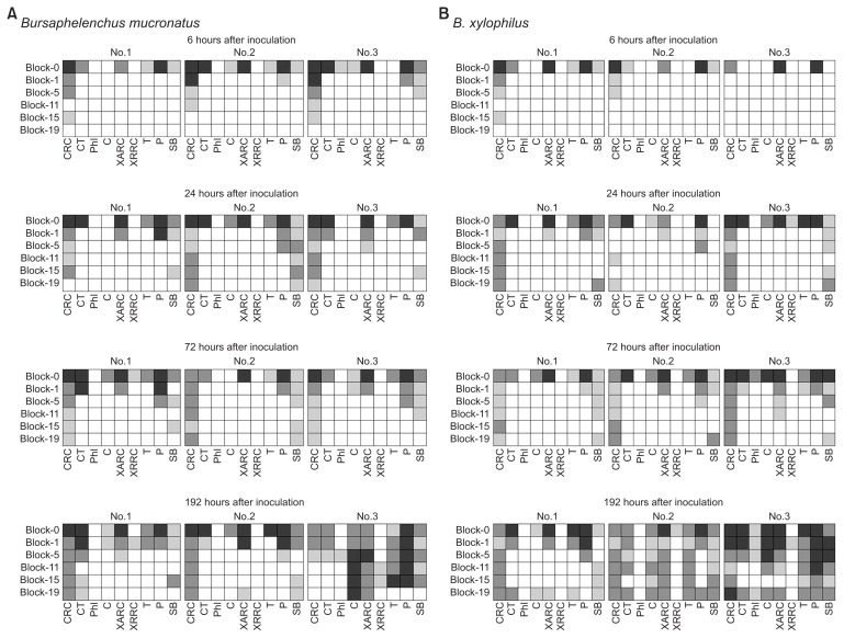
The distributions of B. mucronatus in P. thunbergii stem cuttings are shown in Fig. 1A. B. mucronatus were observed in the cortical resin canals up to block-15 cm and in the xylem resin canals only up to block-0 cm at 6 HAI. At 24 HAI and thereafter, B. mucronatus distribution extended through the cortical resin canals up to block-19 cm. B. mucronatus were present in the xylem axial resin canals up to block-5 cm and block-1 cm at 24 and 72 HAI, respectively. At 192 HAI, their distribution extended up to block-19 cm.
Fig. 1.
Distribution of Bursaphelenchus mucronatus and B. xylophilus inoculated into 20 cm main stem cuttings of 3-year-old Pinus thunbergii seedlings. (A) Distribution of B. mucronatus. (B) Distribution of B. xylophilus. Data from the article of Son et al. (2015) (Forest Pathol. 45:246–253) with permission, conducted at the same time as the experiment in this study. Numbers 1, 2, and 3 at the top side of the columns indicate each of the three different stem cuttings. Abbreviations at the bottom represent cortical resin canals (CRC), cortical tissues (CT), phloem (Phl), cambial layer (C), xylem axial resin canals (XARC), xylem radial resin canals (XRRC), tracheids (T), pith (P), and short branches (SB). Block-0 cm to block-19 cm at the left side indicate the distance from the inoculation surface to the sampling blocks. Each column indicates the average number of nematode segments in five pine thin-sections depending on its color (white, no nematode; light gray, average nematode numbers less than 1; gray, average nematode numbers over 1 but fewer than 10; dark gray, average nematode numbers over 10).
Distribution of dead epithelial cells in the cortical resin canals of P. thunbergii stem cuttings inoculated with B. mucronatus.
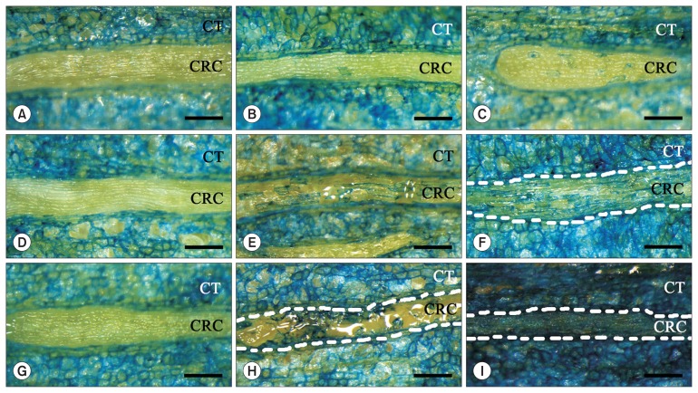
At 1 DAI, no epithelial cells were stained in blue in 21/22 segments from the control stem cuttings (Son et al., 2014; Table 1, Fig. 2A). However, blue-stained epithelial cells were found in 11/24 segments from the B. mucronatus-inoculated stem cuttings (Table 1). More than 50 epithelial cells were stained in blue in one segment, while in other segments the number of stained cells varied from 1 to 18 cells. Blue-stained epithelial cells were distributed sparsely and sporadically in the epithelia of the cortical resin canals (Fig. 2B, C). The relative mean area of the blue-stained epithelial cells in the B. mucronatus-inoculated stem cuttings was 1.15% (Table 1).
Table 1.
The % area of the blue-stained epithelial cells in segments of 20 cm main stem cuttings of 5-year-old Pinus thunbergii inoculated with 1,000 Bursaphelenchus mucronatus shown 1, 3, and 7 days after inoculation (DAI)
| Inoculum | DAI | Sample No. | The % area of the blue-stained epithelial cells in each segment at each distance from inoculation surface (cm) | Mean | |||||||
|---|---|---|---|---|---|---|---|---|---|---|---|
|
| |||||||||||
| 0–2.5 | 2.5–5 | 5–7.5 | 7.5–10 | 10–12.5 | 12.5–15 | 15–17.5 | 17.5–20 | ||||
| Control* | 1 | 1 | 0 | 0 | 0 | 0 | 0 | 0 | 0 | 0 | 0 |
| 2 | 0 | 0 | - | NE | 0 | 0 | 0 | 0 | 0 | ||
| 3 | 0 | 0 | 0.70 | 0 | 0 | 0 | 0 | 0 | 0.09 | ||
| Mean | 0.03 | ||||||||||
| 3 | 1 | - | 0 | 0 | - | 0 | 0 | 0 | - | 0 | |
| 2 | 0 | 0 | 0 | 0 | 0 | 0 | 0 | 0 | 0 | ||
| 3 | 0 | 0 | 0 | 0 | 0 | 0 | 0 | 0 | 0 | ||
| Mean | 0 | ||||||||||
| 7 | 1 | 0 | 2.67 | 0 | 0 | 0 | 0 | 0 | 0 | 0.33 | |
| 2 | - | 0 | - | 0 | 0 | 0 | - | - | 0 | ||
| 3 | 0 | 0 | 0 | 0 | 0 | 0 | 0 | 0 | 0 | ||
| Mean | 0.11 | ||||||||||
| B. mucronatus | 1 | 1 | 0 | 0.34 | 0.37 | 2.65 | 0 | 1.35 | 0 | 0 | 0.59 |
| 2 | 6.03 | 0 | 0.90 | 3.74 | 0 | 1.96 | 4.51 | 0 | 2.14 | ||
| 3 | 5.53 | 0 | 0 | 0 | 0 | 0 | 0 | 0.30 | 0.73 | ||
| Mean | 1.15 | ||||||||||
| 3 | 1 | 25.03 | 37.90 | 15.50 | 11.38 | 5.77 | 32.87 | 9.96 | 19.53 | 19.74 | |
| 2 | 1.20 | 14.04 | 5.99 | 0 | 0 | 1.07 | 11.29 | 30.18 | 7.97 | ||
| 3 | 13.48 | 35.91 | 27.69 | 8.88 | 13.17 | 11.94 | 0.64 | 2.81 | 14.31 | ||
| Mean | 14.01 | ||||||||||
| 7 | 1 | 37.57 | 35.69, e | 24.03, e | 37.52 | 31.39 | 69.24 | 67.17 | 60.41 | 45.38 | |
| 2 | 100 | 85.22 | 100 | 92.61 | 93.64 | 86.13 | 84.97 | 56.37 | 87.37 | ||
| 3 | 18.03 | 20.15 | 26.85 | 70.89 | 52.55 | 17.80 | 95.40 | 69.67 | 46.42 | ||
| Mean | 59.72 | ||||||||||
Values are presented as % of the blue-stained epithelial cell area per whole resin canal area in one section of photograph (area, 1.1 × 1.5 mm).
-, whole epithelial cells covered by resin; NE, no exposed epithelial cell; e, enlarged epithelial cells.
Data from the article of Son et al. (2014) (Nematology 16:663–668) with permission, conducted at the same time as the experiment in this study.
Fig. 2.
Evans blue-stained dead cells in the epithelium of the cortical resin canals in current-year main stem cuttings of 5-year-old Pinus thunbergii. Abbreviations represent cortical resin canals (CRC) and cortical tissues (CT). Scale bars = 0.25 mm. (A, D, G) Stem cuttings inoculated with distilled water shown 1, 3, and 7 days after inoculation (DAI), respectively, and no dead cells were found in the cortical resin canal. Data from the article of Son et al. (2014) (Nematology 16:663–668) with permission, conducted at the same time as the experiment in this study. (B, C) Stem cuttings inoculated with Bursaphelenchus mucronatus, 1 DAI. Note that blue-stained dead cells are sporadically distributed in the resin canal; the area covered by the blue-stained dead cells was 5.53% (C). (E, F) Stem cuttings inoculated with B. mucronatus, 3 DAI. The areas covered by the blue-stained dead cells were 19.53% and 30.18%, respectively. The resin canal is present between the 2 dotted white-lines (F). (H, I) Stem cuttings inoculated with B. mucronatus, 7 DAI. Enlarged epithelial cells are observed between the 2 dotted white-lines (H). The resin canal is also present between the 2 dotted white-lines. All the epithelial cells in the resin canal were dead (I).
At 3 DAI, there were no blue-stained epithelial cells in 21 segments in the control stem cuttings (Son et al., 2014; Table 1, Fig. 2D). Blue-stained cells were found in 22/24 segments from the B. mucronatus-inoculated stem cuttings with 9 segments showing more than 50 blue-stained epithelial cells (Table 1). The distribution of blue-stained cells was still sparse and sporadic, depending on the density (Fig. 2E, F). The relative mean area of the blue-stained epithelial cells in the B. mucronatus-inoculated stem cuttings had increased to 14.01% (Table 1).
At 7 DAI, epithelial cells remained unstained in 19/20 segments from the control stem cuttings (Son et al., 2014; Table 1, Fig. 2G). In contrast, every segment of the B. mucronatus-inoculated stem cuttings contained more than 50 blue-stained epithelial cells. The distribution of the blue-stained cells was sparse and sporadic when the density was relatively low. As the density of blue-stained cells increased, blue-stained cells bridged the gap and eventually filled the whole resin canal (Fig. 2I). The relative mean area of the blue-stained epithelial cells in the B. mucronatus-inoculated stem cuttings was 59.72% (Table 1). In addition, swollen epithelial cells were found in 2/24 segments from the B. mucronatus-inoculated cuttings (Table 1, Fig. 2H).
Discussion
Since it is possible that B. mucronatus in pine stem segments at 72 and 192 HAI showed a population increase (Son and Moon, 2013; Wang et al., 2005), only the distribution data of B. mucronatus at 6 and 24 HAI were used for the migration assessment in this study. B. mucronatus migrated mainly axially through the cortical resin canals to the opposite side of the stem cutting at 6 and 24 HAI. At these time points, the distribution of B. mucronatus coincided with that observed by Son et al. (2015) for B. xylophilus. Based on these results, we suggest that the migrations of B. xylophilus and B. mucronatus described in the studies of Togashi and Matsunaga (2003) and Son and Moon (2013), respectively, occur axially through the cortical resin canals. Therefore, the axial migration abilities of B. xylophilus and B. mucronatus through the cortical resin canals do not appear to differ from each other. However, our study could not explain the conflicting results obtained by Ishida et al. (1993), who suggested that the migration ability of B. mucronatus is much more limited than that of B. xylophilus. Based on the distribution of B. mucronatus at 72 HAI, we suggest that B. mucronatus cannot migrate horizontally from the cortical resin canals to the surrounding tissues. B. xylophilus also showed a similar distribution pattern (Son et al., 2015). Horizontal migration of B. mucronatus was detected at 192 HAI, which coincides with the result obtained for B. xylophilus (Son et al., 2015). Son et al. (2010) indicated that B. xylophilus migrate axially through the cortical and the xylem axial resin canals, and then, migrate horizontally from both resin canals to the surrounding tissues. Our study also showed that B. mucronatus migrate faster through the cortical resin canals, and then, through the xylem axial resin canals, to finally migrate horizontally from both resin canals to the surrounding tissues. Son et al. (2015) also indicated that the xylem axial resin canal of P. thunbergii is an important pathway for B. xylophilus in inducing PWD in the whole pine tree. In the present study, we also detected B. mucronatus in every tissue including in the xylem axial resin canals, meaning that B. mucronatus can also migrate throughout the whole pine tree.
In this study, a sparse and sporadic distribution of dead pine epithelial cells was observed in pine cuttings inoculated with B. mucronatus. This distribution is similar to the one observed for B. xylophilus in a previous study (Son et al., 2014). These results indicate that both nematodes show the same pattern when attacking pine epithelial cells. However, at 1 and 3 DAI, the number and area of dead epithelial cells in pine cuttings inoculated with B. mucronatus were smaller than in those inoculated with B. xylophilus (Son et al., 2014). Based on this result, we suggest that the attacking ability of B. mucronatus is weaker than that of B. xylophilus. B. xylophilus kills pine cells using its stylet together with a cellulase secretion (Kikuchi et al., 2004; Kuroda and Mamiya, 1986; Zhang et al., 2007). However, B. mucronatus secretes a cellulase with weaker activity (Kojima et al., 1994). Therefore, the weaker activity of the cellulase may cause the weaker attacking ability of B. mucronatus. However, the number and area of dead epithelial cells in pine cuttings inoculated with B. mucronatus were larger than in those inoculated with B. xylophilus at 7 DAI (Son et al., 2014). This may be the case because the stainability of dead cell by B. xylophilus was hidden by leaked resin and enlarged epithelial cells. Further study is required to determine why resin canals were covered with resin and had more enlarged epithelial cells inoculated with B. xylophilus than those inoculated with B. mucronatus at 7 DAI.
This study is the first to suggest that the non-pathogenicity of B. mucronatus may be related to their weaker attacking ability. As described in B. xylophilus, B. mucronatus also migrate mainly through the cortical resin canals of pine cuttings, and then, through the xylem axial resin canals, to finally migrate from both resin canals to the surrounding tissues. The migration ability of both nematode species does not seem to differ when the nematodes migrate through pine resin canals. However, when the nematodes attack pine cells, the attacking ability of these nematode species differs due to the weaker attacking ability of B. mucronatus compared to that of B. xylophilus. In conclusion, the present study showed that the non-pathogenicity of B. mucronatus was caused by their weaker attacking ability.
Acknowledgments
Part of this study was supported by a Research Fellowship of the National Institute of Forest Science in 2012.
BRILL license (Number 3827350610933) was obtained for referring ‘Control’ in Table 1 from Nematology (Son et al., 2014).
John Wiley and Sons license (Number 3825590682102) was obtained for referring ‘Fig. 1B’ in Fig. 1 from Forest Pathol. (Son et al., 2015).
Footnotes
Articles can be freely viewed online at www.ppjonline.org.
References
- Fukuda K, Hogetsu T, Suzuki K. Cavitation and cytological changes in xylem of pine seedling inoculated with virulent and avirulent isolates of Bursaphelenchus xylophilus and B. mucronatus. J Jpn For Soc. 1992;74:289–299. [Google Scholar]
- Futai K. Population dynamics of Bursaphelenchus lignicolus (Nematoda: Aphelenchoididae) and B. mucronatus in pine seedlings. Appl Entomol Zool. 1980;15:458–464. [Google Scholar]
- Gaff DF, Okong’o-Ogora O. The use of non-permeating pigments for testing the survival of cells. J Exp Bot. 1971;22:756–758. doi: 10.1093/jxb/22.3.756. [DOI] [Google Scholar]
- Ichihara Y, Fukuda K, Suzuki K. Early symptom development and histological changes associated with migration of Bursaphelenchus xylophilus in seedling tissues of Pinus thunbergii. Plant Dis. 2000;84:675–680. doi: 10.1094/PDIS.2000.84.6.675. [DOI] [PubMed] [Google Scholar]
- Ishida K, Hogetsu T, Fukuda K, Suzuki K. Cortical responses in Japanese black pine attack by the pine wood nematode. Can J Bot. 1993;71:1399–1405. doi: 10.1139/b93-168. [DOI] [Google Scholar]
- Kikuchi T, Jones JT, Aikawa T, Kosaka H, Ogura N. A family of glycosyl hydrolase family 45 cellulases from the pine wood nematode Bursaphelenchus xylophilus. FEBS Lett. 2004;572:201–205. doi: 10.1016/j.febslet.2004.07.039. [DOI] [PubMed] [Google Scholar]
- Kiyohara T, Suzuki K. Nematode population growth and disease development in the pine wilting disease. Eur J For Pathol. 1978;8:285–292. doi: 10.1111/j.1439-0329.1978.tb00641.x. [DOI] [Google Scholar]
- Kojima K, Kamijyo A, Masumori M, Sasaki S. Cellulase activities of pine-wood nematode isolates with different virulences. J Jpn For Soc. 1994;76:258–262. [Google Scholar]
- Komatsu M, Son J, Matsushita N, Hogetsu T. Fluorescein-labeled wheat germ agglutinin stains the pine wood nematode, Bursaphelenchus xylophilus. J For Res. 2008;13:132–136. doi: 10.1007/s10310-008-0065-9. [DOI] [Google Scholar]
- Kuroda K, Mamiya Y. Behavior of the pine wood nematode in pine seedling growing under aseptic conditions. Trans J Jpn For Soc. 1986;97:471–472. (in Japanese) [Google Scholar]
- Mamiya Y, Enda N. Bursaphelenchus mucronatus N. SP. (Nematoda: Aphelenchoididae) from pine wood and its biology and pathogenicity to pine trees. Nematologica. 1979;25:353–361. doi: 10.1163/187529279X00091. [DOI] [Google Scholar]
- Nobuchi T, Tominaga T, Futai K, Harada H. Cytological study of pathological changes in Japanese black pine (Pinus thunbergii) seedlings after inoculation with pinewood nematode (Bursaphelenchus xylophilus) Bull Kyoto Univ For. 1984;56:224–233. [Google Scholar]
- Odani K, Sasaki S, Yamamoto N, Nishiyama Y, Tamura H. Differences in dispersal and multiplication of two associated nematodes, Bursaphelenchus xylophilus and Bursaphelenchus mucronatus in pine seedlings in relation to the pine wilt disease development. J Jpn For Soc. 1985;67:398–403. [Google Scholar]
- Smant G, Stokkermans JP, Yan Y, de Boer JM, Baum TJ, Wang X, Hussey RS, Gommers FJ, Henrissat B, Davis EL, Helder J, Schots A, Bakker J. Endogenous cellulases in animals: isolation of beta-1, 4-endoglucanase genes from two species of plant-parasitic cyst nematodes. Proc Natl Acad Sci U S A. 1998;95:4906–4911. doi: 10.1073/pnas.95.9.4906. [DOI] [PMC free article] [PubMed] [Google Scholar]
- Son JA, Hogetsu T, Moon YS. The process of epithelial cell death in Pinus thunbergii caused by the pine wood nematode, Bursaphelenchus xylophilus. Nematology. 2014;16:663–668. doi: 10.1163/15685411-00002795. [DOI] [Google Scholar]
- Son JA, Komatsu M, Matsushita N, Hogetsu T. Migration of pine wood nematodes in the tissues of Pinus thunbergii. J For Res. 2010;15:186–193. doi: 10.1007/s10310-009-0171-3. [DOI] [Google Scholar]
- Son JA, Matsushita N, Hogetsu T. Migration of Bursaphelenchus xylophilus in cortical and xylem axial resin canals of resistant pines. For Pathol. 2015;45:246–253. [Google Scholar]
- Son JA, Moon YS. Migrations and multiplications of Bursaphelenchus xylophilus and B. mucronatus in Pinus thumbergii in relation to their pathogenicity. Plant Pathol J. 2013;29:116–122. doi: 10.5423/PPJ.OA.06.2012.0091. [DOI] [PMC free article] [PubMed] [Google Scholar]
- Tian X, Cheng X, Mao Z, Chen G, Yang J, Xie B. Composition of bacterial communities associated with a plant-parasitic nematode Bursaphelenchus mucronatus. Curr Microbiol. 2011;62:117–125. doi: 10.1007/s00284-010-9681-7. [DOI] [PubMed] [Google Scholar]
- Togashi K, Matsunaga K. Between-isolate difference in dispersal ability of Bursaphelenchus xylophilus and vulnerability to inhibition by Pinus densiflora. Nematology. 2003;5:559–564. doi: 10.1163/156854103322683274. [DOI] [Google Scholar]
- Wang Y, Yamada T, Sakaue D, Suzuki K. Variations in life history parameters and their influence on rate of population increase of different pathogenic isolates of the pine wood nematode, Bursaphelenchus xylophilus. Nematology. 2005;7:459–467. doi: 10.1163/156854105774355545. [DOI] [Google Scholar]
- Woo KS, Yoon JH, Woo SY, Lee SH, Han SU, Han H, Baek SG, Kim CS. Comparison in disease development and gas exchange rate of Pinus densiflora seedlings artificially inoculated with Bursaphelenchus xylophilus and B. mucronatus. For Sci Technol. 2010;6:110–117. [Google Scholar]
- Zhang Q, Bai G, Cheng J, Yu Y, Yang W. Use of an enhanced green fluorescence protein linked to a single chain fragment variable antibody to localize Bursaphelenchus xylophilus cellulase. Biosci Biotechnol Biochem. 2007;71:1514–1520. doi: 10.1271/bbb.70022. [DOI] [PubMed] [Google Scholar]
- Zhang Q, Bai G, Yang W, Li H, Xiong H. Pathogenic cellulase assay of pine wilt disease and immunological localization. Biosci Biotechnol Biochem. 2006;70:2727–2732. doi: 10.1271/bbb.60330. [DOI] [PubMed] [Google Scholar]
- Zhao BG, Lin F. Mutualistic symbiosis between Bursaphelenchus xylophilus and bacteria of the genus Pseudomonas. For Pathol. 2005;35:339–345. [Google Scholar]
- Zhao BG, Lin F, Guo D, Li R, Li SN, Kulinich O, Ryss A. Pathogenic roles of the bacteria carried by Bursaphelenchus mucronatus. J Nematol. 2009;41:11–16. [PMC free article] [PubMed] [Google Scholar]
- Zhao BG, Liu Y, Lin F. Effects of bacteria associated with pine wood nematode (Bursaphelenchus xylophilus) on development and egg production of the nematode. J Phytopathol. 2007;155:26–30. doi: 10.1111/j.1439-0434.2006.01188.x. [DOI] [Google Scholar]
- Zhao H, Chen C, Liu S, Liu P, Liu Q, Jian H. Aseptic Bursaphelenchus xylophilus does not reduce the mortality of young pine tree. For Pathol. 2013;43:444–454. [Google Scholar]


