Abstract
INTRODUCTION: Ganglion cysts are benign soft tissue swellings commonly found in the wrist. The presence of these cysts in the elbow is uncommon, and few case reports have been reported for this condition at this location. These lesions can compress on the neighbouring structures or cause restriction of the joint movement. The awareness of this entity is a must, to arrive at an early diagnosis.
MATERIALS AND METHOD: We report a patient with swelling in the anterolateral aspect of the elbow which had been causing intermittent pain for the last 13 months. The MRI revealed a fluid-filled cystic swelling which was communicating with the radio-capitellar joint.
RESULTS: The lesion was excised in toto, using anterolateral approach for the elbow, and sent for histopathological examination which confirmed the diagnosis of a ganglion cyst.
CONCLUSION: Thus, due to the infrequent presentation, an awareness of this condition is necessary to prevent a delay in diagnosis and its subsequent management.
Keywords: ganglion, cyst, elbow, diagnosis
Introduction
Ganglion cysts are the most common benign soft tissue swellings, which can be found in any joint of the body, but 60-70% are found in the dorsal aspect of the wrist and communicate with the joint via a pedicle [1]. Occurrence of ganglion cysts in other joints such as the elbow is rare. These are well defined and mobile swellings, which are loosely attached to the sheath of the tendon or the capsule of the joint.
Ganglion cysts around the elbow are rare [2]. Hence, the diagnosis of these lesions may be challenging and often delayed if the awareness of this entity is not known to the clinician. These are usually asymptomatic but sometimes may cause restriction of elbow range of movement or compression of radial or posterior interosseous nerve. Most of these cysts are seen as an incidental finding on Magnetic Resonance Images (MRI) or on ultrasonography (USG). We report the case of a ganglion cyst in the elbow (originating from the radio-capitellar joint), in a middle-aged female where the diagnosis could not be made for more than a year.
Case presentation
A 45-year-old woman presented with a 13-month-history of pain and swelling in the anterior aspect of the right elbow. There was no history of trauma or any other exacerbating factor. She had been taking analgesics for last nine months due to intermittent pain which affected her ability to use the dominant elbow.
Local examination revealed a 3 cm x 3 cm soft tissue swelling on the anterolateral aspect of the elbow, which was tender on palpation. The swelling was mobile in all directions. Elbow range of movement was from zero degrees to 125 degrees. There was no associated erythema around the elbow and over the swelling.
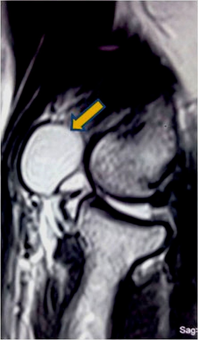
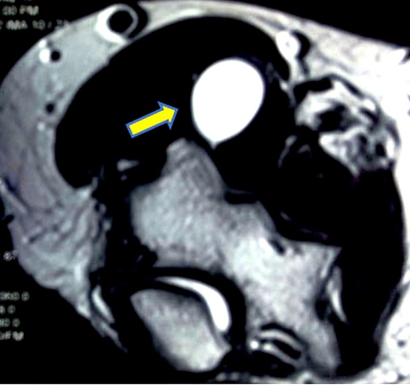
The MRI showed a multiseptate fluid collection measuring 1.73 x 1.32 cm on the lateral aspect of cubital fossa anterior to the radio-capitellar joint space possibly communicating with it showing T1 hypo and T2/STIR hyperintense signals. Anteriorly it was burrowed into the brachioradialis muscle (Figure 1, 2).
Figure 1. MRI T2 weighted saggital view showing hyerintense cyst anterior to radiocapitellar joint.
Figure 2. MRI (T2 weighted) cross section view showing the cyst in the anterolateral aspect of the elbow.
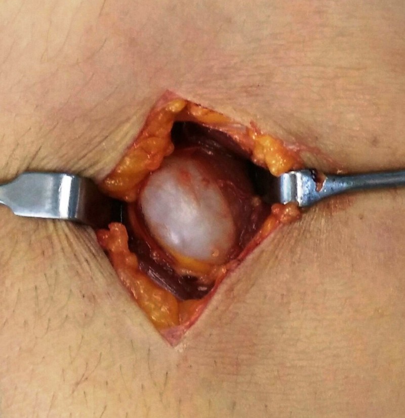
This symptomatic swelling was excised through an anterolateral approach to the elbow joint [3], after making a plane between brachialis and brachioradialis. A cystic swelling of about 2 cm x 2 cm in size was found (Figure 3).
Figure 3. Intraoperative picture showing the cyst.
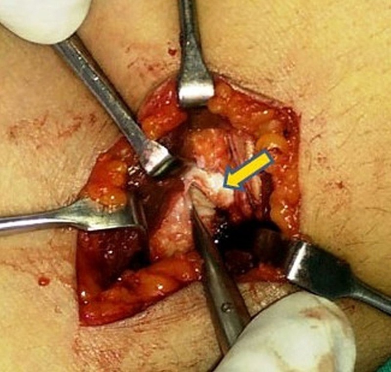
It was carefully isolated from the surrounding soft tissues and excised en-masse. The stalk of the lesion was found to be originating from the radio-capitellar joint (Figure 4).
Figure 4. Intraoperative picture showing rent in the capsule of radiocapitellar joint.
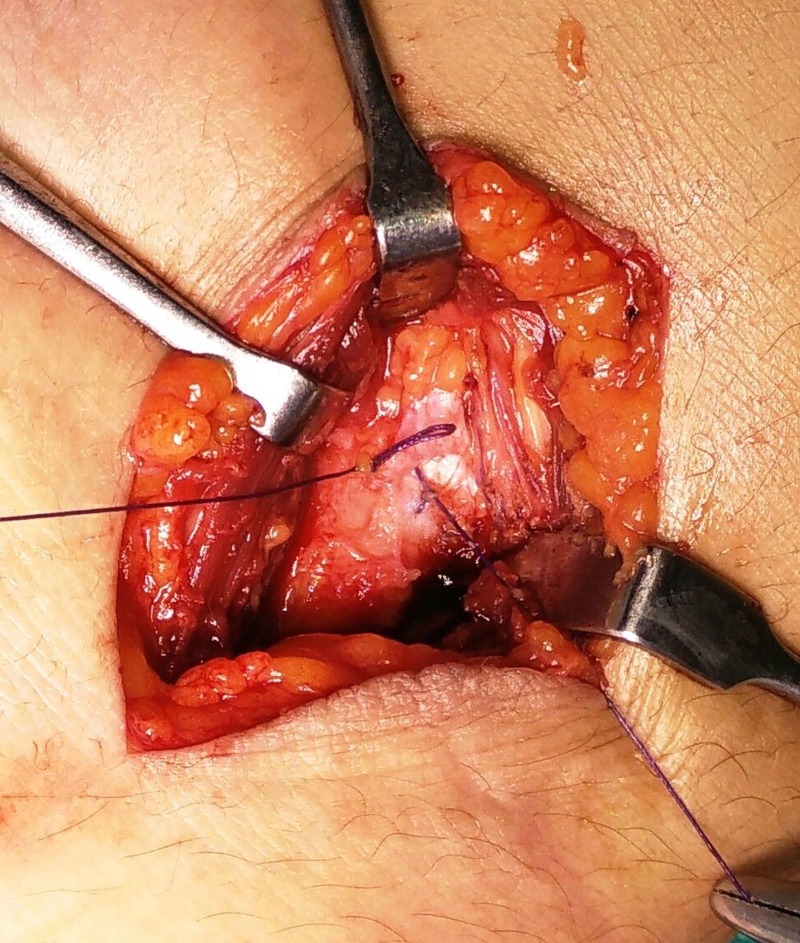
The rent in the capsule was then closed with an absorbable Vicryl 3-0 suture (Figure 5).
Figure 5. Intraoperative picture showing repair of the capsule after excision of the cyst.
The excised lesion was sent for histopathological examination, which confirmed the diagnosis of a ganglion cyst.
Written consent was obtained from the human subject who participated in this case study. Institutional Review Board of Indraprastha Apollo Hospital, India approved the research for this case.
Discussion
The clinical diagnosis of an elbow swelling is often challenging, due to the complex nature of the joint and rarity of the location of the swelling. Such lesions may be a manifestation of a wide variety of conditions like myositis ossificans, ganglion cysts, lipoma, cysticercus cyst, vascular aneurysms, soft tissue sarcoma, septic arthritis, lymphoedema, and pseudogout.
A confirmatory diagnosis can be made using plain radiographs, USG and MRI [4]. We believe that an MRI is the investigation of choice for soft tissue lesions, as one can assess its site of origin, the size of swelling and whether it is compressing on any nerve or vessel around the elbow. Ganglion cyst on MRI has classical features viz. unilocular or multilocular rounded or lobular fluid signal mass (which is hyperintense on T2/STIR and hypointense signals on T1 sequences), adjacent to a joint or tendon which distinguishes it from the other pathologies.
Treatment modalities of ganglion cysts include conservative management by aspiration, injection of steroids, sclerosing agents and hyaluronidase and thread technique. However, most of these modalities are associated with either high recurrence rates or other complications like incomplete resolution and allergic reactions [5]. Steroid therapy is associated with subcutaneous fat atrophy and depigmentation of the skin [6]. Sclerotherapy is known to cause damage to the tendon from which the ganglion cyst arises [7]. The most efficient modality for treating the ganglion is to excise the cyst in toto and repair the rent in the capsule or tendon sheath, which would reduce the rate of recurrence of the tumor [8]. The recurrence rate after excision of the lesion, in current literature, is variable and ranges from 0 - 31.2 %.
There are a few reported cases of ganglion cyst at the elbow, and most of them have been shown to cause compressive neuropathies of the radial nerve or the posterior interosseous nerve [9]. A ganglion cyst in the supinator muscle was reported, which caused compression of the posterior interosseous nerve leading to weakness of wrist extensors [10]. Matsubara et al. reported 8 cases of radial nerve palsy due to ganglions at the elbow, with majority of cases presenting no symptoms [11]. We did not find any evidence of any compressive neuropathy in our case.
Conclusions
Ganglion cysts are common, benign, soft tissue swellings found around the joints. Their presence in the elbow is uncommon, and only few case reports have been noted for this condition at this location. Due to an uncommon presentation in the elbow, the ganglion cysts are often difficult to diagnose. Hence, a delay in diagnosis is inevitable, just as it occured in our case. Hence, an awareness about the rare presentation of a ganglion cyst in the elbow is necessary, so as to arrive at an early diagnosis and treatment.
The content published in Cureus is the result of clinical experience and/or research by independent individuals or organizations. Cureus is not responsible for the scientific accuracy or reliability of data or conclusions published herein. All content published within Cureus is intended only for educational, research and reference purposes. Additionally, articles published within Cureus should not be deemed a suitable substitute for the advice of a qualified health care professional. Do not disregard or avoid professional medical advice due to content published within Cureus.
The authors have declared that no competing interests exist.
Human Ethics
A written consent was obtained from the patient prior to submission of the manuscript. IRB (Indraprastha Apollo Hospital) approved the research for the study. issued approval. COI form signed by Amit Agarwal (corresponding author) on behalf of all the co-authors was included as part of the submission.
References
- 1.Dorsal wrist ganglion: Current review of literature. Meena S, Gupta A. J Clin Orthop Trauma. 2014;5(2):59–64. doi: 10.1016/j.jcot.2014.01.006. [DOI] [PMC free article] [PubMed] [Google Scholar] [Retracted]
- 2.Diagnosis of radial nerve palsy caused by ganglion with use of different imaging techniques. Ogino T, Minami A, Kato H. J Hand Surg Am. 1991;16:230–235. doi: 10.1016/s0363-5023(10)80102-9. [DOI] [PubMed] [Google Scholar]
- 3.Open reduction and internal fixation of capitellar fracture through aterolateral approach with headless double-threaded compression screws: a series of 16 patients. Vaishya R, Vijay V, Jha GK, Agarwal AK. http://www.ncbi.nlm.nih.gov/pubmed/27052272. J Shoulder Elbow Surg. 2016;Apr:1058–2746. doi: 10.1016/j.jse.2016.01.034. [DOI] [PubMed] [Google Scholar]
- 4.Tumour like lesions and benign tumours of the hand and wrist. Plate AM, Lee SJ, Steiner G, et al. J Am Acad orthop Surg. 2003;11(2):129–141. doi: 10.5435/00124635-200303000-00007. [DOI] [PubMed] [Google Scholar]
- 5.Improving the results of ganglion aspiration by the use of hyaluronidase. Paul AS, Sochart DH. Journal of Hand Surgery. 1997;22(2):219–221. doi: 10.1016/s0266-7681(97)80066-6. [DOI] [PubMed] [Google Scholar]
- 6.Varley GW, Needoff M, Davis TRC, Clay NR. Journal of Hand Surgery. 22(5) Davis; 1997. Conservative management of wrist ganglia: aspiration versus steroid infiltration; pp. 636–637. [DOI] [PubMed] [Google Scholar]
- 7. The dangers of sclerotherapy in the treatment of ganglia. Mackie IG, Howard CB, Wilkins P. http://www.sciencedirect.com/science/article/pii/S026676818480025X. Journal of hand surgery. 1984;9(2):181–184. [PubMed] [Google Scholar]
- 8.The dorsal ganglion of the wrist: Its pathogenesis gross and microscopic anatomy, and surgical treatment. Angelides AC, Wallace PF. http://www.sciencedirect.com/science/article/pii/S0363502376800421. Journal of Hand Surgery. 1976;1(3):228–235. doi: 10.1016/s0363-5023(76)80042-1. [DOI] [PubMed] [Google Scholar]
- 9.A ganglion cyst at the elbow causing superficial radial nerve compression: a case report. McFarlane J, Trehan R, Olivera M, et al. Journal of Medical Case Reports. 2008;2:122. doi: 10.1186/1752-1947-2-122. [DOI] [PMC free article] [PubMed] [Google Scholar]
- 10.Posterior interosseous nerve paralysis caused by a ganglion at the elbow. Bowen Tl, Stone KH. http://www.bjj.boneandjoint.org.uk/content/jbjsbr/48-B/4/774.full.pdf. J Bone Joint Surg. 1966;48:774–776. [PubMed] [Google Scholar]
- 11.Radial nerve palsy at the elbow. Matsubara Y, Miyasaka Y, Nobuta SR. Ups J Med Sci. 2006;111:315–320. doi: 10.3109/2000-1967-057. [DOI] [PubMed] [Google Scholar]


