

Abstract
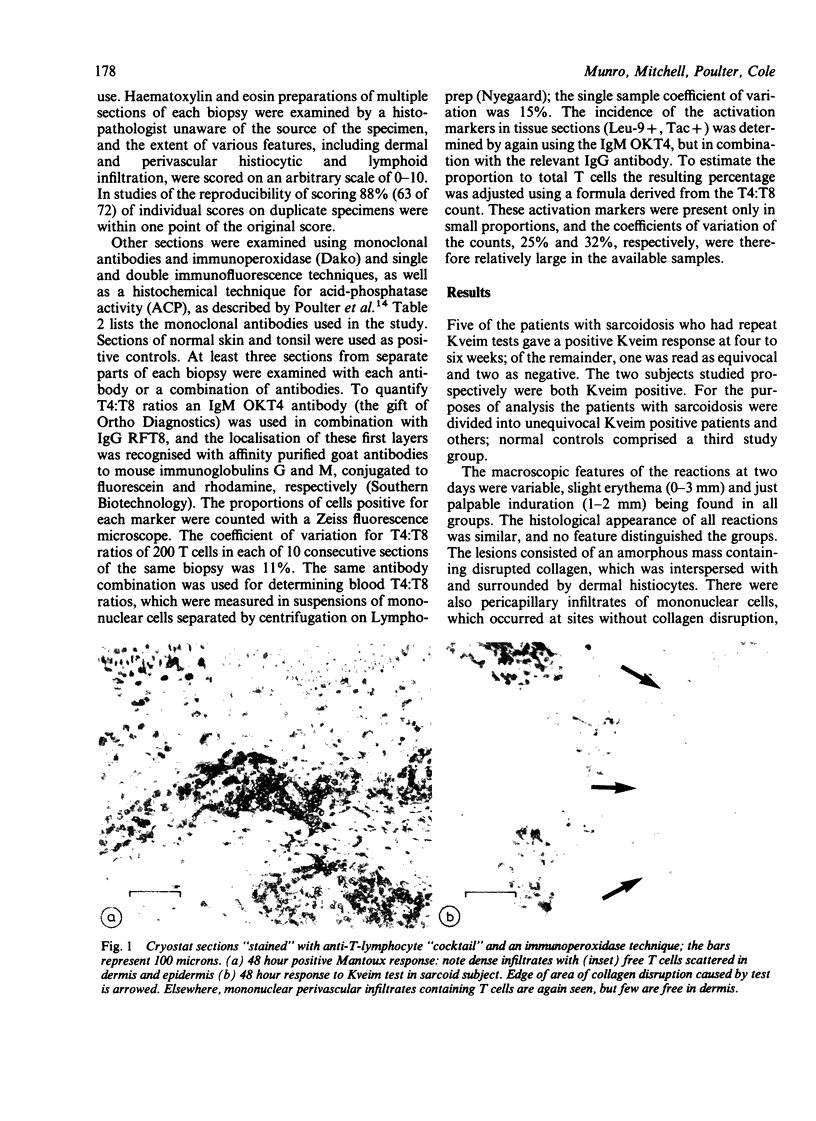
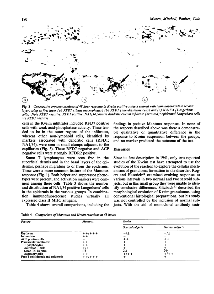
In a detailed controlled study of the cellular response to Kveim suspension in vivo we used immunohistological and histochemical methods to examine cryostat sections of immature Kveim biopsy specimens in subjects with sarcoidosis and normal controls. Changes seen at 48 hours, at which time papular reactions have sometimes been reported, are described. Eight cases of sarcoidosis previously confirmed by a positive Kveim test were studied, in five of whom the test remained positive; plus two subjects with sarcoidosis studied prospectively; and four healthy controls. There were two main features of the 48 hour response: collagen disruption with associated histiocytes, which showed increased acid phosphatase activity; and perivascular infiltrates of lymphocytes and small groups of dendritic cells. The T4:T8 ratios in the infiltrates were similar to those found in the peripheral blood of the subjects, and few lymphocytes showed evidence of activation. T lymphocytes were also seen free in the dermis and migrating to the epidermis. Small juxtacapillary clumps of dendritic cells, identified by NA1/34 (= OKT6; Langerhans' cells) and RFD1 (interdigitating cell) monoclonal antibodies, were found. The Langerhans' cells in the epidermis were, however, normal in number and distribution. These features, which were found in all groups, are not consistent with pre-existing hypersensitivity to Kveim suspension in sarcoidosis. Subsequent differences between sarcoid and normal subjects in the development of granulomas in the Kveim response may therefore relate to the different handling of the foreign material by the cells affected, rather than to differences in the early non-specific recruitment of the cells to the test site.
Full text
PDF


Images in this article

Selected References
These references are in PubMed. This may not be the complete list of references from this article.
- Ando M., Dannenberg A. M., Jr, Shima K. Macrophage accumulation, division, maturation and digestive and microbicidal capacities in tuberculous lesions. II. Rate at which mononuclear cells enter and divide in primary BCG lesions and those of reinfection. J Immunol. 1972 Jul;109(1):8–19. [PubMed] [Google Scholar]
- Bertouch J. V., Roberts-Thomson P. J., Bradley J. Diurnal variation of lymphocyte subsets identified by monoclonal antibodies. Br Med J (Clin Res Ed) 1983 Apr 9;286(6372):1171–1172. doi: 10.1136/bmj.286.6372.1171. [DOI] [PMC free article] [PubMed] [Google Scholar]
- Bradstreet C. M., Dighero M. W., Mitchell D. N. The Kveim test: analysis of results of tests using colindale (K 12) materials. Ann N Y Acad Sci. 1976;278:681–686. doi: 10.1111/j.1749-6632.1976.tb47082.x. [DOI] [PubMed] [Google Scholar]
- Chase M. W. Immunology and experimental dermatology. Annu Rev Immunol. 1985;3:1–29. doi: 10.1146/annurev.iy.03.040185.000245. [DOI] [PubMed] [Google Scholar]
- Fox J. L., Berman B., Teirstein A. S., France D. S., Reed M. L. Quantitation of cutaneous Langerhans cells of sarcoidosis patients. J Invest Dermatol. 1983 Jun;80(6):472–475. doi: 10.1111/1523-1747.ep12534905. [DOI] [PubMed] [Google Scholar]
- Henderson W. R., Fukuyama K., Epstein W. L., Spitler L. E. In vitro demonstration of delayed hypersensitivity in patients with berylliosis. J Invest Dermatol. 1972 Jan;58(1):5–8. doi: 10.1111/1523-1747.ep13077177. [DOI] [PubMed] [Google Scholar]
- Horsmanheimo M., Horsmanheimo A., Fudenberg H. H., Siltzbach L. E., McKee K. T. Leukocyte migration agarose test (LMAT) in sarcoidosis using Kveim test material. Br J Dermatol. 1978 Sep;99(3):263–270. doi: 10.1111/j.1365-2133.1978.tb01995.x. [DOI] [PubMed] [Google Scholar]
- Janossy G., Prentice H. G. T cell subpopulations, monoclonal antibodies and their therapeutic applications. Clin Haematol. 1982 Oct;11(3):631–660. [PubMed] [Google Scholar]
- Link M., Warnke R., Finlay J., Amylon M., Miller R., Dilley J., Levy R. A single monoclonal antibody identifies T-cell lineage of childhood lymphoid malignancies. Blood. 1983 Oct;62(4):722–728. [PubMed] [Google Scholar]
- Mishra B. B., Poulter L. W., Janossy G., James D. G. The distribution of lymphoid and macrophage like cell subsets of sarcoid and Kveim granulomata: possible mechanism of negative PPD reaction in sarcoidosis. Clin Exp Immunol. 1983 Dec;54(3):705–715. [PMC free article] [PubMed] [Google Scholar]
- Modlin R. L., Hofman F. M., Meyer P. R., Sharma O. P., Taylor C. R., Rea T. H. In situ demonstration of T lymphocyte subsets in granulomatous inflammation: leprosy, rhinoscleroma and sarcoidosis. Clin Exp Immunol. 1983 Mar;51(3):430–438. [PMC free article] [PubMed] [Google Scholar]
- NIH conference. Pulmonary sarcoidosis: a disease characterized and perpetuated by activated lung T-lymphocytes. Ann Intern Med. 1981 Jan;94(1):73–94. doi: 10.7326/0003-4819-94-1-73. [DOI] [PubMed] [Google Scholar]
- Pinkston P., Bitterman P. B., Crystal R. G. Spontaneous release of interleukin-2 by lung T lymphocytes in active pulmonary sarcoidosis. N Engl J Med. 1983 Apr 7;308(14):793–800. doi: 10.1056/NEJM198304073081401. [DOI] [PubMed] [Google Scholar]
- Poulter L. W., Collings L. A., Tung K. S., Waters M. F. Parasitism of antigen presenting cells in hyperbacillary leprosy. Clin Exp Immunol. 1984 Mar;55(3):611–617. [PMC free article] [PubMed] [Google Scholar]
- Poulter L. W., Janossy G. The involvement of dendritic cells in chronic inflammatory disease. Scand J Immunol. 1985 May;21(5):401–407. doi: 10.1111/j.1365-3083.1985.tb01825.x. [DOI] [PubMed] [Google Scholar]
- Poulter L. W., Seymour G. J., Duke O., Janossy G., Panayi G. Immunohistological analysis of delayed-type hypersensitivity in man. Cell Immunol. 1982 Dec;74(2):358–369. doi: 10.1016/0008-8749(82)90036-3. [DOI] [PubMed] [Google Scholar]
- ROGERS F. J., HASERICK J. R. Sarcoidosis and the Kveim reaction. J Invest Dermatol. 1954 Nov;23(5):389–406. doi: 10.1038/jid.1954.121. [DOI] [PubMed] [Google Scholar]
- Reinherz E. L., Schlossman S. F. The differentiation and function of human T lymphocytes. Cell. 1980 Apr;19(4):821–827. doi: 10.1016/0092-8674(80)90072-0. [DOI] [PubMed] [Google Scholar]
- SHELLEY W. B., HURLEY H. J. The allergic origin of zirconium deodorant granulomas. Br J Dermatol. 1958 Mar;70(3):75–101. doi: 10.1111/j.1365-2133.1958.tb13297.x. [DOI] [PubMed] [Google Scholar]
- Scheynius A., Klareskog L., Forsum U. In situ identification of T lymphocyte subsets and HLA-DR expressing cells in the human skin tuberculin reaction. Clin Exp Immunol. 1982 Aug;49(2):325–330. [PMC free article] [PubMed] [Google Scholar]
- Semenzato G., Pezzutto A., Chilosi M., Pizzolo G. Redistribution of T lymphocytes in the lymph nodes of patients with sarcoidosis. N Engl J Med. 1982 Jan 7;306(1):48–49. doi: 10.1056/NEJM198201073060114. [DOI] [PubMed] [Google Scholar]
- Thomas J. A., Janossy G., Graham-Brown R. A., Kung P. C., Goldstein G. The relationship between T lymphocyte subsets and Ia-like antigen positive nonlymphoid cells in early stages of cutaneous T cell lymphoma. J Invest Dermatol. 1982 Feb;78(2):169–176. doi: 10.1111/1523-1747.ep12506339. [DOI] [PubMed] [Google Scholar]
- Uchiyama T., Nelson D. L., Fleisher T. A., Waldmann T. A. A monoclonal antibody (anti-Tac) reactive with activated and functionally mature human T cells. II. Expression of Tac antigen on activated cytotoxic killer T cells, suppressor cells, and on one of two types of helper T cells. J Immunol. 1981 Apr;126(4):1398–1403. [PubMed] [Google Scholar]
- Warren K. S., Domingo E. O., Cowan R. B. Granuloma formation around schistosome eggs as a manifestation of delayed hypersensitivity. Am J Pathol. 1967 Nov;51(5):735–756. [PMC free article] [PubMed] [Google Scholar]
- Zweiman B., Israel H. L. Comparative in vitro reactivities of leukocytes from sarcoids and normals to different Kveim preparations. Ann N Y Acad Sci. 1976;278:700–710. doi: 10.1111/j.1749-6632.1976.tb47084.x. [DOI] [PubMed] [Google Scholar]