Abstract
OBJECTIVES: (1) To examine the prevalence and extent of intramural metastasis in squamous cell carcinomas of the oesophagus so as to delineate the resection margins for these tumours; (2) to devise an appropriate method for measurement of these lesions which takes into account of the contraction of the specimens after resection. METHODS: Oesophagectomy specimens were prospectively collected from 96 patients (87 males, nine females) with primary oesophageal squamous cell carcinoma over a two year period. The sizes of the tumours were measured in situ, after resection and after application of muscle relaxant (to regain their in situ length). The specimens were then serially sectioned for histological examination. RESULTS: The sizes of the tumours measured after application of muscle relaxant roughly corresponded to those measured in situ. Intramural metastasis was observed in 26% of the cases. Sixty four per cent (16 cases) of these were on the oral side, 72% (18 cases) on the gastric side, and 25% (nine cases) on both sides of the tumours. The most distant extent of intramural metastasis from the primary tumour was from 0.5 cm to 7.7 cm (mean = 3.4 cm) on the oral side, and 0.5 to 9.5 cm (mean 4 cm) on the gastric aspect of the tumour. Intramural metastasis was seen only in patients in whom the primary cancer had deep muscle infiltration. Multiple neoplastic lesions could be detected in 33% of the patients. Both intramural metastasis and multiple neoplastic lesions were associated with extensive lymph node infiltration. However, they had different histological features and extent of infiltration. CONCLUSIONS: Intramural metastasis was frequently observed in oesophageal squamous cell carcinoma. This implies that excision with wide margins should be considered for local control of the disease.
Full text
PDF
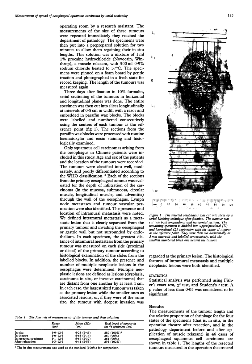
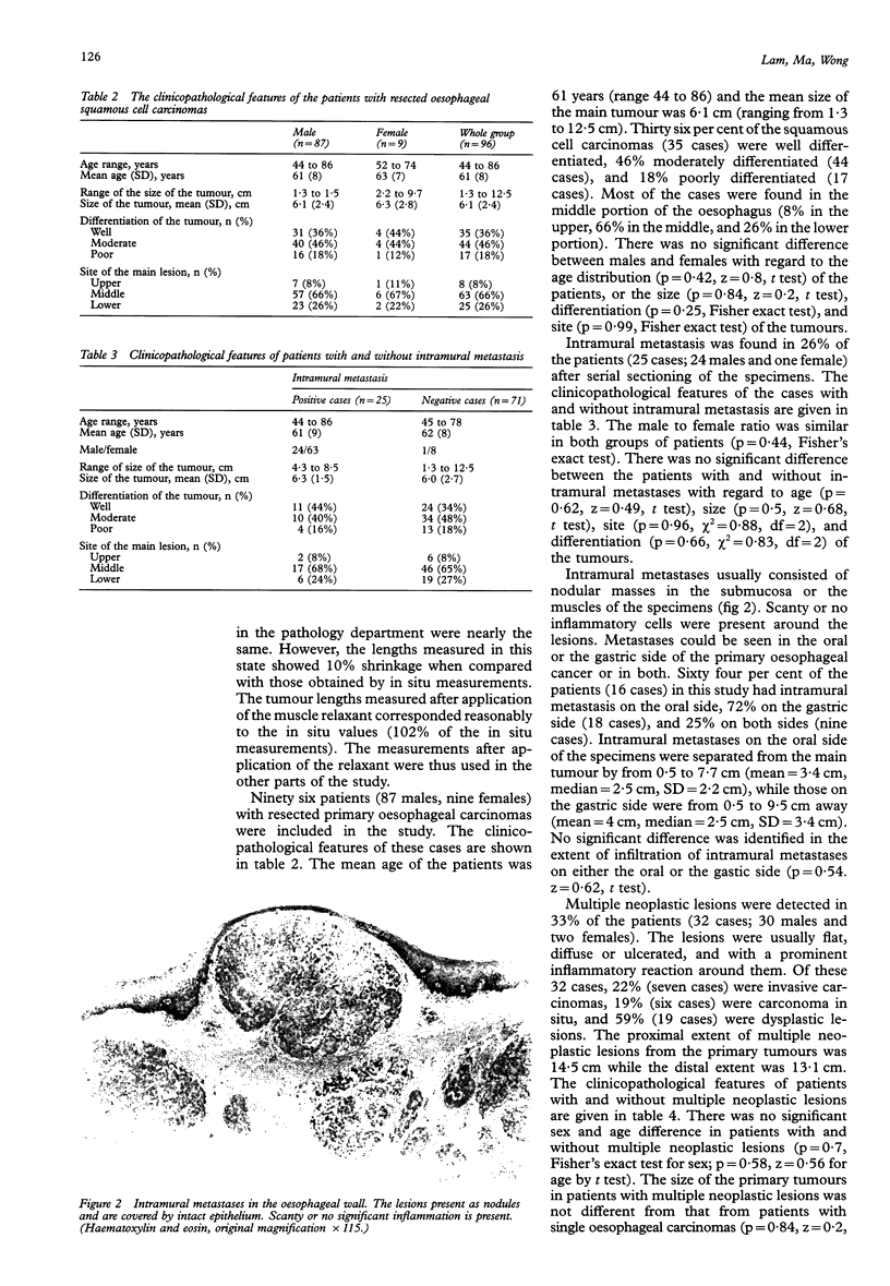

Images in this article
Selected References
These references are in PubMed. This may not be the complete list of references from this article.
- BURGESS H. M., BAGGENSTOSS A. H., MOERSCH H. J., CLAGETT O. T. Carcinoma of the esophagus; a clinicopathologic study. Surg Clin North Am. 1951 Aug;31(4):965–976. [PubMed] [Google Scholar]
- Kato H., Tachimori Y., Watanabe H., Itabashi M., Hirota T., Yamaguchi H., Ishikawa T. Intramural metastasis of thoracic esophageal carcinoma. Int J Cancer. 1992 Jan 2;50(1):49–52. doi: 10.1002/ijc.2910500111. [DOI] [PubMed] [Google Scholar]
- Kuwano H., Ohno S., Matsuda H., Mori M., Sugimachi K. Serial histologic evaluation of multiple primary squamous cell carcinomas of the esophagus. Cancer. 1988 Apr 15;61(8):1635–1638. doi: 10.1002/1097-0142(19880415)61:8<1635::aid-cncr2820610822>3.0.co;2-u. [DOI] [PubMed] [Google Scholar]
- Lam K. Y., Loke S. L., Ma L. T. Histochemistry of mucin secreting components in mucoepidermoid and adenosquamous carcinoma of the oesophagus. J Clin Pathol. 1993 Nov;46(11):1011–1015. doi: 10.1136/jcp.46.11.1011. [DOI] [PMC free article] [PubMed] [Google Scholar]
- Law S. Y., Fok M., Cheng S. W., Wong J. A comparison of outcome after resection for squamous cell carcinomas and adenocarcinomas of the esophagus and cardia. Surg Gynecol Obstet. 1992 Aug;175(2):107–112. [PubMed] [Google Scholar]
- Maeta M., Kondo A., Shibata S., Yamashiro H., Murakami A., Kaibara N. Esophageal cancer associated with multiple cancerous lesions: clinicopathologic comparisons between multiple primary and intramural metastatic lesions. Gastroenterol Jpn. 1993 Apr;28(2):187–192. doi: 10.1007/BF02779219. [DOI] [PubMed] [Google Scholar]
- Mizobuchi S., Kato H., Tachimori Y., Watanabe H., Yamaguchi H., Itabashi M. Multiple primary carcinoma of the oesophagus. Surg Oncol. 1993 Aug;2(4):249–253. doi: 10.1016/0960-7404(93)90014-p. [DOI] [PubMed] [Google Scholar]
- Moses F. M. Squamous cell carcinoma of the esophagus. Natural history, incidence, etiology, and complications. Gastroenterol Clin North Am. 1991 Dec;20(4):703–716. [PubMed] [Google Scholar]
- Parkin D. M., Lärä E., Muir C. S. Estimates of the worldwide frequency of sixteen major cancers in 1980. Int J Cancer. 1988 Feb 15;41(2):184–197. doi: 10.1002/ijc.2910410205. [DOI] [PubMed] [Google Scholar]
- Siu K. F., Cheung H. C., Wong J. Shrinkage of the esophagus after resection for carcinoma. Ann Surg. 1986 Feb;203(2):173–176. doi: 10.1097/00000658-198602000-00011. [DOI] [PMC free article] [PubMed] [Google Scholar]
- Takubo K., Sasajima K., Yamashita K., Tanaka Y., Fujita K. Prognostic significance of intramural metastasis in patients with esophageal carcinoma. Cancer. 1990 Apr 15;65(8):1816–1819. doi: 10.1002/1097-0142(19900415)65:8<1816::aid-cncr2820650825>3.0.co;2-l. [DOI] [PubMed] [Google Scholar]
- Tam P. C., Siu K. F., Cheung H. C., Ma L., Wong J. Local recurrences after subtotal esophagectomy for squamous cell carcinoma. Ann Surg. 1987 Feb;205(2):189–194. doi: 10.1097/00000658-198702000-00014. [DOI] [PMC free article] [PubMed] [Google Scholar]


