Abstract
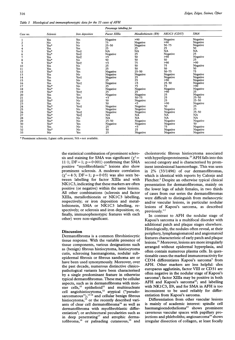
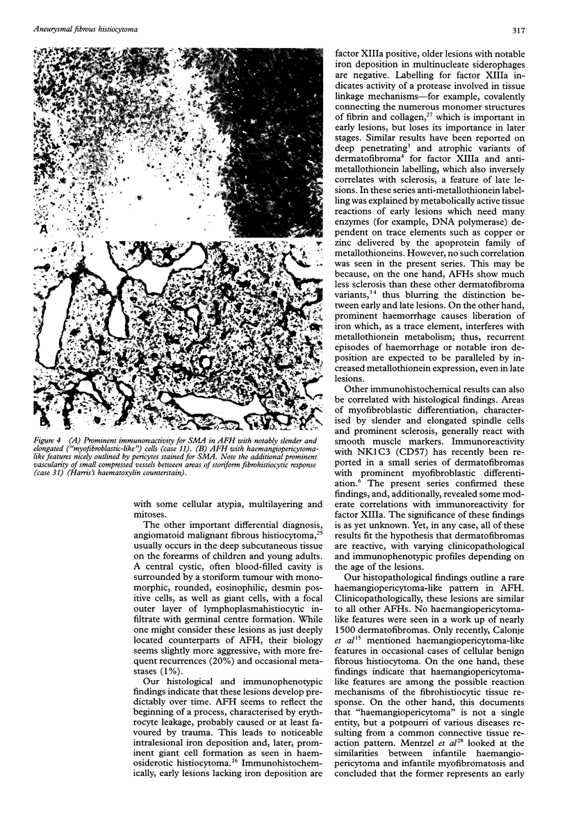
AIM: To describe the clinicopathological features of 33 aneurysmal fibrous histiocytomas (AFH), including five cases with a haemangiopericytoma-like pattern. METHODS: Thirty three cases of AFH were studied by using routine histology and immunohistochemistry for factor XIIIa, the "cell activity marker" E9 (anti-metallothionein), NK1C3 (CD57), smooth muscle actin (SMA), factor VIII, ulex europaeus agglutinin, JC70A (CD31), and QBEND10 (CD34). The time dependent variation in histopathological features was evaluated by statistical methods (Pearson chi 2, likelihood ratio chi 2). RESULTS: Of the AFHs, 29 of 33 occurred on the extremities of adults (age range 30 to 50 years), six of which were associated with rapid growth, probably caused by trauma, and pain. Twenty one lesions were thought to be vascular and/or melanocytic lesions, including two melanomas, because of a bluish-black and/or cystic appearance. Histologically, large areas of haemorrhage, up to 50% of the tumour bulk, lacking an endothelial lining were seen in otherwise typical fibrous histiocytomas. Five cases resembled nodular stages of Kaposi's sarcoma. Variable haemosiderin deposition in histiocytes (18/33) and giant cells (11/33) was suggestive of haemosiderotic histiocytoma. A haemangiopericytoma-like pattern was seen in five otherwise indistinguishable cases. On immunohistochemistry, variable reactivity was seen for factor XIIIa (18/30), with E9 (18/30), NK1C3 (19/30), and for SMA (14/30), but labelling for vascular markers was not detected. Early lesions without iron deposition were factor XIIIa positive; late lesions with iron deposition were factor XIIIa negative. Labelling for SMA correlated with prominent sclerosis. CONCLUSION: AFHs, including a haemangiopericytoma-like variant, have a characteristic time dependent histological and immunophenotypic profile, clearly different from nodular type Kaposi's sarcoma.
Full text
PDF
Images in this article
Selected References
These references are in PubMed. This may not be the complete list of references from this article.
- Calonje E., Mentzel T., Fletcher C. D. Cellular benign fibrous histiocytoma. Clinicopathologic analysis of 74 cases of a distinctive variant of cutaneous fibrous histiocytoma with frequent recurrence. Am J Surg Pathol. 1994 Jul;18(7):668–676. [PubMed] [Google Scholar]
- Enzinger F. M. Angiomatoid malignant fibrous histiocytoma: a distinct fibrohistiocytic tumor of children and young adults simulating a vascular neoplasm. Cancer. 1979 Dec;44(6):2147–2157. doi: 10.1002/1097-0142(197912)44:6<2147::aid-cncr2820440627>3.0.co;2-8. [DOI] [PubMed] [Google Scholar]
- Fukamizu H., Oku T., Inoue K., Matsumoto K., Okayama H., Tagami H. Atypical ("pseudosarcomatous") cutaneous histiocytoma. J Cutan Pathol. 1983 Oct;10(5):327–333. doi: 10.1111/j.1600-0560.1983.tb00335.x. [DOI] [PubMed] [Google Scholar]
- Holden C. A., Spittle M. F., Jones E. W. Angiosarcoma of the face and scalp, prognosis and treatment. Cancer. 1987 Mar 1;59(5):1046–1057. doi: 10.1002/1097-0142(19870301)59:5<1046::aid-cncr2820590533>3.0.co;2-6. [DOI] [PubMed] [Google Scholar]
- Hunt S. J., Santa Cruz D. J., Miller C. W. Cholesterotic fibrous histiocytoma. Its association with hyperlipoproteinemia. Arch Dermatol. 1990 Apr;126(4):506–508. [PubMed] [Google Scholar]
- Jones E. W., Cerio R., Smith N. P. Epithelioid cell histiocytoma: a new entity. Br J Dermatol. 1989 Feb;120(2):185–195. doi: 10.1111/j.1365-2133.1989.tb07782.x. [DOI] [PubMed] [Google Scholar]
- Jones R. R., Spaull J., Spry C., Jones E. W. Histogenesis of Kaposi's sarcoma in patients with and without acquired immune deficiency syndrome (AIDS). J Clin Pathol. 1986 Jul;39(7):742–749. doi: 10.1136/jcp.39.7.742. [DOI] [PMC free article] [PubMed] [Google Scholar]
- Jones W. E., Cerio R., Smith N. P. Multinucleate cell angiohistiocytoma: an acquired vascular anomaly to be distinguished from Kaposi's sarcoma. Br J Dermatol. 1990 May;122(5):651–663. doi: 10.1111/j.1365-2133.1990.tb07287.x. [DOI] [PubMed] [Google Scholar]
- Kamino H., Jacobson M. Dermatofibroma extending into the subcutaneous tissue. Differential diagnosis from dermatofibrosarcoma protuberans. Am J Surg Pathol. 1990 Dec;14(12):1156–1164. doi: 10.1097/00000478-199012000-00008. [DOI] [PubMed] [Google Scholar]
- Leyva W. H., Santa Cruz D. J. Atypical cutaneous fibrous histiocytoma. Am J Dermatopathol. 1986 Dec;8(6):467–471. doi: 10.1097/00000372-198612000-00003. [DOI] [PubMed] [Google Scholar]
- Mentzel T., Calonje E., Nascimento A. G., Fletcher C. D. Infantile hemangiopericytoma versus infantile myofibromatosis. Study of a series suggesting a continuous spectrum of infantile myofibroblastic lesions. Am J Surg Pathol. 1994 Sep;18(9):922–930. doi: 10.1097/00000478-199409000-00007. [DOI] [PubMed] [Google Scholar]
- Nickoloff B. J., Griffiths C. E. Factor XIIIa-expressing dermal dendrocytes in AIDS-associated cutaneous Kaposi's sarcomas. Science. 1989 Mar 31;243(4899):1736–1737. doi: 10.1126/science.2564703. [DOI] [PubMed] [Google Scholar]
- Regezi J. A., Nickoloff B. J., Headington J. T. Oral submucosal dendrocytes: factor XIIIa+ and CD34+ dendritic cell populations in normal tissue and fibrovascular lesions. J Cutan Pathol. 1992 Oct;19(5):398–406. doi: 10.1111/j.1600-0560.1992.tb00612.x. [DOI] [PubMed] [Google Scholar]
- Russell Jones R., Orchard G., Zelger B., Wilson Jones E. Immunostaining for CD31 and CD34 in Kaposi sarcoma. J Clin Pathol. 1995 Nov;48(11):1011–1016. doi: 10.1136/jcp.48.11.1011. [DOI] [PMC free article] [PubMed] [Google Scholar]
- Sanchez R. L. The elusive dermatofibromas. Arch Dermatol. 1990 Apr;126(4):522–523. [PubMed] [Google Scholar]
- Santa Cruz D. J., Kyriakos M. Aneurysmal ("angiomatoid") fibrous histiocytoma of the skin. Cancer. 1981 Apr 15;47(8):2053–2061. doi: 10.1002/1097-0142(19810415)47:8<2053::aid-cncr2820470825>3.0.co;2-a. [DOI] [PubMed] [Google Scholar]
- Schmid K. W., Ellis I. O., Gee J. M., Darke B. M., Lees W. E., Kay J., Cryer A., Stark J. M., Hittmair A., Ofner D. Presence and possible significance of immunocytochemically demonstrable metallothionein over-expression in primary invasive ductal carcinoma of the breast. Virchows Arch A Pathol Anat Histopathol. 1993;422(2):153–159. doi: 10.1007/BF01607167. [DOI] [PubMed] [Google Scholar]
- Schwob V. S., Santa Cruz D. J. Palisading cutaneous fibrous histiocytoma. J Cutan Pathol. 1986 Dec;13(6):403–407. doi: 10.1111/j.1600-0560.1986.tb01083.x. [DOI] [PubMed] [Google Scholar]
- Tamada S., Ackerman A. B. Dermatofibroma with monster cells. Am J Dermatopathol. 1987 Oct;9(5):380–387. doi: 10.1097/00000372-198710000-00003. [DOI] [PubMed] [Google Scholar]
- Weiss S. W., Enzinger F. M. Spindle cell hemangioendothelioma. A low-grade angiosarcoma resembling a cavernous hemangioma and Kaposi's sarcoma. Am J Surg Pathol. 1986 Aug;10(8):521–530. [PubMed] [Google Scholar]
- Zelger B. W., Calonje E., Sepp N., Fink F. M., Zelger B. G., Schmid K. W. Monophasic cellular variant of infantile myofibromatosis. An unusual histopathologic pattern in two siblings. Am J Dermatopathol. 1995 Apr;17(2):131–138. doi: 10.1097/00000372-199504000-00004. [DOI] [PubMed] [Google Scholar]
- Zelger B. W., Ofner D., Zelger B. G. Atrophic variants of dermatofibroma and dermatofibrosarcoma protuberans. Histopathology. 1995 Jun;26(6):519–527. doi: 10.1111/j.1365-2559.1995.tb00270.x. [DOI] [PubMed] [Google Scholar]
- Zelger B., Hittmair A., Schir M., Ofner C., Ofner D., Fritsch P. O., Böcker W., Jasani B., Schmid K. W. Immunohistochemically demonstrated metallothionein expression in malignant melanoma. Histopathology. 1993 Sep;23(3):257–263. doi: 10.1111/j.1365-2559.1993.tb01198.x. [DOI] [PubMed] [Google Scholar]
- Zelger B., Sidoroff A., Stanzl U., Fritsch P. O., Ofner D., Zelger B., Jasani B., Schmid K. W. Deep penetrating dermatofibroma versus dermatofibrosarcoma protuberans. A clinicopathologic comparison. Am J Surg Pathol. 1994 Jul;18(7):677–686. doi: 10.1097/00000478-199407000-00003. [DOI] [PubMed] [Google Scholar]


