

Abstract
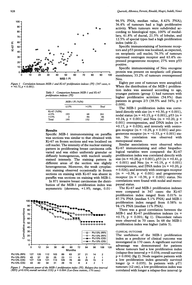
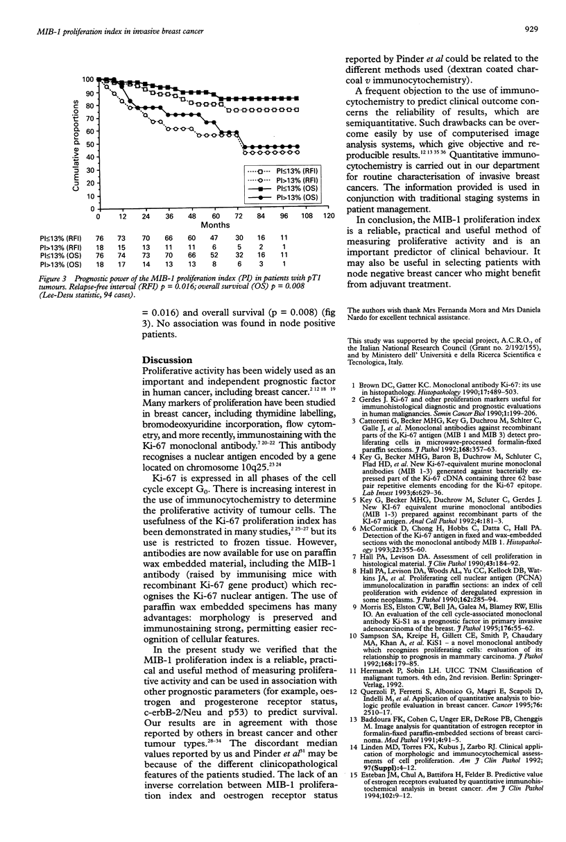
AIMS: To determine cell proliferation in infiltrating breast carcinomas. METHODS: Using the MIB-1 monoclonal antibody, the proliferation index was measured in paraffin wax sections of 871 breast cancers. The MIB-1 proliferation index was compared with other markers of disease progression: size, lymph node status, histotype, oestrogen and progesterone receptor status, expression of p53 and Neu, and DNA ploidy. All parameters were measured using image analysis. In 347 tumours, the MIB-1 and Ki-67 proliferation indexes were compared. Follow up data were available for 170 cases (median 66.5 months). RESULTS: Of the tumours, 314 (36%) had a high proliferation index. The MIB-1 proliferation index was correlated directly with size, nodal status, overexpression of p53 and Neu, and the DNA index; and inversely with oestrogen and progesterone receptor status. The correlation between MIB-1 and Ki-67 proliferation indexes was statistically significant. In patients with pT1 tumours, a low proliferation index correlated with a longer relapse-free interval and overall survival; node negative patients with a low proliferation index had a longer overall survival. CONCLUSIONS: The MIB-1 proliferation index is a reliable, practical and useful method of measuring proliferative activity and is an important predictor of clinical behaviour.
Full text
PDF
Selected References
These references are in PubMed. This may not be the complete list of references from this article.
- Bacus S. S., Goldschmidt R., Chin D., Moran G., Weinberg D., Bacus J. W. Biological grading of breast cancer using antibodies to proliferating cells and other markers. Am J Pathol. 1989 Nov;135(5):783–792. [PMC free article] [PubMed] [Google Scholar]
- Barbareschi M., Girlando S., Mauri F. M., Forti S., Eccher C., Mauri F. A., Togni R., Dalla Palma P., Doglioni C. Quantitative growth fraction evaluation with MIB1 and Ki67 antibodies in breast carcinomas. Am J Clin Pathol. 1994 Aug;102(2):171–175. doi: 10.1093/ajcp/102.2.171. [DOI] [PubMed] [Google Scholar]
- Brown D. C., Gatter K. C. Monoclonal antibody Ki-67: its use in histopathology. Histopathology. 1990 Dec;17(6):489–503. doi: 10.1111/j.1365-2559.1990.tb00788.x. [DOI] [PubMed] [Google Scholar]
- Cattoretti G., Becker M. H., Key G., Duchrow M., Schlüter C., Galle J., Gerdes J. Monoclonal antibodies against recombinant parts of the Ki-67 antigen (MIB 1 and MIB 3) detect proliferating cells in microwave-processed formalin-fixed paraffin sections. J Pathol. 1992 Dec;168(4):357–363. doi: 10.1002/path.1711680404. [DOI] [PubMed] [Google Scholar]
- Dodd R. Y. Hepatitis C virus, antibodies, and infectivity. Paradox, pragmatism, and policy. Am J Clin Pathol. 1992 Jan;97(1):4–6. doi: 10.1093/ajcp/97.1.4. [DOI] [PubMed] [Google Scholar]
- Esteban J. M., Ahn C., Battifora H., Felder B. Predictive value of estrogen receptors evaluated by quantitative immunohistochemical analysis in breast cancer. Am J Clin Pathol. 1994 Oct;102(4 Suppl 1):S9–12. [PubMed] [Google Scholar]
- Fonatsch C., Duchrow M., Rieder H., Schlüter C., Gerdes J. Assignment of the human Ki-67 gene (MK167) to 10q25-qter. Genomics. 1991 Oct;11(2):476–477. doi: 10.1016/0888-7543(91)90163-9. [DOI] [PubMed] [Google Scholar]
- Gerdes J. Ki-67 and other proliferation markers useful for immunohistological diagnostic and prognostic evaluations in human malignancies. Semin Cancer Biol. 1990 Jun;1(3):199–206. [PubMed] [Google Scholar]
- Gerdes J., Li L., Schlueter C., Duchrow M., Wohlenberg C., Gerlach C., Stahmer I., Kloth S., Brandt E., Flad H. D. Immunobiochemical and molecular biologic characterization of the cell proliferation-associated nuclear antigen that is defined by monoclonal antibody Ki-67. Am J Pathol. 1991 Apr;138(4):867–873. [PMC free article] [PubMed] [Google Scholar]
- Hall P. A., Levison D. A. Review: assessment of cell proliferation in histological material. J Clin Pathol. 1990 Mar;43(3):184–192. doi: 10.1136/jcp.43.3.184. [DOI] [PMC free article] [PubMed] [Google Scholar]
- Hall P. A., Levison D. A., Woods A. L., Yu C. C., Kellock D. B., Watkins J. A., Barnes D. M., Gillett C. E., Camplejohn R., Dover R. Proliferating cell nuclear antigen (PCNA) immunolocalization in paraffin sections: an index of cell proliferation with evidence of deregulated expression in some neoplasms. J Pathol. 1990 Dec;162(4):285–294. doi: 10.1002/path.1711620403. [DOI] [PubMed] [Google Scholar]
- Hall P. A., Woods A. L. Immunohistochemical markers of cellular proliferation: achievements, problems and prospects. Cell Tissue Kinet. 1990 Nov;23(6):505–522. doi: 10.1111/j.1365-2184.1990.tb01343.x. [DOI] [PubMed] [Google Scholar]
- Jensen V., Ladekarl M., Holm-Nielsen P., Melsen F., Soerensen F. B. The prognostic value of oncogenic antigen 519 (OA-519) expression and proliferative activity detected by antibody MIB-1 in node-negative breast cancer. J Pathol. 1995 Aug;176(4):343–352. doi: 10.1002/path.1711760405. [DOI] [PubMed] [Google Scholar]
- Kerns B. J., Jordan P. A., Faerman L. L., Berchuck A., Bast R. C., Jr, Layfield L. J. Determination of proliferation index with MIB-1 in advanced ovarian cancer using quantitative image analysis. Am J Clin Pathol. 1994 Feb;101(2):192–197. doi: 10.1093/ajcp/101.2.192. [DOI] [PubMed] [Google Scholar]
- Keshgegian A. A., Cnaan A. Proliferation markers in breast carcinoma. Mitotic figure count, S-phase fraction, proliferating cell nuclear antigen, Ki-67 and MIB-1. Am J Clin Pathol. 1995 Jul;104(1):42–49. doi: 10.1093/ajcp/104.1.42. [DOI] [PubMed] [Google Scholar]
- Key G., Becker M. H., Baron B., Duchrow M., Schlüter C., Flad H. D., Gerdes J. New Ki-67-equivalent murine monoclonal antibodies (MIB 1-3) generated against bacterially expressed parts of the Ki-67 cDNA containing three 62 base pair repetitive elements encoding for the Ki-67 epitope. Lab Invest. 1993 Jun;68(6):629–636. [PubMed] [Google Scholar]
- Lee E. T., Desu M. M. A computer program for comparing K samples with right-censored data. Comput Programs Biomed. 1972 Nov;2(4):315–321. doi: 10.1016/0010-468x(72)90019-0. [DOI] [PubMed] [Google Scholar]
- McCormick D., Chong H., Hobbs C., Datta C., Hall P. A. Detection of the Ki-67 antigen in fixed and wax-embedded sections with the monoclonal antibody MIB1. Histopathology. 1993 Apr;22(4):355–360. doi: 10.1111/j.1365-2559.1993.tb00135.x. [DOI] [PubMed] [Google Scholar]
- Morris E. S., Elston C. W., Bell J. A., Galea M., Blamey R. W., Ellis I. O. An evaluation of the cell cycle-associated monoclonal antibody Ki-S1 as a prognostic factor in primary invasive adenocarcinoma of the breast. J Pathol. 1995 May;176(1):55–62. doi: 10.1002/path.1711760109. [DOI] [PubMed] [Google Scholar]
- Onda K., Davis R. L., Shibuya M., Wilson C. B., Hoshino T. Correlation between the bromodeoxyuridine labeling index and the MIB-1 and Ki-67 proliferating cell indices in cerebral gliomas. Cancer. 1994 Oct 1;74(7):1921–1926. doi: 10.1002/1097-0142(19941001)74:7<1921::aid-cncr2820740716>3.0.co;2-9. [DOI] [PubMed] [Google Scholar]
- Pinder S. E., Wencyk P., Sibbering D. M., Bell J. A., Elston C. W., Nicholson R., Robertson J. F., Blamey R. W., Ellis I. O. Assessment of the new proliferation marker MIB1 in breast carcinoma using image analysis: associations with other prognostic factors and survival. Br J Cancer. 1995 Jan;71(1):146–149. doi: 10.1038/bjc.1995.30. [DOI] [PMC free article] [PubMed] [Google Scholar]
- Querzoli P., Ferretti S., Albonico G., Magri E., Scapoli D., Indelli M., Nenci I. Application of quantitative analysis to biologic profile evaluation in breast cancer. Cancer. 1995 Dec 15;76(12):2510–2517. doi: 10.1002/1097-0142(19951215)76:12<2510::aid-cncr2820761216>3.0.co;2-q. [DOI] [PubMed] [Google Scholar]
- Quinn C. M., Wright N. A. The clinical assessment of proliferation and growth in human tumours: evaluation of methods and applications as prognostic variables. J Pathol. 1990 Feb;160(2):93–102. doi: 10.1002/path.1711600202. [DOI] [PubMed] [Google Scholar]
- Sahin A. A., Ro J., Ro J. Y., Blick M. B., el-Naggar A. K., Ordonez N. G., Fritsche H. A., Smith T. L., Hortobagyi G. N., Ayala A. G. Ki-67 immunostaining in node-negative stage I/II breast carcinoma. Significant correlation with prognosis. Cancer. 1991 Aug 1;68(3):549–557. doi: 10.1002/1097-0142(19910801)68:3<549::aid-cncr2820680318>3.0.co;2-j. [DOI] [PubMed] [Google Scholar]
- Sampson S. A., Kreipe H., Gillett C. E., Smith P., Chaudary M. A., Khan A., Wicks K., Parwaresch R., Barnes D. M. KiS1--a novel monoclonal antibody which recognizes proliferating cells: evaluation of its relationship to prognosis in mammary carcinoma. J Pathol. 1992 Oct;168(2):179–185. doi: 10.1002/path.1711680205. [DOI] [PubMed] [Google Scholar]
- Silvestrini R., Benini E., Daidone M. G., Veneroni S., Boracchi P., Cappelletti V., Di Fronzo G., Veronesi U. p53 as an independent prognostic marker in lymph node-negative breast cancer patients. J Natl Cancer Inst. 1993 Jun 16;85(12):965–970. doi: 10.1093/jnci/85.12.965. [DOI] [PubMed] [Google Scholar]
- Veronese S. M., Gambacorta M. Detection of Ki-67 proliferation rate in breast cancer. Correlation with clinical and pathologic features. Am J Clin Pathol. 1991 Jan;95(1):30–34. doi: 10.1093/ajcp/95.1.30. [DOI] [PubMed] [Google Scholar]
- Veronese S. M., Gambacorta M., Gottardi O., Scanzi F., Ferrari M., Lampertico P. Proliferation index as a prognostic marker in breast cancer. Cancer. 1993 Jun 15;71(12):3926–3931. doi: 10.1002/1097-0142(19930615)71:12<3926::aid-cncr2820711221>3.0.co;2-2. [DOI] [PubMed] [Google Scholar]