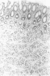
Abstract
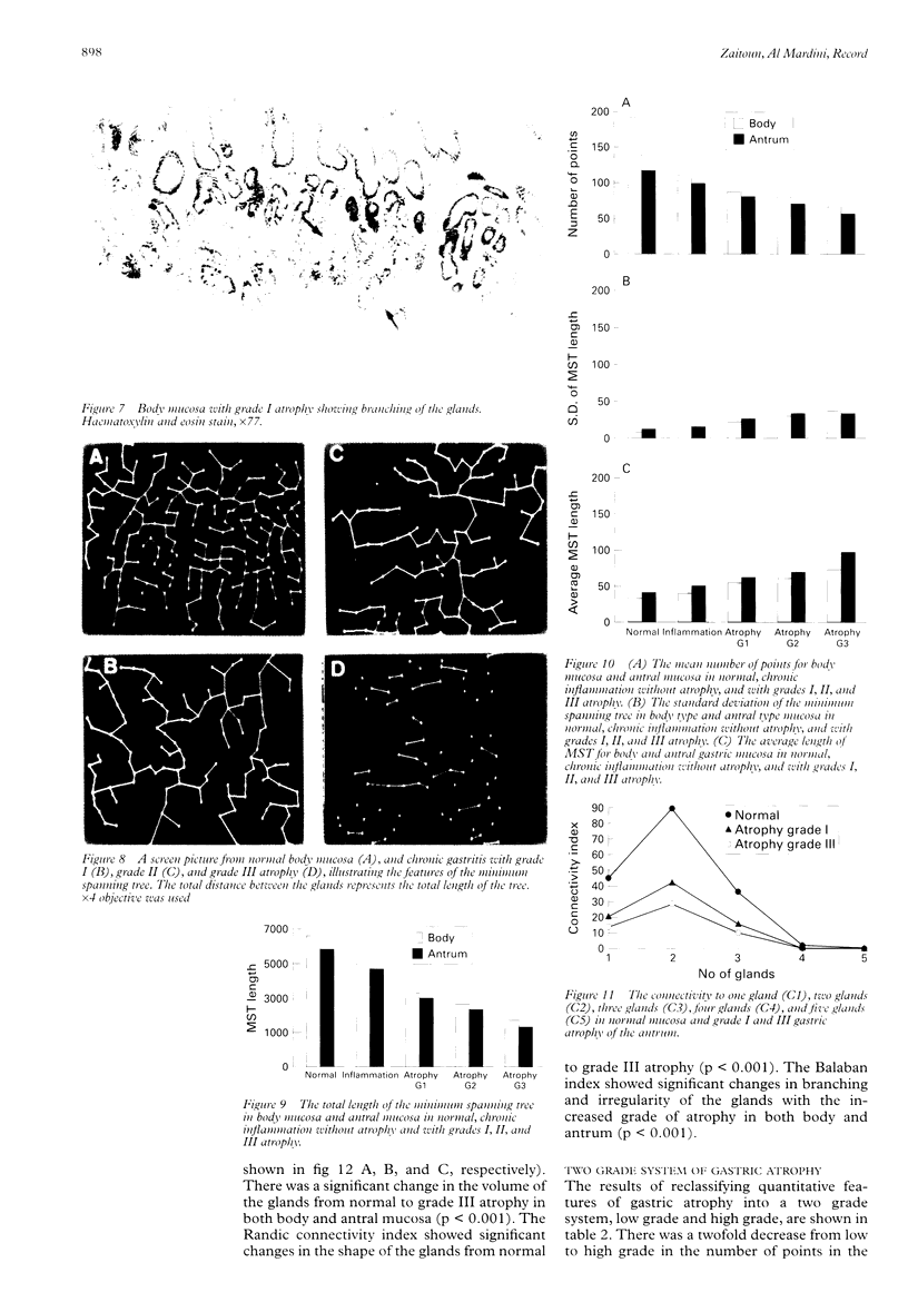
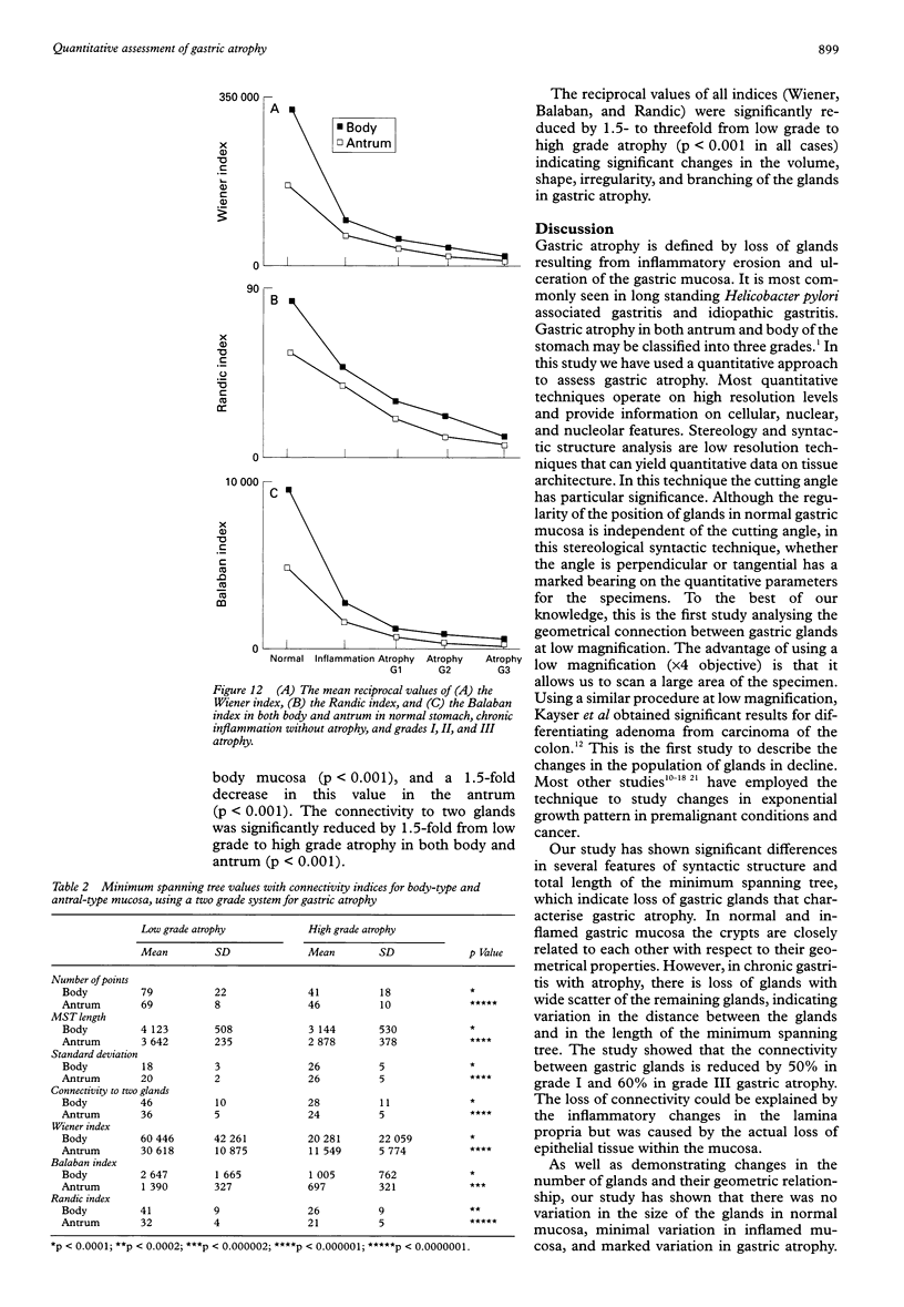
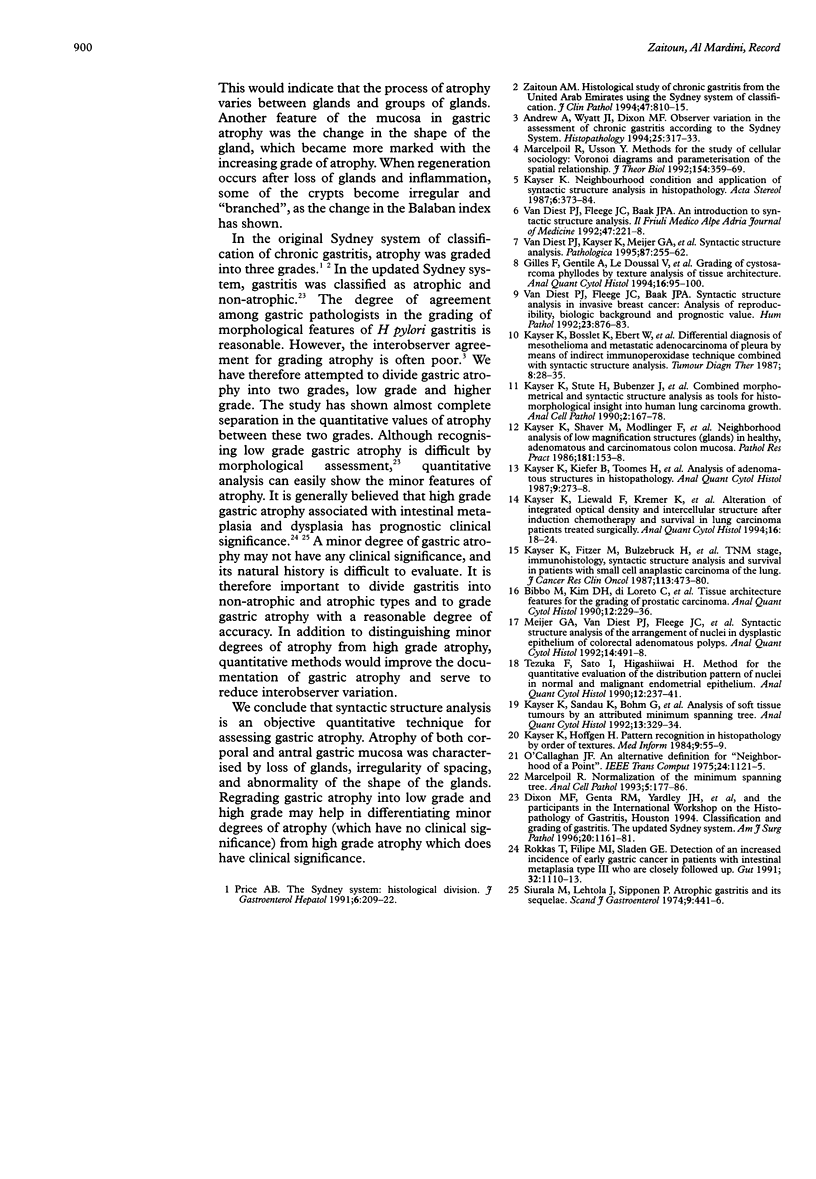
AIM: To assess the topographical relation between gastric glands, using the minimum spanning tree (MST), to derive both a model of neighbourhood and quantitative representation of the tissue's architecture, to assess the characteristic features of gastric atrophy, and to assess the grades of gastric atrophy. METHODS: Haematoxylin and eosin stained sections from corporal and antral biopsy specimens (n = 139) from normal patients and from patients with nonatrophic gastritis and atrophic gastritis of grades 1, 2, and 3 (Sydney system) were assessed by image analysis system (Prodit 5.2) and 11 syntactic structure features were derived. These included both line and connectivity features. RESULTS: Syntactic structure analysis was correlated with the semiquantitative grading system of gastric atrophy. The study showed significant reductions in the number of points and the length of MST in both body and antrum. The standard deviation of the length of MST was significantly increased in all grades of atrophy. The connectivity to two glands was the highest and most affected by the increased grade of atrophy. The reciprocal values of the Wiener, Randic, and Balaban indices showed significant changes in the volume of gland, abnormality in the shape of glands, and changes in irregularity and branching of the glands in both types of gastric mucosa. There was a complete separation in the MST, connectivity, and index values between low grade and high grade gastric atrophy. CONCLUSIONS: (1) Gastric atrophy was characterised by loss of the gland, variation in the volume, reduction in the neighbourhood, irregularity in spacing, and abnormality in the shape of the glands. (2) Syntactic structure analysis significantly differentiated minor changes in gastric gland (low grade atrophy) from high grade atrophy of clinical significance. (3) Syntactic structure analysis is a simple, fast, and highly reproducible technique and appears a promising method for quantitative assessment of atrophy.
Full text
PDF
Images in this article
Selected References
These references are in PubMed. This may not be the complete list of references from this article.
- Andrew A., Wyatt J. I., Dixon M. F. Observer variation in the assessment of chronic gastritis according to the Sydney system. Histopathology. 1994 Oct;25(4):317–322. doi: 10.1111/j.1365-2559.1994.tb01349.x. [DOI] [PubMed] [Google Scholar]
- Bibbo M., Kim D. H., di Loreto C., Dytch H. E., Galera-Davidson H., Thompson D., Richards D. L., Bartels H. G., Bartels P. H. Tissue architectural features for the grading of prostatic carcinoma. Anal Quant Cytol Histol. 1990 Aug;12(4):229–236. [PubMed] [Google Scholar]
- Dixon M. F., Genta R. M., Yardley J. H., Correa P. Classification and grading of gastritis. The updated Sydney System. International Workshop on the Histopathology of Gastritis, Houston 1994. Am J Surg Pathol. 1996 Oct;20(10):1161–1181. doi: 10.1097/00000478-199610000-00001. [DOI] [PubMed] [Google Scholar]
- Gilles F., Gentile A., Le Doussal V., Bertrand F., Kahn E. Grading of cystosarcoma phyllodes by texture analysis of tissue architecture. Anal Quant Cytol Histol. 1994 Apr;16(2):95–100. [PubMed] [Google Scholar]
- Kayser K. K., Kiefer B., Toomes H., Burkhard H. U. Analysis of adenomatous structures in histopathology. Anal Quant Cytol Histol. 1987 Jun;9(3):273–278. [PubMed] [Google Scholar]
- Kayser K., Fitzer M., Bülzebruck H., Bosslet K., Drings P. TNM stage, immunohistology, syntactic structure analysis and survival in patients with small cell anaplastic carcinoma of the lung. J Cancer Res Clin Oncol. 1987;113(5):473–480. doi: 10.1007/BF00390042. [DOI] [PubMed] [Google Scholar]
- Kayser K., Höffgen H. Pattern recognition in histopathology by orders of textures. Med Inform (Lond) 1984 Jan-Mar;9(1):55–59. doi: 10.3109/14639238409010938. [DOI] [PubMed] [Google Scholar]
- Kayser K., Liewald F., Kremer K., Tacke M., Storck M., Faber P., Bonomi P. Alteration of integrated optical density and intercellular structure after induction chemotherapy and survival in lung carcinoma patients treated surgically. Anal Quant Cytol Histol. 1994 Feb;16(1):18–24. [PubMed] [Google Scholar]
- Kayser K., Sandau K., Böhm G., Kunze K. D., Paul J. Analysis of soft tissue tumors by an attributed minimum spanning tree. Anal Quant Cytol Histol. 1991 Oct;13(5):329–334. [PubMed] [Google Scholar]
- Kayser K., Shaver M., Modlinger F., Postl K., Moyers J. J. Neighborhood analysis of low magnification structures (glands) in healthy, adenomatous, and carcinomatous colon mucosa. Pathol Res Pract. 1986 May;181(2):153–158. doi: 10.1016/s0344-0338(86)80004-8. [DOI] [PubMed] [Google Scholar]
- Kayser K., Stute H., Bubenzer J., Paul J. Combined morphometrical and syntactic structure analysis as tools for histomorphological insight into human lung carcinoma growth. Anal Cell Pathol. 1990 Apr;2(3):167–178. [PubMed] [Google Scholar]
- Marcelpoil R. Normalization of the minimum spanning tree. Anal Cell Pathol. 1993 May;5(3):177–186. [PubMed] [Google Scholar]
- Meijer G. A., van Diest P. J., Fleege J. C., Baak J. P. Syntactic structure analysis of the arrangement of nuclei in dysplastic epithelium of colorectal adenomatous polyps. Anal Quant Cytol Histol. 1992 Dec;14(6):491–498. [PubMed] [Google Scholar]
- Rokkas T., Filipe M. I., Sladen G. E. Detection of an increased incidence of early gastric cancer in patients with intestinal metaplasia type III who are closely followed up. Gut. 1991 Oct;32(10):1110–1113. doi: 10.1136/gut.32.10.1110. [DOI] [PMC free article] [PubMed] [Google Scholar]
- Siurala M., Lehtola J., Ihamäki T. Atrophic gastritis and its sequelae. Results of 19-23 years' follow-up examinations. Scand J Gastroenterol. 1974;9(5):441–446. [PubMed] [Google Scholar]
- Tezuka F., Sato I., Higashiiwai H., Endo N., Ito K., Kasai M. Method for the quantitative evaluation of the distribution pattern of nuclei in normal and malignant endometrial epithelia. Anal Quant Cytol Histol. 1990 Aug;12(4):237–241. [PubMed] [Google Scholar]
- Zaitoun A. M. Histological study of chronic gastritis from the United Arab Emirates using the Sydney system of classification. J Clin Pathol. 1994 Sep;47(9):810–815. doi: 10.1136/jcp.47.9.810. [DOI] [PMC free article] [PubMed] [Google Scholar]
- van Diest P. J., Fleege J. C., Baak J. P. Syntactic structure analysis in invasive breast cancer: analysis of reproducibility, biologic background, and prognostic value. Hum Pathol. 1992 Aug;23(8):876–883. doi: 10.1016/0046-8177(92)90398-m. [DOI] [PubMed] [Google Scholar]
- van Diest P. J., Kayser K., Meijer G. A., Baak J. P. Syntactic structure analysis. Pathologica. 1995 Jun;87(3):255–262. [PubMed] [Google Scholar]


