Abstract
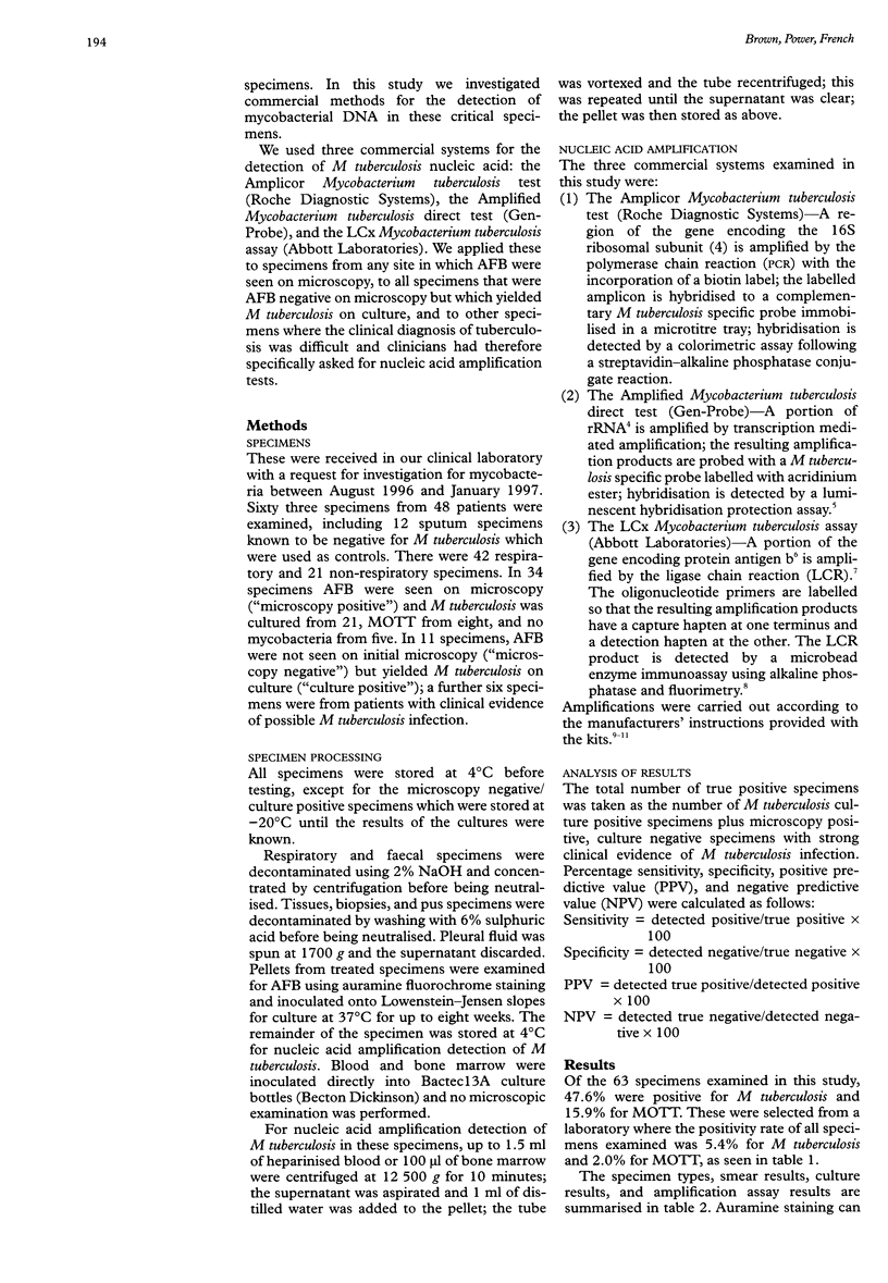
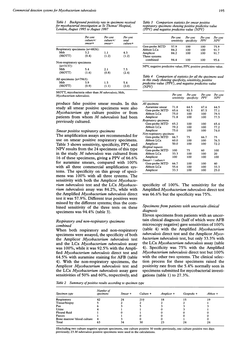
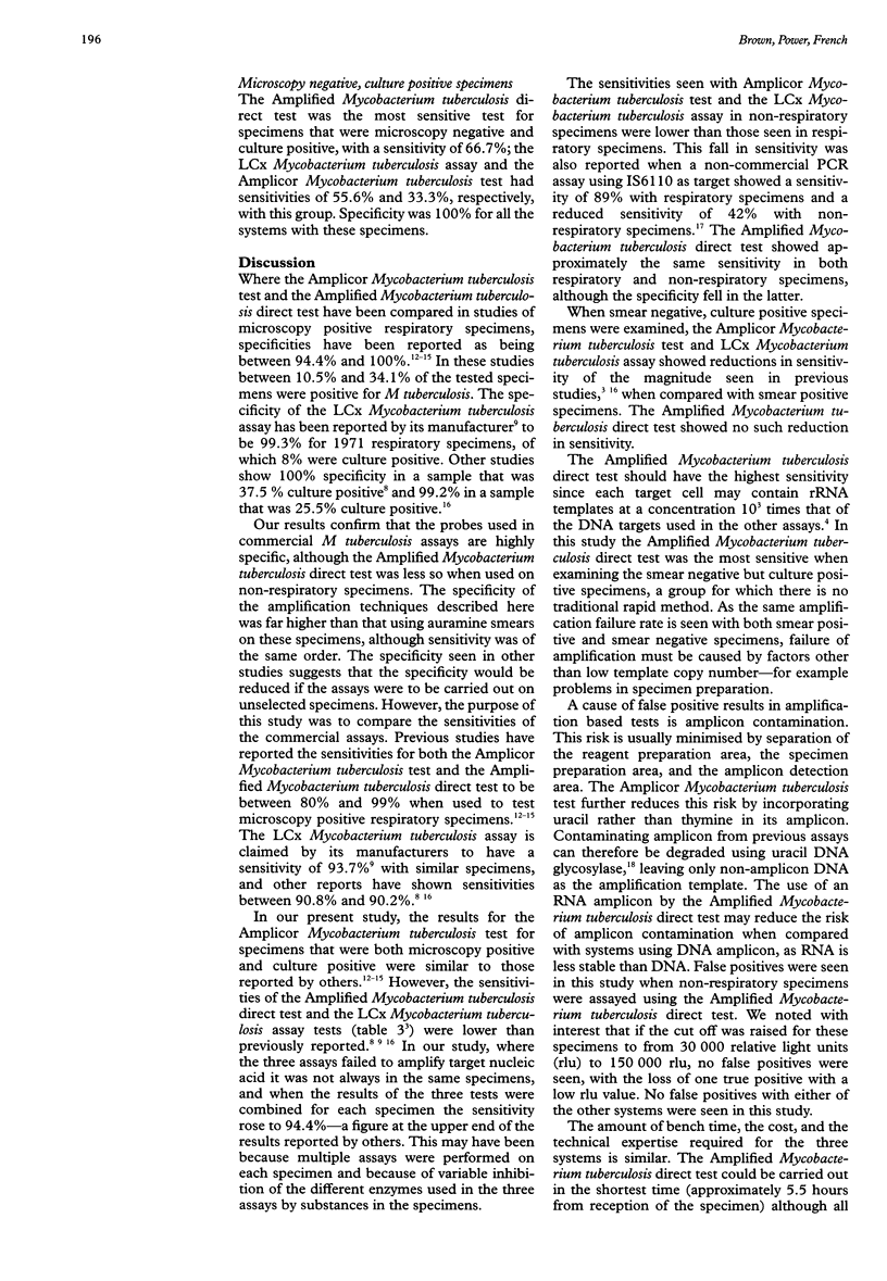
AIMS: To assess the performance of three commercially available Mycobacterium tuberculosis detection systems employing nucleic acid amplification, when applied directly to respiratory and non-respiratory specimens from patients where the diagnosis of tuberculosis is difficult using clinical and traditional bacteriological methods. METHODS: 42 respiratory and 21 non-respiratory specimens were concentrated, examined with auramine staining, and cultured on Lowenstein-Jensen slopes. These specimens were also assayed using the Amplicor Mycobacterium tuberculosis test (AM) (Roche Diagnostic Systems), the Amplified Mycobacterium tuberculosis direct test (AMD) (Gen-Probe), and the LCx Mycobacterium tuberculosis assay (LMA) (Abbott Laboratories). RESULTS: All three amplification systems used in this study gave specificities of 100% when used on respiratory specimens. When used on non-respiratory specimens, AM and LMA gave specificities of 100% and AMD 75%. With respiratory specimens the AM, AMD, and LMA systems gave sensitivities of 75%, 65.2%, and 79.2%, respectively. With non-respiratory specimens the sensitivities were 50%, 66.7%, and 60%, while with smear negative, culture positive specimens they were 33.3%, 66.7%, and 55.6%. Positive predictive values of 100% were seen with all specimens except non-respiratory specimens assayed using AMD where the value was 66.7%. CONCLUSIONS: The manufacturers of these systems recommend that they should only be used for the direct analysis of respiratory specimens, and the US Food and Drug Administration has approved them for use only with smear positive specimens. This study confirms that sensitivities are lower for non-respiratory and smear negative specimens, but positive predictive values are high. Provided they are interpreted with caution, positive results with these tests in respiratory and non-respiratory specimens are useful in tuberculous patients who are otherwise difficult to diagnose. Each amplification has advantages and disadvantages compared with the others.
Full text
PDF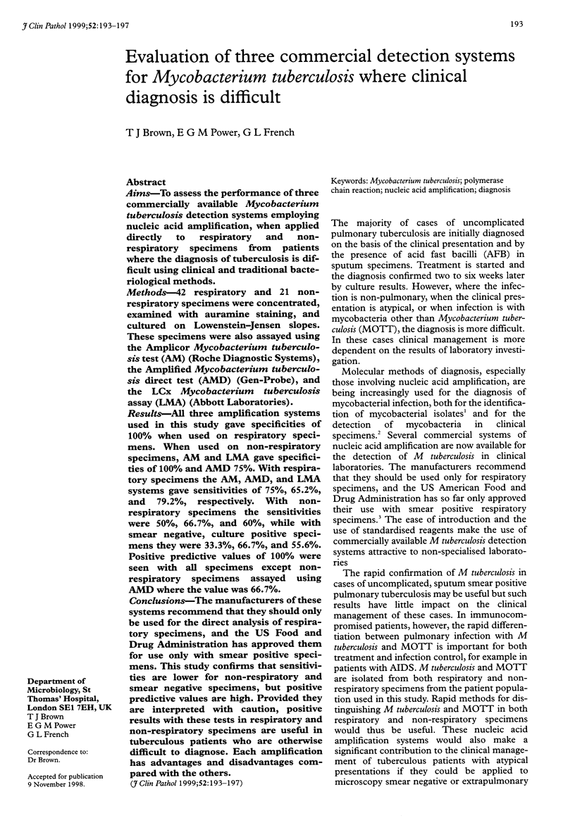
Selected References
These references are in PubMed. This may not be the complete list of references from this article.
- Andersen A. B., Hansen E. B. Structure and mapping of antigenic domains of protein antigen b, a 38,000-molecular-weight protein of Mycobacterium tuberculosis. Infect Immun. 1989 Aug;57(8):2481–2488. doi: 10.1128/iai.57.8.2481-2488.1989. [DOI] [PMC free article] [PubMed] [Google Scholar]
- Ausina V., Gamboa F., Gazapo E., Manterola J. M., Lonca J., Matas L., Manzano J. R., Rodrigo C., Cardona P. J., Padilla E. Evaluation of the semiautomated Abbott LCx Mycobacterium tuberculosis assay for direct detection of Mycobacterium tuberculosis in respiratory specimens. J Clin Microbiol. 1997 Aug;35(8):1996–2002. doi: 10.1128/jcm.35.8.1996-2002.1997. [DOI] [PMC free article] [PubMed] [Google Scholar]
- Böddinghaus B., Rogall T., Flohr T., Blöcker H., Böttger E. C. Detection and identification of mycobacteria by amplification of rRNA. J Clin Microbiol. 1990 Aug;28(8):1751–1759. doi: 10.1128/jcm.28.8.1751-1759.1990. [DOI] [PMC free article] [PubMed] [Google Scholar]
- Chan C. M., Yuen K. Y., Chan K. S., Yam W. C., Yim K. H., Ng W. F., Ng M. H. Single-tube nested PCR in the diagnosis of tuberculosis. J Clin Pathol. 1996 Apr;49(4):290–294. doi: 10.1136/jcp.49.4.290. [DOI] [PMC free article] [PubMed] [Google Scholar]
- Dalovisio J. R., Montenegro-James S., Kemmerly S. A., Genre C. F., Chambers R., Greer D., Pankey G. A., Failla D. M., Haydel K. G., Hutchinson L. Comparison of the amplified Mycobacterium tuberculosis (MTB) direct test, Amplicor MTB PCR, and IS6110-PCR for detection of MTB in respiratory specimens. Clin Infect Dis. 1996 Nov;23(5):1099–1108. doi: 10.1093/clinids/23.5.1099. [DOI] [PubMed] [Google Scholar]
- Ichiyama S., Iinuma Y., Tawada Y., Yamori S., Hasegawa Y., Shimokata K., Nakashima N. Evaluation of Gen-Probe Amplified Mycobacterium Tuberculosis Direct Test and Roche PCR-microwell plate hybridization method (AMPLICOR MYCOBACTERIUM) for direct detection of mycobacteria. J Clin Microbiol. 1996 Jan;34(1):130–133. doi: 10.1128/jcm.34.1.130-133.1996. [DOI] [PMC free article] [PubMed] [Google Scholar]
- Lebrun L., Espinasse F., Poveda J. D., Vincent-Levy-Frebault V. Evaluation of nonradioactive DNA probes for identification of mycobacteria. J Clin Microbiol. 1992 Sep;30(9):2476–2478. doi: 10.1128/jcm.30.9.2476-2478.1992. [DOI] [PMC free article] [PubMed] [Google Scholar]
- Lindbråthen A., Gaustad P., Hovig B., Tønjum T. Direct detection of Mycobacterium tuberculosis complex in clinical samples from patients in Norway by ligase chain reaction. J Clin Microbiol. 1997 Dec;35(12):3248–3253. doi: 10.1128/jcm.35.12.3248-3253.1997. [DOI] [PMC free article] [PubMed] [Google Scholar]
- Longo M. C., Berninger M. S., Hartley J. L. Use of uracil DNA glycosylase to control carry-over contamination in polymerase chain reactions. Gene. 1990 Sep 1;93(1):125–128. doi: 10.1016/0378-1119(90)90145-h. [DOI] [PubMed] [Google Scholar]
- Vuorinen P., Miettinen A., Vuento R., Hällström O. Direct detection of Mycobacterium tuberculosis complex in respiratory specimens by Gen-Probe Amplified Mycobacterium Tuberculosis Direct Test and Roche Amplicor Mycobacterium Tuberculosis Test. J Clin Microbiol. 1995 Jul;33(7):1856–1859. doi: 10.1128/jcm.33.7.1856-1859.1995. [DOI] [PMC free article] [PubMed] [Google Scholar]
- Zolnir-Dovc M., Poljak M., Seme K., Rus A., Avsic-Zupanc T. Evaluation of two commercial amplification assays for detection of Mycobacterium tuberculosis complex in respiratory specimens. Infection. 1995 Jul-Aug;23(4):216–221. doi: 10.1007/BF01781200. [DOI] [PubMed] [Google Scholar]


