Abstract
Closely-related, and otherwise morphologically similar insect species frequently show striking divergence in the shape and/or size of male genital structures, a phenomenon thought to be driven by sexual selection. Comparative interspecific studies can help elucidate the evolutionary forces acting on genital structures to drive this rapid differentiation. However, genetic dissection of sexual trait divergence between species is frequently hampered by the difficulty generating interspecific recombinants. Intraspecific variation can be leveraged to investigate the genetics of rapidly-evolving sexual traits, and here we carry out a genetic analysis of variation in the posterior lobe within D. melanogaster. The lobe is a male-specific process emerging from the genital arch of D. melanogaster and three closely-related species, is essential for copulation, and shows radical divergence in form across species. There is also abundant variation within species in the shape and size of the lobe, and while this variation is considerably more subtle than that seen among species, it nonetheless provides the raw material for QTL mapping. We created an advanced intercross population from a pair of phenotypically-different inbred strains, and after phenotyping and genotyping-by-sequencing the recombinants, mapped several QTL contributing to various measures of lobe morphology. The additional generations of crossing over in our mapping population led to QTL intervals that are smaller than is typical for an F2 mapping design. The intervals we map overlap with a pair of lobe QTL we previously identified in an independent mapping cross, potentially suggesting a level of shared genetic control of trait variation. Our QTL additionally implicate a suite of genes that have been shown to contribute to the development of the posterior lobe. These loci are strong candidates to harbor naturally-segregating sites contributing to phenotypic variation within D. melanogaster, and may also be those contributing to divergence in lobe morphology between species.
Introduction
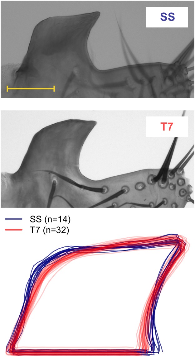
The posterior lobe is a male-specific elaboration of the genital arch (Fig 1) present in all four species of the melanogaster clade of Drosophila (i.e., D. mauritiana, D. melanogaster, D. sechellia, and D. simulans), and absent in other Drosophilid species, including those of the related yakuba clade [1]. The lobe is an evolutionarily novel cuticular structure used by the male to grasp the female ovipositor during mating [1], and both specific laser manipulation of lobe morphology in D. simulans [2] and genetic ablation of the lobe in D. melanogaster [3], have indicated the lobe is essential for copulation to occur.
Fig 1. Posterior lobes of parental lines.
A representative image of a single posterior lobe is shown for each parental strain. The yellow horizontal scale bar represents 0.05mm. In addition, post-elliptic Fourier analysis reconstructed outlines of all parental lobes are overlaid to highlight the differences in morphology.
The four species of the melanogaster complex are extremely similar morphologically, but differ markedly in the shape and size of the posterior lobe [4, 5]. As such the lobe represents the primary character used to establish species identity in the group. This striking diversity in the lobe mirrors observations from numerous other insect taxa, demonstrating that male genitalia traits are frequently subject to rapid evolution, likely as a result of sexual selection [6, 7]. The precise mechanism(s) by which sexual selection has influenced posterior lobe morphology is unclear. However, behavioral genetics investigations have revealed a range of pre- and post-copulatory effects acting through variation in lobe morphology, suggesting sexual selection may play a role at multiple levels to influence the phenotype [2, 3, 8].
One additional approach to understand the selective forces acting on a trait is to dissect the genetic basis of phenotypic variation to the level of the underlying causative alleles. Describing the genetic architecture of trait variation in this way provides estimates of the effects and frequencies of alleles at quantitative trait loci, QTL [9], from which one can make predictions about the types of process acting to maintain heritability [10, 11]. Cloning the precise causative alleles will ultimately allow exploration of the selective forces acting at causative loci through the application of comparative genome sequencing and tests for patterns of selection [12–14].
Several studies have mapped QTL contributing to the radical divergence among species in posterior lobe morphology using pairwise intercrosses of D. mauritiana, D. sechellia, and D. simulans [15–20]. Exploring genetic differences between D. melanogaster and its relatives is not possible given the evolutionary divergence between taxa [21], and the resulting inability to generate interspecific recombinants [22]. A challenge for these interspecific mapping studies is the difficulty directly validating putative candidate genes in the species under test, relying instead on functional tests in the D. melanogaster model system [19]. We and others have instead interrogated standing genetic variation within D. melanogaster to identify loci contributing to segregating intraspecific variation in lobe shape and size [23, 24]. By using D. melanogaster we can leverage the wealth of information on gene function in the system to uncover potential candidates [25], and ultimately use sophisticated tools to validate the functional roles of potential lobe loci in the same species in which the QTL were isolated. Under the assumption that the loci contributing to within and between species lobe variation are the same, genes identified within D. melanogaster as harboring alleles contributing to intraspecific lobe variation, could also be those that have fixed for allelic differences between species (see [26]).
In our previous work we identified three QTL contributing to variation in posterior lobe morphology in D. melanogaster using a cross between a pair of phenotypically different inbred strains [23]. Here we attempt to replicate these QTL using an independent mapping cross. We take an different pair of highly-inbred, naturally-derived lines showing a difference in lobe morphology, intercross for several generations to expand the genetic map, and identify several QTL that collectively explain a large fraction of the variation in the cross. Two of the QTL mapped by McNeil et al. [23] overlap with QTL mapped in the present study, potentially indicating common genetic underpinnings. In addition, mapped QTL intervals include loci implicated by a genomewide association study, GWAS [24], and genes that have been functionally implicated in the control of genital morphology in D. melanogaster [19, 27, 28]. These loci are attractive candidates to harbor segregating variation contributing to posterior lobe variation.
Materials and Methods
Drosophila stocks
We used a pair of highly-inbred D. melanogaster stocks in this study. Strain "SS" is a version of the iso1 reference genome sequenced strain, Bloomington Drosophila Stock Center number 5027 [29], that was purged of Wolbachia via tetracycline treatment. Strain "T7" is a version of Drosophila Species Stock Center strain 14021–0231.7 that was originally collected in 1968 from Taiwan, is naturally Wolbachia-free, and was further inbred in the Macdonald lab via 18 generations of brother-sister mating [30]. Both SS and T7 are free of P-elements, i.e., have the M cytotype.
Mapping population
We generated F1 males and females from both reciprocal crosses, i.e., male SS × virgin female T7 (cross A), and male T7 × virgin female SS (cross B). We then generated F2 males and females by intercrossing F1 animals in all pairwise combinations (i.e., male cross A progeny × virgin female cross A progeny, male cross A progeny × virgin female cross B progeny, male cross B progeny × virgin female cross A progeny, and male cross B progeny × virgin female cross B progeny). All F2 animals were mixed, re-distributed into 12 replicate vials to lay eggs, and adults were cleared to maintain roughly similar egg density across vials. Each subsequent generation was treated similarly, using 18–29 replicate vials per generation. No effort was made to collect virgin flies after the F2 generation, and generations (egg to adult) were 12–14 days. At the F13 generation several hundred recombinant male progeny were frozen at –20°C for phenotyping and genotyping.
All fly maintenance was conducted at 25°C and 50% relative humidity under a 12 hour light: 12 hour dark cycle. Flies were reared in narrow polystyrene vials (25 × 95 mm) containing 10 ml of cornmeal-molasses-yeast medium.
Lobe dissection and phenotype acquisition
Full details of the dissection and imaging procedures followed are described in McNeil et al. [23], except that in the current study we employed PVA mounting medium (BioQuip catalog number 6371A). Carcasses of dissected males were stored at –20°C in preparation for genotyping.
All high-quality posterior lobe images were manually flipped/rotated to ensure images were of the same handedness, with the "point" of the lobe pointing clockwise. We then employed ImageJ [31] to manually outline the lobe, closing the outline with an artificial baseline that extends from the point at which the lateral plate connects to the lobe. Each lobe is thus represented by an outline described by a set of Cartesian coordinates. The coordinates for each lobe were then rotated so that the baseline is horizontal, and translated to center the coordinate series at the centroid of the outline (S1 Fig). This serves to standardize lobes with respect to handedness, orientation, and relative location, allowing comparison of shape across genotypes.
In common with previous investigators [16–18, 23] we employed elliptic Fourier analysis, EFA [32, 33], to describe the shape of each posterior lobe as a series of Fourier coefficients. Since we had already placed each lobe in a standard configuration the additional coefficient normalization routines described in Kuhl & Giardina [33] were not employed. We then used 100 Fourier coefficients for each of the successfully imaged lobes from SS, T7, and the F13 recombinant genotypes in a principal components analysis (PCA) to encapsulate shape variation in a small number of quantitative metrics. PCA was carried out using the 'prcomp' function in R (r-project.org), and the principal components (PCs) used as phenotypes for QTL mapping. In addition to PC-based phenotypes we calculated area, height and width (measured as the length of the vertical or horizontal line passing through the centroid and intersecting the outline), and the height:width ratio for each lobe.
Genotyping-by-sequencing
We extracted DNA from the carcasses of 192 recombinant F13 males, and from the SS and T7 parental strains, using the Puregene cell and tissue kit (Qiagen) following the manufacturer's protocol, and subsequently used two different strategies to collect genomewide genotyping data.
Half of the phenotyped F13 males, along with both parental genotypes, were genotyped via low pass whole genome sequencing (WGS). Briefly, we mechanically sheared genomic DNA isolated from each sample (Covaris S220), quantified the amount of sheared DNA using the Qubit high sensitivity double-stranded DNA kit (ThermoFisher), and generated indexed sequencing libraries from ~60ng of sheared DNA (New England Biolabs, NEBNext E6040L). Libraries were combined into four 24-plex pools, and each pool was run over a single lane of an Illumina HiSeq2500 sequencer (KU Genome Sequencing Core facility) generating paired-end 100bp reads. Data is available on Dryad (doi:10.5061/dryad.gc182).
The other half of the recombinant males, along with additional samples from both parental genotypes, were genotyped by the MSG (multiplexed shotgun genotyping) method of Andolfatto et al. [34]. Briefly, 10ng of genomic DNA from each fly was digested using a restriction enzyme, and barcoded adaptors were ligated onto each fragmented sample. Multiple samples were then pooled, and each pool was purified, size-selected, and PCR amplified to generate an indexed, sequenceable library. Individual samples are then jointly distinguished by an "in line barcode" (the first 6 nucleotides of the Read1, or "forward" sequencing read) and a standard index read sequence. For reasons unrelated to the current project, we subjected the same 96 samples to MSG using three experimental protocols, using different restriction enzymes (MseI and NdeI) and reaction conditions. Reads resulting from all three regimes were pooled for each individual prior to genotype calling. MSG libraries were sequenced over a fraction of a HiSeq2500 lane resulting in single-end 100bp reads. Data is available on Dryad (doi:10.5061/dryad.gc182).
There was no association between sample phenotype and the method employed to collect genotypes. Simple t-tests contrasting the trait values of those recombinants subjected to WGS- or MSG-based genotyping were not significant in most cases (p > 0.1 for lobe area, height, width, height-to-width ratio, PC2, and PC3), and only nominally significant in one case that does not survive correction for multiple testing (p = 0.045 for PC1).
Genotype calling
Sequencing reads were first de-multiplexed via indices (WGS and MSG datasets) and barcode sequences (MSG dataset only), and raw reads were preprocessed using Scythe (version 0.991, github.com/vsbuffalo/scythe) to remove adaptors, and Sickle (version 1.200, github.com/najoshi/sickle) to trim low-quality bases/reads. Filtering resulted in a median of ~572,000 single-end reads for each sample subjected to MSG, and ~6.2 million paired reads for most WGS samples (eight WGS samples experienced problems during sequencing resulting in the collection of only single-end data).
Filtered reads were assembled to a repeatmasked version of the D. melanogaster genome (release 6.03) consisting solely of the major chromosome arms (X, 2L, 2R, 3L, 3R, 4) using BWA with default settings [35]. Next, data from all genotyped samples—all recombinants, and both parental strains SS and T7—was passed through GATK [36] to identify putative SNP differences between the parents that segregate in the mapping panel.
GATK yielded a set of 692,603 putative SNPs in a VCF file, and we used a custom Python script to retain only those SNPs that passed a series of quality filters (adapted from [37]). Specifically, SNPs were retained when (1) only two bases segregated in the panel, (2) both parental strains SS and T7 had read data for the site, (3) parental strains were both homozygous (defined as having a sample frequency of at least 0.9 for the most common base in each strain), (4) parents were fixed for different alleles, (5) at least 48 recombinant individuals yielded a genotype call, and (6) the frequency of heterozygous calls for the SNP was at most 75%. Individual genotype calls based on >500 reads were ignored when assessing SNP quality.
The number of SNPs surviving all filters was 571,937. Given low sequencing coverage, confidence in the calls for individual SNPs is low. Since F13 recombinant animals are expected to have long contiguous stretches of the genome with the same genotype (homozygous for either SS or T7, or heterozygous), we summed the SS and T7 allele read counts for the set of filtered SNPs present within non-overlapping blocks of 250 kb throughout the genome (S1 Table). We then converted the window-based read counts for each individual into called genotypes (i.e., SS/SS, SS/T7, or T7/T7) following a series of rules; (a) To call a genotype the minimum number of reads for the window must be 20 for autosomal windows or 10 for windows on the X chromosome (applied because of the difference in dosage between the X chromosome and the autosomes in males), (b) to call a homozygote, the number of SS reads must be at least 10 times greater than the number of T7 reads, or vice versa, and (c) to call a heterozygote the frequency of the T7 allele must be 0.3–0.7. Subsequently, we eliminated recombinant animals from the dataset if less than 40% of the window markers yielded a called genotype. We also masked entire genotype windows from the dataset if (i) fewer than 90% of the individuals were called for the window, and (ii) the minor allele frequency for the window was less than 5%. As a result, 426 markers were used for QTL mapping (X = 70, 2L = 86, 2R = 78, 3L = 87, 3R = 105; S2 Fig).
QTL mapping
Following genotype filtering, 181 phenotyped F13 male recombinants remained. For those animals where both lobes were successfully measured (58%) we randomly selected one of the two lobes for QTL mapping. Both map estimation and QTL mapping were carried out within r/qtl [38]. Mapping was carried out using multiple imputation [39], stepping through the genome in 1 cM increments, and statistical significance was determined via 1000 permutations [40], estimating thresholds for the X and autosomes separately [41].
Confidence intervals on true QTL genetic locations were defined by a 2-LOD drop from each peak. We know the physical positions (in bp) of the markers, and their genetic positions (in cM) are estimated in r/qtl, therefore we can convert all genetic positions along the map to physical positions by virtue of data for the flanking markers. In turn, these physical positions were converted to cytological locations using the map conversion files available on FlyBase [25].
Results and Discussion
Recombinant genotyping
We queried over half a million SNPs in an F13 recombinant mapping panel derived from a pair of inbred strains of D. melanogaster (SS and T7), using low-pass whole genome sequencing for half the animals, and reduced-representation genotyping-by-sequencing [34] for the other half (S2 Table). Given the relatively low read coverage at each variant in each individual, and the mosaic haplotype structure of the recombinants, we elected to bin read data for neighboring SNPs in a set of 250 kb non-overlapping windows across the genome (see Materials and Methods). In contrast to individual SNP calls these consensus window genotype marker have high information content, and give an accurate picture of the genotypes of the recombinants (S2 Fig).
We tested whether the window-based genotypes were influenced by the genotyping method employed (MSG or WGS). We used the genomewide genotypes to generate a dissimilarity matrix for all pairs of recombinants, and using hierarchical clustering ('hclust' function in R) found no evidence that samples cluster based on the method used to obtain genotypes (S3 Fig). Thus, either genotyping method appears to work equivalently for the purposes of providing markers for the type of two-parent QTL mapping study we carry out here.
One concern with our window-based genotyping approach relates to those 250 kb windows for a given individual that contain a crossover. In such cases the genotype call will depend on the true position of the crossover, and the information available in the reads covering variable sites within the window. The crossover-containing window can receive the same genotype as that of the window immediately up- or downstream, either way slightly mis-estimating the true crossover position. Alternatively, if the mixture of genotypes at the SNPs up- and downstream of the crossover event within the window lead to read counts that fail to meet our thresholds (see Materials and Methods), the window will receive a no call. Examination of the genotypes of the recombinants (S2 Fig) suggests that in many cases a transition between segments having a different genotype is frequently accompanied by an intervening window lacking a genotype call, suggesting window markers harboring a crossover may often not receive a genotype. Methods using hidden Markov models (HMMs) have been implemented to directly use the low information SNP calls to estimate the genotype structure of recombinants (for example [34]). We anticipate an HMM method would yield similar estimates of crossover positions to our binning approach for the current population since it contains modest numbers of crossover events. HMM-based methods may be more beneficial with a highly recombinant population where intervals between adjacent crossovers are physically small.
We chose to generate an F13 advanced intercross mapping population in order to achieve enhanced QTL mapping resolution over an F2 design [42, 43]. We did not attempt to control breeding through the generations, and as such both selection and drift are likely to have played a role in shifting allele frequencies away from the expected ratio (50:50 SS:T7), as has been seen in previous advanced intercross populations of Drosophila [23, 30]. S4 Fig highlights three principal regions where allele frequency has skewed towards high T7 (right, telomeric end of 2R; left, telomeric end of X) or high SS (right, telomeric end of 3R). Regardless of the mechanism responsible for such allele frequency skew, it is difficult to identify QTL in regions dominated by a single allele, and indeed we ignored all those markers present in the 3.5 Mb region at the tip of the X for mapping, since all F13 recombinants were homozygous for T7 in this region. Since the posterior lobe is itself under sexual selection [2, 3, 8], selection could conceivably have acted through the trait of interest, making some lobe QTL more difficult to identify. Controlled breeding using random, single pair crosses, and ensuring parents contribute equal numbers of progeny to subsequent generations may lessen the allele frequency variation we observe [44], and minimize the action of certain selective forces (e.g., pre-copulatory sexual selection). However, relatively large numbers of crosses are needed to avoid the effects of drift, and given the trivial nature of uncontrolled fly breeding the additional work may simply not be practical in most cases.
Genetic map
The standard D. melanogaster genetic map is 276 cM [25], while the map in our F13 population is 822 cM, an overall expansion of around 3X. This is approximately in line with theoretical expectations [45, 46]; For a two-parent advanced intercross Fs population, autosomal map expansion is s/2. Given that crossing over in flies occurs only in the female germline, s is equal to half the number of generations the population has experienced. Thus, map expansion for autosomes in an F13 panel is expected to be s/2 = (13/2)/2 = 3.3. On the X chromosome expected expansion is 2/3 of this value, or 2.2. Generally consistent with these values, observed map expansion was 2.8, 2.8, and 3.3 on chromosomes X, 2, and 3, respectively. Further increasing the number of generations of intercrossing would clearly provide additional gains in mapping resolution, but potentially at a cost of even greater allele frequency skew (for instance [30]).
Phenotypic variation among genotypes
Previous surveys of posterior lobe variation within D. melanogaster have revealed abundant size and shape variation [23, 24], although the variation observed is substantially less pronounced than is seen across the four species of the melanogaster complex [4, 5], where male lobe shape is a principal marker of species identity. The two inbred strains selected for the current study—SS and T7—have different lobe morphologies (Fig 1); T7 individuals have narrower lobes than SS individuals, and the posterior section of T7 lobes (opposite the "point") is more curved than in SS lobes.
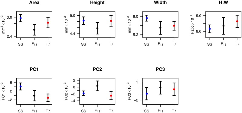
Since among-individual morphological variation for complex structures such as the posterior lobe is not completely captured by simple descriptors (e.g., width), we additionally used a combination of elliptic Fourier analysis and PCA to describe variation across all lobes in the study (i.e., SS, T7, and F13) in terms of principal components (PCs). The top three PCs collectively explain nearly 90% of the morphological variation in the dataset (PC1 = 54.6%, PC2 = 24.4%, PC3 = 10.5%). Fig 2 highlights the differences among the two parental strains, and the recombinant animals considered as a single class, for three measures of lobe size (area, height, width), a simple measure of lobe shape (ratio of height to width, or H:W), and the first three PCs. The parental strains can be discriminated by area (Welch's t-test, p = 0.001), width (p < 10−7), H:W (p < 10−4), PC1 (p < 10−9), PC2 (p = 0.02), and PC3 (p = 0.03), but not by height (p = 0.9). For area, height, and PC2 the set of recombinant F13 animals have phenotypes that, on average, fall outside the range of the parental strains, potentially indicative of transgressive segregation for these phenotypes. We note that any lobe size (area, height, width) variation is not necessarily a result of overall body size variation, since previous investigators have found that measures of body size are not predictive of lobe size [16–18, 47].
Fig 2. Summary of posterior lobe size and shape phenotypes in parental strains and recombinants.
Points represent the mean phenotype across lobes for each class, and whiskers represent 1-SD. Sample sizes are 14, 303, and 32 lobes for SS, recombinants, and T7, respectively. The total number of independent individuals scored was 8, 192, and 21.
Since the first three PCs explain much of the shape variation among individuals in this study, and PC1 is highly significantly different between the parent strains used for mapping (Fig 2), we sought to define the aspects of lobe phenotype the PCs describe. First, we used a single lobe from each of the F13 individuals to carry out correlations among phenotypes (Table 1). PC1 is strongly positively correlated with lobe width (Pearson's r = 0.59, p < 10−10), strongly negatively correlated with H:W (r = –0.58, p < 10−10), and has a very minor negative correlation with lobe height (r = –0.16, p = 0.03). This implies that the relatively simplistic H:W ratio describes a substantial fraction of the shape variation among lobes (see also [23]), with much of the PC1 variation being effectively described as changes in lobe width. Nonetheless, inspection of variation among lobes ordered by PC1 suggests that other phenotypes are changing along with width and H:W (S5 Fig), although it is not particularly clear how to describe these additional shape changes. This underlies the benefit and the cost of using EFA-PCA to describe shape changes for outlines lacking clear landmarks; One can capture variation that is difficult to define, but one lacks a clear definition that may be helpful to understand the phenotype. PC2 is tightly negatively correlated with lobe area (r = –0.89), and PC2 can be considered a proxy for overall lobe size (Table 1). It is not clear what feature of lobe morphology PC3 explains, and given the limited contribution this measure makes to overall lobe variation (10.5%), we do not focus on this PC further.
Table 1. Correlations among posterior lobe size and shape phenotypes.
| Area | Height | Width | H:W | PC1 | PC2 | PC3 | |
| Area | — | 0.69 | 0.73 | –0.16 | 0.31 | –0.89 | –0.08 |
| Height | *** | — | 0.07 | 0.58 | –0.16 | –0.60 | –0.54 |
| Width | *** | ns | — | –0.77 | 0.59 | –0.64 | 0.40 |
| H:W | * | *** | *** | — | –0.58 | 0.15 | –0.67 |
| PC1 | ** | * | *** | *** | — | –0.02 | 0.01 |
| PC2 | *** | *** | *** | * | ns | — | –0.17 |
| PC3 | ns | *** | ** | *** | ns | * | — |
Correlations among traits (Pearson's r) above the diagonal, and significance of the correlation tests below the diagonal (*** p < 10−10; ** p < 0.0001; * p < 0.05; ns, p > 0.05). PCs are orthogonal, and by definition correlations among them will be zero using the full dataset. However, only a single lobe from each individual is used for these tests, so non-zero correlations can be observed.
QTL mapping of posterior lobe variation
Previous studies have detected loci contributing to intraspecific posterior lobe variation within D. melanogaster using both QTL mapping [23] and a GWAS [24] using 155 of the sequenced inbred strains of the Drosophila genetic reference panel, DGRP [48, 49]. The difference in power between linkage-based QTL studies and population-based association studies is well known. Loci mapped in QTL studies routinely explain large fractions of the heritable variation in the cross (for example [50, 51]), while association studies generally struggle to identify causative variants explaining anything more than a fraction of the heritability for the trait [52, 53]. It is also very clear that association approaches can have much greater mapping resolution than traditional F2 QTL mapping studies due to a radical difference in the number of crossover events in the history of the mapping panels. Here, we sought to use a linkage-based, advanced intercross mapping panel to benefit from the power of a linkage-based design, but garner greater mapping resolution by including more crossover events [42, 54], although obviously falling short of the resolution possible with a GWAS.
We note that one can increase crossover number, and thus QTL mapping resolution, by simply increasing the number of F2 (or backcross) individuals tested, rather than by generating an advanced generation population. However, for any given level of resolution desired, a larger sample size is required for a population that has undergone fewer generations of recombination [55]. Therefore, the appropriate mapping design needs to weigh the additional phenotyping burden of a very large F2 study against any difficulty generating an advanced intercross population.
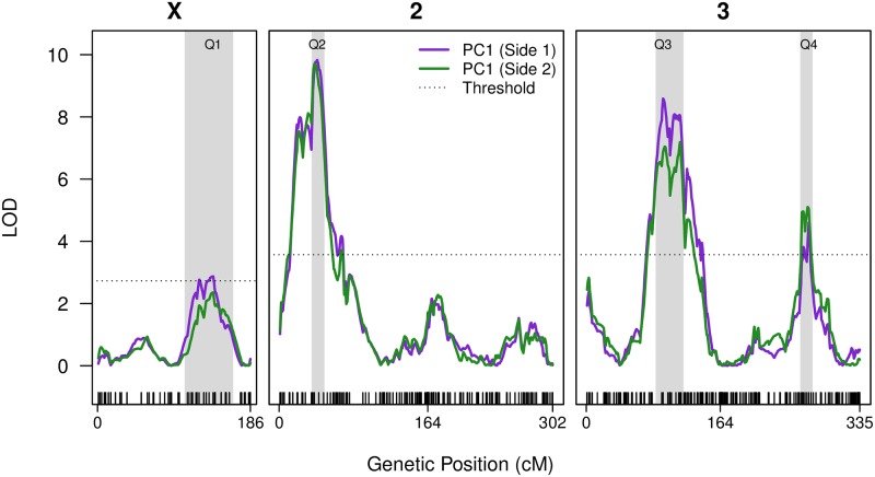
We mapped variation for three posterior lobe size traits (area, height, width) and four lobe shape traits (H:W, PC1, PC2, PC3) in the bi-parental F13 mapping population using r/qtl [38]. We provide the r/qtl input file as S3 Table and the LOD score output as S4 Table. Fig 3 shows QTL mapping results for the principal measure of morphology, PC1, for which we map four QTL, one on the X chromosome, and three on the autosomes. For those recombinant animals were we successfully scored both lobes (58%), only one of the two was initially used for mapping (Fig 3, purple curve). Nevertheless, we identify the same peaks regardless of which lobe side is chosen (Fig 3, compare purple and green curves), consistent with strong bilateral symmetry observed for this trait: The correlation between lobe sides for PC1 is r = 0.75 (p = 10−10). Using various routines in r/qtl [38] we explored the properties of the four PC1 QTL further. The additive model, y = Q1+Q2+Q3+Q4 explains 41.6% of the PC1 variation among the F13 individuals (using the 'fitqtl' function). Including all possible interaction terms in the model (i.e., y = Q1×Q2×Q3×Q4) increases the fraction of the variance explained to 54.5%, but allowing pairwise interactions among these four QTL did not significantly improve the fit of the model (using 'addint'). Additionally, the 'scantwo' function, which allows two-dimensional scans for multiple QTL, did not show clear evidence for interactions among the four PC1 loci. Finally, we tested whether there was evidence for additional QTL given the presence of Q1–Q4 using 'addqtl'. There was some suggestion of another QTL at the very tip of 3L (at 0 cM on chromosome 3, Fig 3), although evidence was not strong, and we do not consider this potential peak further.
Fig 3. Four QTL mapped for PC1 lobe shape variation.
The two solid curves present evidence for mapped QTL. The purple curve is derived from a dataset using one randomly-selected lobe from each of the recombinants were both lobes were successfully scored, and the only lobe present for the remaining animals. The green curve uses the other lobe for the 58% of animals where both were scored. The horizontal dotted lines are the permutation-derived, genomewide 5% statistical thresholds (X; LOD = 2.73, Autosomes; LOD = 3.57). The positions of the markers are presented as ticks along the inside of the x-axis. The 2-LOD drop support intervals for each QTL are highlighted as gray bars.
We performed similar routines to examine the pair of QTL mapped to chromosome 3L that contribute to PC2 (Fig 4, second panel from the bottom), a measure of lobe morphological variation that is highly negatively correlated with lobe area. The model y = Q1+Q2 explains 25.2% of the variation in PC2, and no additional QTL or interaction terms improved the fit of the model.
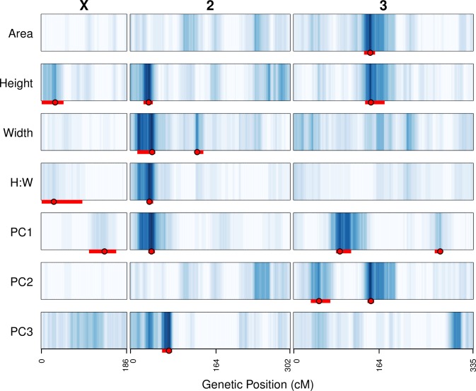
Fig 4. Positions of QTL mapped for all lobe shape and size traits.
The LOD scores at each test position along the genome have been converted to blue-scale, with darker blue corresponding to higher LOD scores. LOD score color-encoding was carried out separately for each trait, such that the maximum LOD score for every trait was given the same dark blue color, facilitating comparisons across LOD curves with varying maxima. Intensities on the X chromosome are lower given the lower statistical thresholds for QTL detection on the X. The peak positions for each QTL are presented as solid red points, and 2-LOD drop intervals are shown as red bars.
Table 2 provides details of the four PC1 and two PC2 QTL, and S6 Fig shows the effect of each QTL genotype on phenotype in the F13 panel. All QTL explain appreciable fractions of the variation, with 4/6 explaining >10% of the variation in the traits (Table 2). Single, relatively large-effect loci causative variants may underlie these QTL, although given the size of the implicated intervals (Table 2), multiple linked variants may be responsible for each QTL (see [56]).
Table 2. Details of QTL mapped for PC1 and PC2 lobe morphology descriptors.
| PC1-Q1 | PC1-Q2 | PC1-Q3 | PC1-Q4 | PC2-Q1 | PC2-Q2 | |
|---|---|---|---|---|---|---|
| Chromosome | X | 2L | 3L | 3R | 3L | 3L |
| LOD score | 2.9 | 9.8 | 8.6 | 4.6 | 4.3 | 6.0 |
| Variance explained (%) a | 1.6 | 13.7 | 10.1 | 2.5 | 11.1 | 15.0 |
| Additive effect a | 3.4 | 13.3 | –10.9 | 1.4 | –6.5 | 2.2 |
| Dominance effect a | — | –0.4 | 1.7 | –7.4 | 1.4 | –9.4 |
| Interval (cM) b | 106–165 | 36–50 | 85–119 | 262–277 | 34–72 | 146–155 |
| Physical position (Mb) b | 13.6–19.4 | 4.1–5.6 | 10.4–13.7 | 21.7–23.7 | 4.5–9.5 | 18.7–21.2 |
| Cytology c | 12A-18C | 24D-25F | 67E-70C | 93F-95A | 64B-67B | 75D-78C |
| Number of genes d | 619 | 172 | 377 | 251 | 580 | 331 |
a The fraction of the phenotypic variance explained, and the additive and dominance effects, are derived from the r/qtl function 'fitqtl'. The effects have been multiplied by 104 for clarity.
b QTL intervals are defined by 2-LOD drops from the peaks. Genetic distance is given on the expanded map (map distances on the typical D. melanogaster genetic map will be approximately 3 times shorter.) Physical distance is provided relative to Release 6 of the genome.
c Cytological positions were derived from the physical positions (in bp) using the conversion files on FlyBase [25].
d Only the numbers of protein-coding genes are provided.
At PC1-Q1 and PC1-Q2 replacing a T7 allele with an SS allele increases the phenotype in a largely additive fashion (Table 2), making the lobe more SS-like (S5 Fig). PC1-Q3 shows the opposite effect; SS alleles decrease the phenotype, making the lobe less parental SS-like. Since the parental inbred lines were not directionally-selected for lobe morphology, there is no expectation QTL will act in a consistent direction. Finally, PC1-Q4 principally shows an overdominant effect (Table 2) with heterozygous genotypes having a lower phenotype compared to either homozygous genotypic class (S6 Fig). PC2-Q1 acts additively, while PC2-Q2 also shows an overdominant effect (Table 2; S6 Fig). The combination of additive and dominant QTL action is consistent with our previous QTL mapping work employing an independent pair of starting parental strains [23].
We also obtained genotypes for window markers across the small fourth chromosome. For each individual, all genotype calls at windows across the chromosome were consistent (S1 Table), allowing us to generate a single, consensus chromosome 4 genotype for each recombinant. For those 144 recombinants with a called genotype we carried out linear regressions of genotype on phenotype for each of the seven lobe traits using the 'lm' function in R. In no case was there an association between fourth chromosome genotype and phenotype (p > 0.53 in all cases).
Overlap of QTL describing different lobe shape/size metrics
Fig 4 depicts the positions of QTL mapped for all seven measures of posterior lobe size and shape (see also S5 Table). If we consider QTL to coincide when the 2-LOD drop intervals overlap we identify a total of nine independent QTL, although without considerable further work we cannot be completely confident the underlying genetic basis of overlapping QTL is the same. The largest effect PC1 QTL on 2L, PC1-Q2 is located in the same position as QTL mapped for lobe height, width, and H:W, suggesting this locus has robust effects on several metrics of lobe morphology. Neither of the other three PC1 QTL overlap with QTL found for other traits, suggesting they confer effects on aspects of lobe shape that are not easily defined with simple measures of trait morphology. The major PC2 QTL, PC2-Q2 overlaps with QTL for area and height (Fig 4), consistent with the strong correlation between PC2 and area (Table 1). The other PC2 QTL, PC2-Q1 and the sole PC3 QTL do not overlap any loci implicated for other aspects of lobe morphological variation, again suggesting they are responsible for morphological variation that is difficult to describe without PCs. Generating introgression lines, specifically isolating QTL alleles from each parent in the genetic background of the other, would be a fruitful approach to help define the morphological change conferred by the QTL, while additionally helping resolve the causative variants.
Overlap of posterior lobe loci among studies
The advanced intercross design of our mapping population succeeded in mapping QTL to smaller regions than is possible with an F2 mapping population with a modest sample size (e.g., [23]). On the unexpanded D. melanogaster genetic map PC1 and PC2 QTL are mapped to 2–18 cM, corresponding to physical intervals of 1.5–5.8 Mb (Table 2). Nonetheless, these QTL intervals still implicate fairly large numbers of protein-coding genes (Table 2 and S6 Table). Since other work has mapped loci contributing to posterior lobe variation within D. melanogaster [23, 24], and studies have used expression analysis or RNAi to define plausible candidate posterior lobe genes [19, 27, 28], we sought to investigate any overlap among QTL and functional candidates in D. melanogaster. See S1 Data for a summary of intervals, genes, and sites previously implicated in posterior lobe development or variation in D. melanogaster.
McNeil at al. [23] mapped three autosomal QTL for a measure of posterior lobe shape defined by EFA-PCA in an F17 cross between the inbred lines Samarkand ry506 and b3852, and two of these QTL coincide with regions mapped in the present study. The QTL mapped to cytological location 66B–69B by McNeil at al. [23] overlaps with both PC1-Q3 and PC2-Q1 mapped here (Table 2). Given that PC axes are defined relative to a specific dataset, there is no necessity that the shape/size metric represented by a given PC is consistent across studies. The observation that two independent QTL intervals overlap the larger interval implicated in McNeil et al. [23] could imply multiple loci underlie this previously-mapped QTL. In addition, the QTL mapped to 75F–86C by McNeil et al. [23] overlaps PC2-Q2 identified here, although PC2-Q2 implicates a much smaller region of the genome (2.5 Mb versus 12.0 Mb, Table 2). Under the assumption the underlying genetic basis of these loci is the same, combining the two QTL studies can help to resolve the genes (S6 Table) underlying phenotypic variation in the two different crosses.
GWAS designs can provide additional mapping resolution over linkage-based studies. In flies, where linkage disequilibrium extends over relatively short distances [49, 57], a significantly associated site would be expected to reside very close to the actual causative variant (and potentially be the causative variant). A GWAS for posterior lobe morphology found no nucleotide variants formally associated with any of the tested lobe shape/size phenotypes after correction for multiple tests [24], a result of the lack of power to find modest effect causative loci in the DGRP design given the small number of genotypes and the severe correction for multiple tests that must be applied [52]. However, a number of nominally significant sites were identified, and as a class one might anticipate this group to be enriched for variants truly associated with lobe variation. Although the actual level of enrichment of true positives is unknown, an appealing possibility is that one could combine high-powered linkage-based approaches to broadly map QTL, and low-powered, but high-resolution population-based association approaches to fine map within QTL intervals, thereby reducing the burden of multiple testing. Thus, we examined whether any of the nominally-associated loci implicated by Takahara & Takahashi [24] are present within our QTL intervals. Nine genes within the six PC1/PC2 we map in this study (Table 2) additionally contain GWAS variants with p-values below 10−5 (S1 Data); hang (PC1-Q1), Hel25E (PC1-Q2), bru-3, CAH2, CG43693, and Nrx-IV (PC1-Q3), Sec13 (PC1-Q4), GluRIB and unc-13-4A (PC2-Q1). Evidence for the role of these genes in genital morphology or development is currently lacking [25]. However, specific validation of these genes using quantitative complementation tests [58, 59], reciprocal hemizygosity tests [60], or CRISPR (swapping putatively-functional alternative alleles into a common genetic background), may be able to identify candidate genes contributing to lobe morphological variation. Simultaneously, such tests may help validate whether using a combination of QTL and association mapping can aid in the dissection of complex phenotypes to the nucleotide level.
A number of studies have used functional tests or expression measurements in the developing male genitalia to identify plausible candidate genes contributing to lobe morphogenesis and/or adult morphology. Chatterjee et al. [27] identified a set of 22 genes showing consistent differences between male and female larval genital discs, the primordia of the adult genitalia. Three of these genes—toe, eyg, and caup—are within the PC1-Q3 interval (Fig 3 and Table 2), suggesting—as we have previously [23]—that genes with sex-based gene expression in the developing genitalia may be plausible candidates to harbor functional variation contributing to lobe morphology. Glassford et al. [28] provide a number of genes involved in lobe development. Two of these are present within QTL mapped here; upd1, a gene found to be expressed in the developing lobe (figure four in [28]) is within PC1-Q1, and RhoGEF64C, that when knocked down specifically in the developing male genitalia leads to reduced lobe size (figure four in [28]), resides within PC2-Q1. Finally, Tanaka et al. [19] employed RNAi targeted to wing, leg, and genital discs in D. melanogaster to test the effects of multiple genes implicated in the differences in lobe morphology between D. simulans and D. mauritiana. All these genes are located in short introgressed regions that have significant effects on morphological differentiation between species in either the morphology of the lobe or number of bristles on the clasper (another genital structure critical for mating success). A number of genes with significant RNAi effects reside within QTL we map in the present study; CG11652, CG14130, CG32081, CG32082, Mob2, and wls (PC1-Q3), CG6673, CG14835, CG14838, dally, Mcm7, msl-3, Prm, and Surf1 (PC2-Q1). Of particular interest with respect to this gene set is the potential for it to contain one or more loci that harbor allelic variation contributing to both intra- and interspecific differences in posterior lobe morphology.
Conclusion
Dissecting the genetic basis of intraspecific variation in the morphology of genital structures that exhibit rapid evolution among species can provide information on the loci that are the targets of sexual selection. We used a pair of genetically and phenotypically distinct inbred lines of D. melanogaster to generate an advanced intercross population enabling powerful, and relatively high resolution genetic mapping of variation in the posterior lobe, a structure that is essential for mating in the species. By making use of genotyping-by-sequencing technologies, and a fairly simple analytical pipeline employing standard genomics software (e.g., bwa, GATK), we were able to efficiently collect genomewide genotypes for a set of highly-recombinant individuals. We identified several, modest-effect loci influencing lobe morphology, some of which overlap with previously-identified QTL, and with candidates from functional genetics studies. We are obviously some way from our final goal of identifying the precise series of naturally-segregating causative variants contributing to posterior lobe variation. Nonetheless, by combining disparate datasets we can home in on likely candidates, paving the way for functional validation of causative genes, and testing for the action of selective forces at these loci.
Supporting Information
Information about QTL and loci previously implicated in the genetic control of posterior lobe development and adult morphology in Drosophila melanogaster.
(PDF)
(PDF)
Each of the recombinant individuals (rows) is given a genotype for a set of non-overlapping 250 kb windows along the genome (columns). Windows where no genotype call was made are shown in white. The top two rows of the image represent the homozygous parental T7 and SS parental strains, which receive homozygous calls (red and blue, respectively) for all windows genotyped. The thick black line at the bottom of the image depicts which genotyped windows were used as markers for QTL mapping.
(PDF)
The number of differences in genotype was calculated between every pair of recombinants for the set of window-based markers on the five major chromosome arms (X, 2L, 2R, 3L, 3R). This dissimilarity, or mismatch matrix was then subjected to hierarchical clustering using the 'hclust' function in R (GenoTree <- hclust(as.dist(MismatchMatrix),method = "average")). The plot depicts the resulting dendrogram, with recombinants highlighted based on genotyping data collection method (black, MSG; red, WGS). There is no clear clustering of animals genotyped with the same method.
(PDF)
Each point depicts the frequency of the T7 allele in the panel of 181 recombinant individuals at one of the 250kb markers. Windows with T7 allele frequency above 0.8 or below 0.2 are highlighted in red.
(PDF)
Four lobes from each parental strain are presented (SS, blue; T7, red), along with 88 lobes from recombinant individuals (black). The number within each image is the PC1 (× 104) value assigned to the lobe.
(PDF)
The phenotype data for the recombinants is plotted against their genotypes at the six QTL peaks. For each genotypic class, the mean (+/– 1-SE) phenotype is also presented.
(PDF)
For each individual (see "ID" column) genotyped by either MSG or low-pass WGS (see "GenotypeMethod" column) the sum of the number of reads consistent with either the T7 ("T7reads") or SS ("SSreads") allele at all SNPs within a series of non-overlapping windows of 250 kb throughout the genome is presented. Thus, each window is presented as a pair of columns.
(TXT)
A list of the sample IDs for all 181 recombinants, along with a column stating whether the individual was genotyped with low pass whole genome sequencing (WGS) or reduced representation multiplexed shotgun genotyping (MSG).
(TXT)
This file can be directly read into r/qtl using 'read.cross'. Recombinant individuals are defined using an internal lab ID. Markers are given genetic distances calculated within r/qtl. Genotypes are given using the following format: A = T7 homozygote, H = heterozygote, B = SS homozygote, NA = no call.
(CSV)
The "Name" column is the name of the test position, which is either a marker (format similar to "Chr2L.1") or an intermarker position (format similar to "c2.loc1"), "Chr" is the chromosome, and "Position_cM" is the genetic position of the marker estimated via r/qtl. Columns "PositionR6_Lower_Mb" and "PositionR6_Upper_Mb" give the upper and lower boundaries of the 250kb marker genotype windows, and the equivalent interpolated boundaries for intermarker positions. The "*.LOD" columns give the LOD associated with hypothesis tests for the presence of a QTL for each of the seven lobe shape/size traits.
(TXT)
(PDF)
The "Phenotype" column is the trait the QTL is mapped for, the "QTL" column is the name of the QTL (corresponding to the names in Table 2), the "Cytology" and "PhysicalPos_R6" columns are the cytological locations and the physical positions (in genome Release 6 coordinates) of the genes, respectively, and "GeneSymbol" and "FBgn" provide information on the gene names.
(TXT)
Acknowledgments
We thank John Kelly for providing analytical support and code that helped us genotype samples, Sophia Loschky for help with dissections, and Justin Blumenstiel for comments on an earlier version of this manuscript. We also thank the KU Genome Sequencing Core facility for sequencing services, and the K-INBRE project for supporting our bioinformatics efforts.
Data Availability
All relevant data are within the paper and its Supporting Information files. Sequence data are available on Dryad (doi:10.5061/dryad.gc182).
Funding Statement
This work was funded by NIH R01 GM085260 to SJM. The KU Genome Sequencing Core is supported by NIH P20 GM103638, and the KINBRE Bioinformatics Core is supported by NIH P20 GM103418. The funders had no role in study design, data collection and analysis, decision to publish, or preparation of the manuscript.
References
- 1.Jagadeeshan S, Singh RS. A time-sequence functional analysis of mating behaviour and genital coupling in Drosophila: role of cryptic female choice and male sex-drive in the evolution of male genitalia. J Evol Biol. 2006;19(4):1058–70. 10.1111/j.1420-9101.2006.01099.x . [DOI] [PubMed] [Google Scholar]
- 2.LeVasseur-Viens H, Polak M, Moehring AJ. No evidence for external genital morphology affecting cryptic female choice and reproductive isolation in Drosophila. Evolution. 2015;69(7):1797–807. 10.1111/evo.12685 . [DOI] [PubMed] [Google Scholar]
- 3.Frazee SR, Masly JP. Multiple sexual selection pressures drive the rapid evolution of complex morphology in a male secondary genital structure. Ecol Evol. 2015;5(19):4437–50. 10.1002/ece3.1721 [DOI] [PMC free article] [PubMed] [Google Scholar]
- 4.Coyne JA. Genetic basis of differences in genital morphology among three sibling species of Drosophila. Evolution. 1983;37(6):1101–18. [DOI] [PubMed] [Google Scholar]
- 5.Coyne JA, Kreitman M. Evolutionary Genetics of two Sibling Species, Drosophila-Simulans and Drosophila-Sechellia. Evolution. 1986;40(4):673–91. 10.2307/2408455. WOS:A1986D533500001. [DOI] [PubMed] [Google Scholar]
- 6.Eberhard WG. Sexual Selection and Animal Genitalia. Cambridge, MA: Harvard University Press; 1985. [Google Scholar]
- 7.Hosken DJ, Stockley P. Sexual selection and genital evolution. Trends Ecol Evol. 2004;19(2):87–93. 10.1016/j.tree.2003.11.012 . [DOI] [PubMed] [Google Scholar]
- 8.Masly JP, Kamimura Y. Asymmetric mismatch in strain-specific genital morphology causes increased harm to Drosophila females. Evolution. 2014;68(8):2401–11. 10.1111/evo.12436 . [DOI] [PubMed] [Google Scholar]
- 9.Macdonald SJ, Long AD. Joint estimates of quantitative trait locus effect and frequency using synthetic recombinant populations of Drosophila melanogaster. Genetics. 2007;176(2):1261–81. 10.1534/genetics.106.069641 [DOI] [PMC free article] [PubMed] [Google Scholar]
- 10.Barton NH, Keightley PD. Understanding quantitative genetic variation. Nat Rev Genet. 2002;3(1):11–21. 10.1038/nrg700 . [DOI] [PubMed] [Google Scholar]
- 11.Barton NH, Turelli M. Evolutionary Quantitative Genetics—How Little Do We Know. Annu Rev Genet. 1989;23:337–70. 10.1146/annurev.genet.23.1.337. WOS:A1989CE68800014. [DOI] [PubMed] [Google Scholar]
- 12.Ellegren H, Sheldon BC. Genetic basis of fitness differences in natural populations. Nature. 2008;452(7184):169–75. 10.1038/nature06737 . [DOI] [PubMed] [Google Scholar]
- 13.Mitchell-Olds T, Willis JH, Goldstein DB. Which evolutionary processes influence natural genetic variation for phenotypic traits? Nat Rev Genet. 2007;8(11):845–56. 10.1038/nrg2207 . [DOI] [PubMed] [Google Scholar]
- 14.Schielzeth H, Husby A. Challenges and prospects in genome-wide quantitative trait loci mapping of standing genetic variation in natural populations. Ann N Y Acad Sci. 2014;1320:35–57. 10.1111/nyas.12397 . [DOI] [PubMed] [Google Scholar]
- 15.Laurie CC, True JR, Liu J, Mercer JM. An introgression analysis of quantitative trait loci that contribute to a morphological difference between Drosophila simulans and D. mauritiana. Genetics. 1997;145(2):339–48. [DOI] [PMC free article] [PubMed] [Google Scholar]
- 16.Liu J, Mercer JM, Stam LF, Gibson GC, Zeng ZB, Laurie CC. Genetic analysis of a morphological shape difference in the male genitalia of Drosophila simulans and D. mauritiana. Genetics. 1996;142(4):1129–45. [DOI] [PMC free article] [PubMed] [Google Scholar]
- 17.Macdonald SJ, Goldstein DB. A quantitative genetic analysis of male sexual traits distinguishing the sibling species Drosophila simulans and D. sechellia. Genetics. 1999;153(4):1683–99. [DOI] [PMC free article] [PubMed] [Google Scholar]
- 18.Masly JP, Dalton JE, Srivastava S, Chen L, Arbeitman MN. The genetic basis of rapidly evolving male genital morphology in Drosophila. Genetics. 2011;189(1):357–74. 10.1534/genetics.111.130815 [DOI] [PMC free article] [PubMed] [Google Scholar]
- 19.Tanaka KM, Hopfen C, Herbert MR, Schlotterer C, Stern DL, Masly JP, et al. Genetic architecture and functional characterization of genes underlying the rapid diversification of male external genitalia between Drosophila simulans and Drosophila mauritiana. Genetics. 2015;200(1):357–69. 10.1534/genetics.114.174045 [DOI] [PMC free article] [PubMed] [Google Scholar]
- 20.Zeng ZB, Liu J, Stam LF, Kao CH, Mercer JM, Laurie CC. Genetic architecture of a morphological shape difference between two Drosophila species. Genetics. 2000;154(1):299–310. [DOI] [PMC free article] [PubMed] [Google Scholar]
- 21.Lachaise D, Cariou ML, David JR, Lemeunier F, Tsacas L, Ashburner M. Historical Biogeography of the Drosophila-Melanogaster Species Subgroup. Evol Biol. 1988;22:159–225. WOS:A1988M960600004. [Google Scholar]
- 22.Barbash DA. Ninety years of Drosophila melanogaster hybrids. Genetics. 2010;186(1):1–8. 10.1534/genetics.110.121459 [DOI] [PMC free article] [PubMed] [Google Scholar]
- 23.McNeil CL, Bain CL, Macdonald SJ. Multiple Quantitative Trait Loci Influence the Shape of a Male-Specific Genital Structure in Drosophila melanogaster. G3 (Bethesda). 2011;1(5):343–51. 10.1534/g3.111.000661 [DOI] [PMC free article] [PubMed] [Google Scholar]
- 24.Takahara B, Takahashi KH. Genome-Wide Association Study on Male Genital Shape and Size in Drosophila melanogaster. PLoS One. 2015;10(7):e0132846 10.1371/journal.pone.0132846 [DOI] [PMC free article] [PubMed] [Google Scholar]
- 25.Attrill H, Falls K, Goodman JL, Millburn GH, Antonazzo G, Rey AJ, et al. FlyBase: establishing a Gene Group resource for Drosophila melanogaster. Nucleic Acids Res. 2016;44(D1):D786–92. 10.1093/nar/gkv1046 [DOI] [PMC free article] [PubMed] [Google Scholar]
- 26.Wittkopp PJ, Stewart EE, Arnold LL, Neidert AH, Haerum BK, Thompson EM, et al. Intraspecific polymorphism to interspecific divergence: genetics of pigmentation in Drosophila. Science. 2009;326(5952):540–4. 10.1126/science.1176980 . [DOI] [PubMed] [Google Scholar]
- 27.Chatterjee SS, Uppendahl LD, Chowdhury MA, Ip PL, Siegal ML. The female-specific doublesex isoform regulates pleiotropic transcription factors to pattern genital development in Drosophila. Development. 2011;138(6):1099–109. 10.1242/dev.055731 . [DOI] [PubMed] [Google Scholar]
- 28.Glassford WJ, Johnson WC, Dall NR, Smith SJ, Liu Y, Boll W, et al. Co-option of an Ancestral Hox-Regulated Network Underlies a Recently Evolved Morphological Novelty. Dev Cell. 2015;34(5):520–31. 10.1016/j.devcel.2015.08.005 [DOI] [PMC free article] [PubMed] [Google Scholar]
- 29.Adams MD, Celniker SE, Holt RA, Evans CA, Gocayne JD, Amanatides PG, et al. The genome sequence of Drosophila melanogaster. Science. 2000;287(5461):2185–95. . [DOI] [PubMed] [Google Scholar]
- 30.King EG, Merkes CM, McNeil CL, Hoofer SR, Sen S, Broman KW, et al. Genetic dissection of a model complex trait using the Drosophila Synthetic Population Resource. Genome Res. 2012;22(8):1558–66. 10.1101/gr.134031.111 [DOI] [PMC free article] [PubMed] [Google Scholar]
- 31.Schneider CA, Rasband WS, Eliceiri KW. NIH Image to ImageJ: 25 years of image analysis. Nat Methods. 2012;9(7):671–5. . [DOI] [PMC free article] [PubMed] [Google Scholar]
- 32.Ferson S, Rohlf FJ, Koehn RK. Measuring Shape Variation of Two-Dimensional Outlines. Syst Zool. 1985;34(1):59–68. 10.2307/2413345. WOS:A1985AJD0600005. [DOI] [Google Scholar]
- 33.Kuhl FP G CR. Elliptic Fourier features of a closed contour. Computer Graphics and Image Processing. 1982;18:236–58. [Google Scholar]
- 34.Andolfatto P, Davison D, Erezyilmaz D, Hu TT, Mast J, Sunayama-Morita T, et al. Multiplexed shotgun genotyping for rapid and efficient genetic mapping. Genome Res. 2011;21(4):610–7. 10.1101/gr.115402.110 [DOI] [PMC free article] [PubMed] [Google Scholar]
- 35.Li H, Durbin R. Fast and accurate short read alignment with Burrows-Wheeler transform. Bioinformatics. 2009;25(14):1754–60. 10.1093/bioinformatics/btp324 [DOI] [PMC free article] [PubMed] [Google Scholar]
- 36.McKenna A, Hanna M, Banks E, Sivachenko A, Cibulskis K, Kernytsky A, et al. The Genome Analysis Toolkit: a MapReduce framework for analyzing next-generation DNA sequencing data. Genome Res. 2010;20(9):1297–303. 10.1101/gr.107524.110 [DOI] [PMC free article] [PubMed] [Google Scholar]
- 37.Holeski LM, Monnahan P, Koseva B, McCool N, Lindroth RL, Kelly JK. A high-resolution genetic map of yellow monkeyflower identifies chemical defense QTLs and recombination rate variation. G3 (Bethesda). 2014;4(5):813–21. 10.1534/g3.113.010124 [DOI] [PMC free article] [PubMed] [Google Scholar]
- 38.Broman KW S S. A Guide to QTL Mapping with R/qtl. New York: Springer; 2009. [Google Scholar]
- 39.Sen S, Churchill GA. A statistical framework for quantitative trait mapping. Genetics. 2001;159(1):371–87. [DOI] [PMC free article] [PubMed] [Google Scholar]
- 40.Churchill GA, Doerge RW. Empirical threshold values for quantitative trait mapping. Genetics. 1994;138(3):963–71. [DOI] [PMC free article] [PubMed] [Google Scholar]
- 41.Broman KW, Sen S, Owens SE, Manichaikul A, Southard-Smith EM, Churchill GA. The X chromosome in quantitative trait locus mapping. Genetics. 2006;174(4):2151–8. 10.1534/genetics.106.061176 [DOI] [PMC free article] [PubMed] [Google Scholar]
- 42.Cheng R, Lim JE, Samocha KE, Sokoloff G, Abney M, Skol AD, et al. Genome-wide association studies and the problem of relatedness among advanced intercross lines and other highly recombinant populations. Genetics. 2010;185(3):1033–44. 10.1534/genetics.110.116863 [DOI] [PMC free article] [PubMed] [Google Scholar]
- 43.Darvasi A, Soller M. Advanced intercross lines, an experimental population for fine genetic mapping. Genetics. 1995;141(3):1199–207. [DOI] [PMC free article] [PubMed] [Google Scholar]
- 44.Rockman MV, Kruglyak L. Breeding designs for recombinant inbred advanced intercross lines. Genetics. 2008;179(2):1069–78. 10.1534/genetics.107.083873 [DOI] [PMC free article] [PubMed] [Google Scholar]
- 45.Broman KW. Haplotype probabilities in advanced intercross populations. G3 (Bethesda). 2012;2(2):199–202. 10.1534/g3.111.001818 [DOI] [PMC free article] [PubMed] [Google Scholar]
- 46.Liu SC, Kowalski SP, Lan TH, Feldmann KA, Paterson AH. Genome-wide high-resolution mapping by recurrent intermating using Arabidopsis thaliana as a model. Genetics. 1996;142(1):247–58. [DOI] [PMC free article] [PubMed] [Google Scholar]
- 47.Shingleton AW, Estep CM, Driscoll MV, Dworkin I. Many ways to be small: different environmental regulators of size generate distinct scaling relationships in Drosophila melanogaster. Proc Biol Sci. 2009;276(1667):2625–33. 10.1098/rspb.2008.1796 [DOI] [PMC free article] [PubMed] [Google Scholar]
- 48.Huang W, Massouras A, Inoue Y, Peiffer J, Ramia M, Tarone AM, et al. Natural variation in genome architecture among 205 Drosophila melanogaster Genetic Reference Panel lines. Genome Res. 2014;24(7):1193–208. 10.1101/gr.171546.113 [DOI] [PMC free article] [PubMed] [Google Scholar]
- 49.Mackay TF, Richards S, Stone EA, Barbadilla A, Ayroles JF, Zhu D, et al. The Drosophila melanogaster Genetic Reference Panel. Nature. 2012;482(7384):173–8. 10.1038/nature10811 [DOI] [PMC free article] [PubMed] [Google Scholar]
- 50.Bloom JS, Ehrenreich IM, Loo WT, Lite TL, Kruglyak L. Finding the sources of missing heritability in a yeast cross. Nature. 2013;494(7436):234–7. 10.1038/nature11867 [DOI] [PMC free article] [PubMed] [Google Scholar]
- 51.Valdar W, Solberg LC, Gauguier D, Burnett S, Klenerman P, Cookson WO, et al. Genome-wide genetic association of complex traits in heterogeneous stock mice. Nat Genet. 2006;38(8):879–87. 10.1038/ng1840 . [DOI] [PubMed] [Google Scholar]
- 52.Long AD, Macdonald SJ, King EG. Dissecting complex traits using the Drosophila Synthetic Population Resource. Trends Genet. 2014;30(11):488–95. 10.1016/j.tig.2014.07.009 [DOI] [PMC free article] [PubMed] [Google Scholar]
- 53.Manolio TA, Collins FS, Cox NJ, Goldstein DB, Hindorff LA, Hunter DJ, et al. Finding the missing heritability of complex diseases. Nature. 2009;461(7265):747–53. 10.1038/nature08494 [DOI] [PMC free article] [PubMed] [Google Scholar]
- 54.Parker CC, Carbonetto P, Sokoloff G, Park YJ, Abney M, Palmer AA. High-resolution genetic mapping of complex traits from a combined analysis of F2 and advanced intercross mice. Genetics. 2014;198(1):103–16. 10.1534/genetics.114.167056 [DOI] [PMC free article] [PubMed] [Google Scholar]
- 55.Darvasi A. Experimental strategies for the genetic dissection of complex traits in animal models. Nat Genet. 1998;18(1):19–24. 10.1038/ng0198-19 . [DOI] [PubMed] [Google Scholar]
- 56.Flint J, Mackay TF. Genetic architecture of quantitative traits in mice, flies, and humans. Genome Res. 2009;19(5):723–33. 10.1101/gr.086660.108 [DOI] [PMC free article] [PubMed] [Google Scholar]
- 57.Long AD, Lyman RF, Langley CH, Mackay TF. Two sites in the Delta gene region contribute to naturally occurring variation in bristle number in Drosophila melanogaster. Genetics. 1998;149(2):999–1017. [DOI] [PMC free article] [PubMed] [Google Scholar]
- 58.Long AD, Mullaney SL, Mackay TF, Langley CH. Genetic interactions between naturally occurring alleles at quantitative trait loci and mutant alleles at candidate loci affecting bristle number in Drosophila melanogaster. Genetics. 1996;144(4):1497–510. [DOI] [PMC free article] [PubMed] [Google Scholar]
- 59.Turner TL. Fine-mapping natural alleles: quantitative complementation to the rescue. Mol Ecol. 2014;23(10):2377–82. 10.1111/mec.12719 [DOI] [PMC free article] [PubMed] [Google Scholar]
- 60.Stern DL. Identification of loci that cause phenotypic variation in diverse species with the reciprocal hemizygosity test. Trends Genet. 2014;30(12):547–54. 10.1016/j.tig.2014.09.006 . [DOI] [PubMed] [Google Scholar]
Associated Data
This section collects any data citations, data availability statements, or supplementary materials included in this article.
Supplementary Materials
Information about QTL and loci previously implicated in the genetic control of posterior lobe development and adult morphology in Drosophila melanogaster.
(PDF)
(PDF)
Each of the recombinant individuals (rows) is given a genotype for a set of non-overlapping 250 kb windows along the genome (columns). Windows where no genotype call was made are shown in white. The top two rows of the image represent the homozygous parental T7 and SS parental strains, which receive homozygous calls (red and blue, respectively) for all windows genotyped. The thick black line at the bottom of the image depicts which genotyped windows were used as markers for QTL mapping.
(PDF)
The number of differences in genotype was calculated between every pair of recombinants for the set of window-based markers on the five major chromosome arms (X, 2L, 2R, 3L, 3R). This dissimilarity, or mismatch matrix was then subjected to hierarchical clustering using the 'hclust' function in R (GenoTree <- hclust(as.dist(MismatchMatrix),method = "average")). The plot depicts the resulting dendrogram, with recombinants highlighted based on genotyping data collection method (black, MSG; red, WGS). There is no clear clustering of animals genotyped with the same method.
(PDF)
Each point depicts the frequency of the T7 allele in the panel of 181 recombinant individuals at one of the 250kb markers. Windows with T7 allele frequency above 0.8 or below 0.2 are highlighted in red.
(PDF)
Four lobes from each parental strain are presented (SS, blue; T7, red), along with 88 lobes from recombinant individuals (black). The number within each image is the PC1 (× 104) value assigned to the lobe.
(PDF)
The phenotype data for the recombinants is plotted against their genotypes at the six QTL peaks. For each genotypic class, the mean (+/– 1-SE) phenotype is also presented.
(PDF)
For each individual (see "ID" column) genotyped by either MSG or low-pass WGS (see "GenotypeMethod" column) the sum of the number of reads consistent with either the T7 ("T7reads") or SS ("SSreads") allele at all SNPs within a series of non-overlapping windows of 250 kb throughout the genome is presented. Thus, each window is presented as a pair of columns.
(TXT)
A list of the sample IDs for all 181 recombinants, along with a column stating whether the individual was genotyped with low pass whole genome sequencing (WGS) or reduced representation multiplexed shotgun genotyping (MSG).
(TXT)
This file can be directly read into r/qtl using 'read.cross'. Recombinant individuals are defined using an internal lab ID. Markers are given genetic distances calculated within r/qtl. Genotypes are given using the following format: A = T7 homozygote, H = heterozygote, B = SS homozygote, NA = no call.
(CSV)
The "Name" column is the name of the test position, which is either a marker (format similar to "Chr2L.1") or an intermarker position (format similar to "c2.loc1"), "Chr" is the chromosome, and "Position_cM" is the genetic position of the marker estimated via r/qtl. Columns "PositionR6_Lower_Mb" and "PositionR6_Upper_Mb" give the upper and lower boundaries of the 250kb marker genotype windows, and the equivalent interpolated boundaries for intermarker positions. The "*.LOD" columns give the LOD associated with hypothesis tests for the presence of a QTL for each of the seven lobe shape/size traits.
(TXT)
(PDF)
The "Phenotype" column is the trait the QTL is mapped for, the "QTL" column is the name of the QTL (corresponding to the names in Table 2), the "Cytology" and "PhysicalPos_R6" columns are the cytological locations and the physical positions (in genome Release 6 coordinates) of the genes, respectively, and "GeneSymbol" and "FBgn" provide information on the gene names.
(TXT)
Data Availability Statement
All relevant data are within the paper and its Supporting Information files. Sequence data are available on Dryad (doi:10.5061/dryad.gc182).


