Abstract
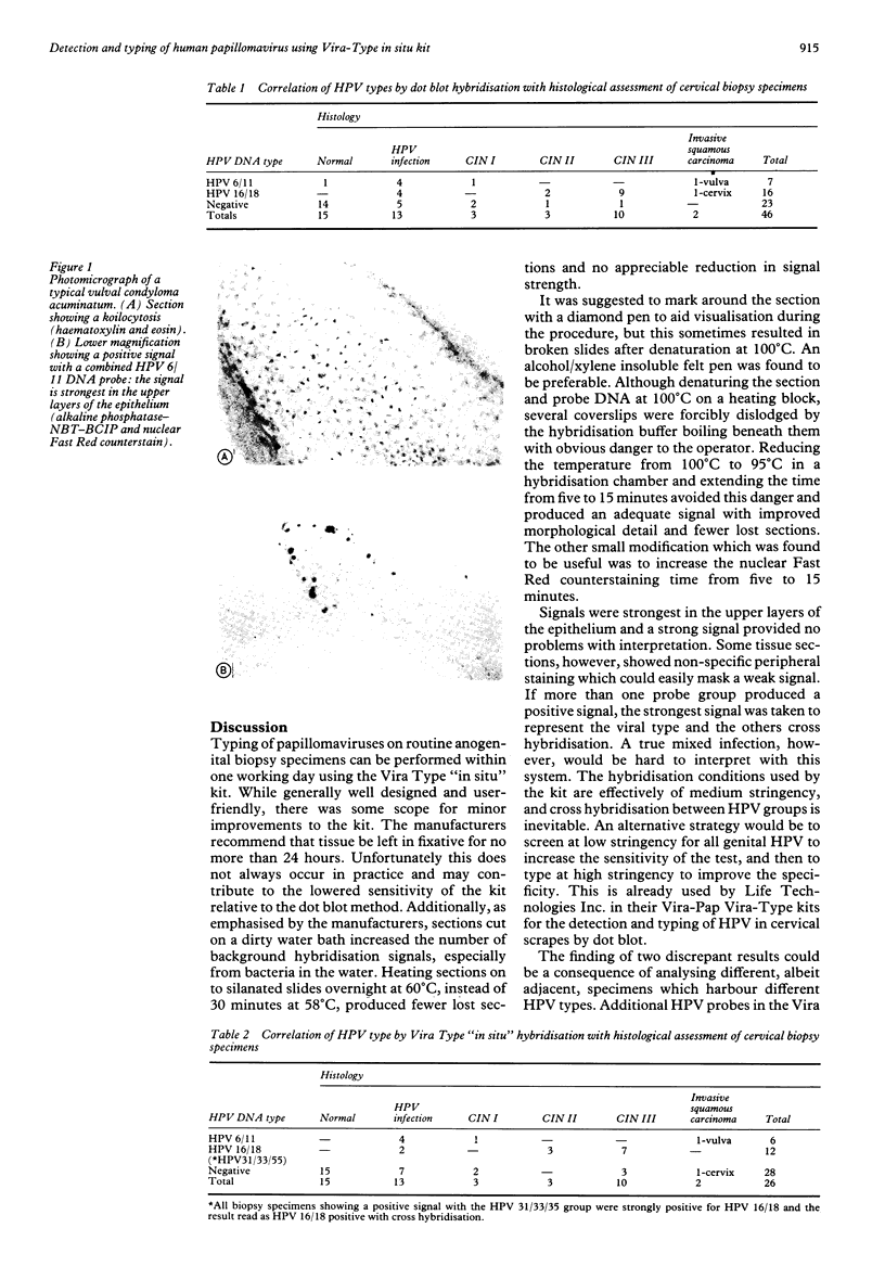
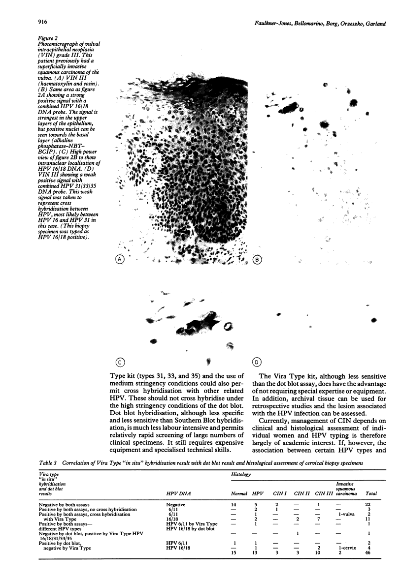
A new commercial kit (Vira Type "in situ", Life Technologies, Inc., Molecular Diagnostics Division, Guithersburg, Maryland, USA) for the detection of human papillomavirus (HPV) types 6, 11, 16, 18, 31, 33 and 35 in routinely processed human anogenital tissue was compared with a conventional dot blot assay for HPV 6, 11, 16 and 18. Both systems use double-stranded genomic DNA probes for the detection of type specific HPV DNA. The probes used on the dot blots were labelled with 32P and visualised autoradiographically. The Vira Type probes were labelled with biotin and visualised using a streptavidin-alkaline phosphatase conjugate with NBT-BCIP substrate. Biopsy specimens from the cervix, vagina, and vulva of 46 women were processed by both methods and compared. The histological diagnoses ranged from benign changes, to dysplasia, and invasive carcinoma. Overall, 50% of biopsy specimens were positive for HPV DNA by dot blot hybridisation; only 39% were positive by Vira Type in situ hybridisation. Three of the specimens positive by the Vira Type "in situ" kit showed no cross hybridisation and were the same HPV type as the dot blot. A further 13 showed hybridisation, but the showed cross hybridisation, but the to the dot blot results. One biopsy specimen was positive for different HPV types by the two tests and one was positive by Vira Type and negative by dot blot. Six biopsy specimens were negative by Vira Type but positive by dot blot. It is concluded that the Vira Type "in situ" kit has a similar specificity but lower sensitivity than the dot blot hybridisation method for the detection of HPV DNA.
Full text
PDF

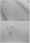
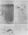
Images in this article
Selected References
These references are in PubMed. This may not be the complete list of references from this article.
- Coleridge J. C., Coleridge H. M. Afferent vagal C fibre innervation of the lungs and airways and its functional significance. Rev Physiol Biochem Pharmacol. 1984;99:1–110. doi: 10.1007/BFb0027715. [DOI] [PubMed] [Google Scholar]
- Feinberg A. P., Vogelstein B. A technique for radiolabeling DNA restriction endonuclease fragments to high specific activity. Anal Biochem. 1983 Jul 1;132(1):6–13. doi: 10.1016/0003-2697(83)90418-9. [DOI] [PubMed] [Google Scholar]
- Lörincz A. T. Detection of human papillomavirus infection by nucleic acid hybridization. Obstet Gynecol Clin North Am. 1987 Jun;14(2):451–469. [PubMed] [Google Scholar]
- McCance D. J. Cervical cancer. News on papillomaviruses. Nature. 1988 Oct 27;335(6193):765–766. doi: 10.1038/335765a0. [DOI] [PubMed] [Google Scholar]
- McIndoe W. A., McLean M. R., Jones R. W., Mullins P. R. The invasive potential of carcinoma in situ of the cervix. Obstet Gynecol. 1984 Oct;64(4):451–458. [PubMed] [Google Scholar]
- Syrjänen K., Mäntyjärvi R., Väyrynen M., Syrjänen S., Parkkinen S., Yliskoski M., Saarikoski S., Castrén O. Evolution of human papillomavirus infections in the uterine cervix during a long-term prospective follow-up. Appl Pathol. 1987;5(2):121–135. [PubMed] [Google Scholar]


