Abstract
Trophoblast cells from bovine somatic cell nuclear transfer (SCNT) conceptuses express major histocompatibility complex class I (MHC-I) proteins early in gestation, and this may be one cause of the significant first-trimester embryonic mortality observed in these pregnancies. MHC-I homozygous-compatible (n = 9), homozygous-incompatible (n = 8), and heterozygous-incompatible (n = 5) SCNT pregnancies were established. The control group consisted of eight pregnancies produced by artificial insemination. Uterine and placental samples were collected on Day 35 ± 1 of pregnancy, and expression of MHC-I, leukocyte markers, and cytokines were examined by immunohistochemistry. Trophoblast cells from all SCNT pregnancies expressed MHC-I, while trophoblast cells from age-matched control pregnancies were negative for MHC-I expression. Expression of MHC-I antigens by trophoblast cells from SCNT pregnancies was associated with lymphocytic infiltration in the endometrium. Furthermore, MHC-I-incompatible conceptuses, particularly the heterozygous-incompatible ones, induced a more pronounced lymphocytic infiltration than MHC-I-compatible conceptuses. Cells expressing cluster of differentiation (CD) 3, gamma/deltaTCR, and MHC-II were increased in the endometrium of SCNT pregnancies compared to the control group. CD4+ lymphocytes were increased in MHC-I-incompatible pregnancies compared to MHC-I-compatible and control pregnancies. CD8+, FOXP3+, and natural killer cells were increased in MHC-I heterozygous-incompatible SCNT pregnancies compared to homozygous SCNT and control pregnancies.
Keywords: cattle, immunohistochemistry, miscarriage, pregnancy, somatic cell nuclear transfer
INTRODUCTION
Although the birth of Dolly, the first offspring produced by somatic cell nuclear transfer (SCNT), happened more than 15 yr ago [1], SCNT is still a very inefficient process, with less than 10% of transferred embryos resulting in a live offspring [1–4]. In cattle, Day 28–32 pregnancy rates in SCNT and in vitro fertilization (IVF)-produced embryos are comparable (approximately 40%) [5–7]. While loss of pregnancies produced by IVF is approximately 13%–16% between Days 30 and 90 of gestation, and, in in vivo-produced embryos, pregnancy loss during this time period is about 5%, pregnancy losses range from 50% to 100% in SCNT pregnancies [8].
Abnormal placentation appears to be the main cause of these embryonic losses [9]. During the first trimester of SCNT pregnancies, hypoplastic, partially developed placentae with rudimentary cotyledons have been described [9]. In the later stages of gestation, placentae are grossly abnormal with membranes that are thickened and edematous, and placentomes that are reduced in number and hypertrophic [8].
One of the mechanisms cattle and other species use to escape from maternal immune attack is to downregulate the expression of trophoblast major histocompatibility complex class I (MHC-I) proteins during the first trimester of pregnancy. This is a mechanism to protect the semiallogeneic conceptus from attack by the maternal immune system. Unlike other mammals, in cattle, placental MHC-I expression is temporally and regionally regulated [10]. In the interplacentomal region, a significant number of trophoblast cells are positive for classical MHC-I (MHC-Ia) and nonclassical MHC-I (MHC-Ib) proteins from the sixth month of pregnancy on, while the placentomal villous trophoblast cells, the area of intimate contact between fetal cells and the maternal epithelium, are negative throughout gestation [10, 11].
Immunological recognition of allogeneic MHC-I proteins facilitates successful detachment of the bovine placenta at parturition. An increased incidence of placental retention in normal gestations has been associated with MHC-I compatibility between the mother and the fetus [12–14]. Davies et al. [13] found that MHC-I-compatible pregnancies had decreased numbers of trophoblast and epithelial cells undergoing apoptosis, decreased expression of IL2 at the fetal-maternal interface, and incomplete degranulation of binucleate cells.
While MHC-I incompatibility seems to be beneficial for placental detachment at term, the expression of MHC-I proteins by SCNT embryos during the first trimester of pregnancy appears to be detrimental to embryonic survival [13, 15]. In SCNT pregnancies from 34 to 63 days, trophoblast cells were found to express MHC-I proteins, while age-matched control pregnancies were negative for MHC-I [15]. This abnormal MHC-I expression likely results from incomplete reprogramming of somatic cell nuclei during the SCNT process. In the same study, SCNT pregnancies had an increased number of endometrial cluster of differentiation (CD)3+ T lymphocytes that was likely a response to the abnormal MHC-I expression [15]. A maternal immune response against trophoblast MHC-I proteins is likely one of the causes of the high first-trimester embryonic mortality rate observed in SCNT pregnancies.
The present study investigated the expression of MHC-I proteins by trophoblast cells, endometrial lymphocyte populations, and endometrial cytokines at Day 35 of gestation in MHC-I-compatible and -incompatible pregnancies. Our hypothesis is that inappropriate placental MHC-I expression by SCNT conceptuses induces a uterine immune response against the placenta in MHC-I-incompatible pregnancies, while conceptuses that are MHC-I compatible or established by artificial insemination (AI) do not trigger such a response.
MATERIALS AND METHODS
Materials
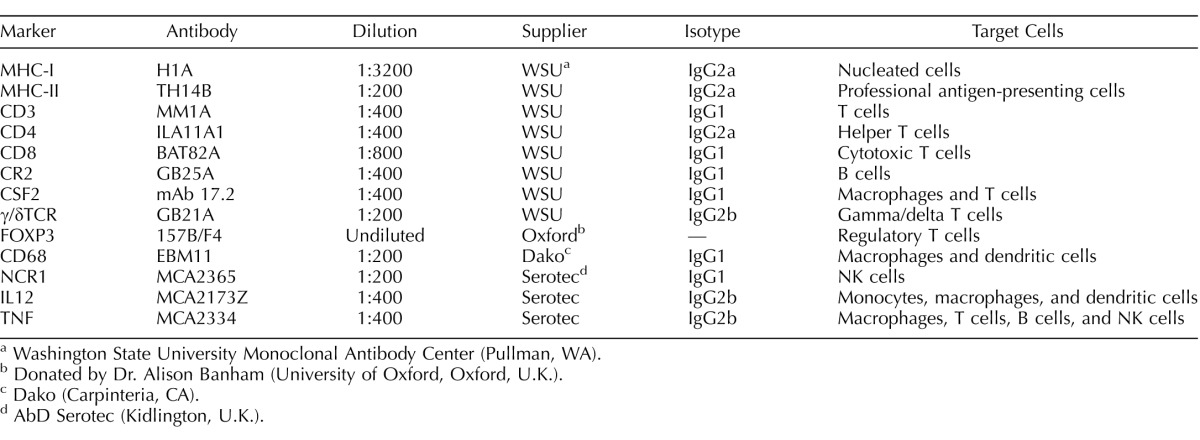
PBS was purchased from Fisher Scientific (Fair Lawn, NJ), as were hydrogen peroxide and bovine serum albumin (BSA). Normal goat serum, aminoethyl carbazole (AEC) substrate kits, and streptavidin peroxidase were obtained from Invitrogen (Camarillo, CA). Detailed information about the monoclonal antibodies is shown in Table 1. Monoclonal antibodies against bovine MHC-I, MHC-II, CD3, CD4, CD8, CR2 (previously known as CD21), γ/δTCR, CSF2 (previously known as GM-CSF), and their respective isotype-matched negative controls were purchased from the Washington State University Monoclonal Antibody Center (Pullman, WA). The FOXP3 monoclonal antibody (clone 157B/F4) was a generous donation from Dr. Alison Banham at the University of Oxford [16]. The mouse anti-human CD68 antibody (clone EBM11, ascites, 2.3 μg/ml) was purchased from Dako (Carpinteria, CA). Monoclonal antibodies against NCR1 (previously known as NKp46), IL12, and TNF (previously known as TNFα) were purchased from AbD Serotec (Kidlington, U.K.). The biotinylated anti-mouse IgG secondary antibody was purchased from Vector Laboratories (Burlingame, CA). Fluoromount-G mounting medium was purchased from Southern Biotech (Birmingham, AL).
TABLE 1.
Monoclonal antibodies used for immunohistochemistry.
Washington State University Monoclonal Antibody Center (Pullman, WA).
Donated by Dr. Alison Banham (University of Oxford, Oxford, U.K.).
Dako (Carpinteria, CA).
AbD Serotec (Kidlington, U.K.).
Animals
The use of animals for this study was approved by the Institutional Animal Care and Use Committee at Utah State University (protocol #1171). All of the cattle used in this study, including the donors of the SCNT cell line, were typed for MHC-I and MHC-II genes by massively parallel pyrosequencing using a Roche 454 Genome Sequencer FLX (Roche Diagnostics, Bradford, CT), as previously described [14]. The MHC typing data were used to establish MHC-I homozygous-compatible, MHC-I homozygous-incompatible, and MHC-I heterozygous-incompatible pregnancies (Supplemental Table S1; Supplemental Data are available online at www.biolreprod.org). Homozygous or heterozygous refers to the MHC genes of the SCNT cell line and conceptus. In MHC-compatible pregnancies, the surrogate dam expressed all of the MHC-I isoforms expressed by the conceptus, while, in MHC-incompatible pregnancies, the conceptus expressed MHC-I isoforms that would be recognized as foreign antigens by the maternal immune system. Oocytes for SCNT were harvested from ovaries collected at a local abattoir. Recipient females were beef cows from the J.R. Simplot Company Cattle Reproduction Facility in Emmett, ID. To test our hypothesis, MHC-I homozygous-compatible (n = 9), homozygous-incompatible (n = 8), and heterozygous-incompatible (n = 5) pregnancies were produced by SCNT, as described previously [17]. Day 7 embryos were transferred to cows synchronized ±1 day to the stage of the embryos. Control pregnancies (n = 8) were established by AI. Pregnancy was diagnosed by transrectal ultrasonography on Pregnancy Day 28 by the presence of a conceptus with a heartbeat. Concepti were humanely harvested on Day 35 ± 1 of pregnancy at the Utah State University, USDA-inspected Meat Laboratory.
Tissues
Uterine and placental tissues were harvested and processed immediately after the cows were killed. Fetal crown-to-rump length and fetal weight were measured for assessment of fetal development. Death of the embryo at the time of tissue collection was determined by the presence of hemorrhagic amniotic fluid and/or evidence of placental or embryonic degeneration. In addition, direct evidence of embryonic death was always correlated with lack of visible blood-filled placental blood vessels. For immunohistochemistry, endometrium and placenta from two areas (3 × 2 cm) of the contra- and ipsilateral horns to the pregnancy were frozen in Tissue-Tek optimal cutting temperature compound. Sections (8-μm thick) were acquired using a cryostat microtome, put on precleaned Superfrost Plus Microscope Slides, fixed in ice-cold acetone for 5 min, and air-dried and frozen at −80°C for long-term storage. Two sections from each tissue sample were stained with each antibody.
Immunohistochemistry
Slides containing frozen sections were allowed to thaw at room temperature and then were rehydrated in two changes of PBS for 10 min. Sections were treated with 0.3% hydrogen peroxide in PBS for 10 min to block endogenous peroxidase activity. All incubations were done at room temperature in a humidity chamber and slides were washed in three changes of PBS between incubations, except between the blocking solution and primary antibody incubation. Nonspecific binding sites were blocked with PBS containing 1% BSA and 2% normal goat serum for 20 min. Immediately after treatment with blocking solution, sections were incubated with monoclonal primary antibodies specific to the protein of interest and at the appropriate concentration, as previously determined by titration, for 1 h (Table 1). Sections were then treated with biotinylated anti-mouse IgG secondary antibody for 20 min followed by streptavidin peroxidase for 20 min. Slides were incubated with AEC for 5 min and then excess AEC was removed by washing in distilled water. Sections were counterstained with hematoxylin for 1 min and excess was removed by washing in distilled water. Slides were mounted using water-soluble Fluoromount-G mounting medium. Stained sections were analyzed using a Zeiss Axio Observer microscope (Zeiss, Gottingen, Germany) with a 10× objective. Digital images were acquired using AxioVision software (Zeiss) and a high-resolution AxioCam HRC digital camera.
In order to quantify the percent of endometrial stroma occupied by the cells of interest (MHC-II+, CD3+, CD4+, CD8+, CR2+, γ/δTCR+, FOXP3+, CD68+, and NCR1+ cells) and the percent of trophoblast cells positive for MHC-I, 15 ± 3 images per section were acquired, a region of interest was drawn around the endometrial stroma or the trophoblast, respectively, and the AutoMeasure module of the AxioVision software was used to set intensity threshold values and measure the area occupied by the cells of interest. Lymphoid nodules were excluded from the region of the endometrium assessed, because the number of nodules varied greatly between pregnancies from the same experimental group and their inclusion would have biased the results. The percent of the surface area occupied by the cells of interest was calculated by dividing the area occupied by positively stained cells by the area of the frame. Observations were conducted without knowledge of treatment group.
Statistical Analyses
Analyses of embryonic length, weight, and area of positive cells employed ANOVA models using the MIXED procedure of SAS (SAS for Windows, version 9.3; SAS Institute Inc., Cary, NC) with treatment, embryonic viability, and their interaction as fixed effects and the random effect of animal nested within treatment. Significant differences between treatments were determined by Student t-test with the pdiff option and Tukey adjustment. Treatment effects on embryonic mortality rate from Days 28 to 35 of pregnancy were examined by chi-square analysis using the FREQ procedure of SAS. Logistic regression performed using the LOGISTIC procedure of SAS was used to assess the relationship between embryonic mortality and trophoblast MHC-I expression, and embryonic mortality and the presence of endometrial lymphocytes (SAS). Effects were considered to be significant when the P value was equal to or below 0.05.
The effect of embryonic viability was only significant for the area of CD4+ cells. Therefore, the analysis of area of CD4+ cells included treatment, embryonic viability, and the interaction between treatment and embryonic viability in the model (see Results section). For the other variables related to the area of positive cells, embryonic viability and its interaction with treatment were not significant and, consequently, these effects were removed from the model.
RESULTS
Gross Morphology and Embryonic Survival
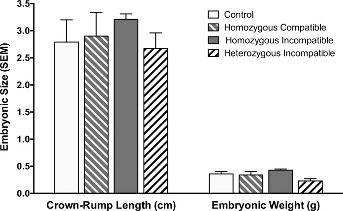
Embryonic mortality rates between 28 and 35 days were significantly increased in the MHC-I heterozygous-incompatible pregnancies compared with the MHC-I homozygous-compatible and -incompatible, and control pregnancies (100%, 11.1%, 12.5%, and 12.5%, respectively; P < 0.001; Table 2). There was no significant difference between the MHC-I homozygous-compatible, homozygous-incompatible, and control pregnancies for embryonic mortality rates. The presence of grossly visible placental vasculature, readily seen as blood vessels filled with bright red blood, was completely correlated with embryonic viability (Table 2 and Supplemental Fig. S1). Embryonic crown-to-rump length and weight of SCNT embryos did not significantly differ between groups (P = 0.32; Fig. 1).
TABLE 2.
Comparison of embryonic mortality rates and placental vascularization.
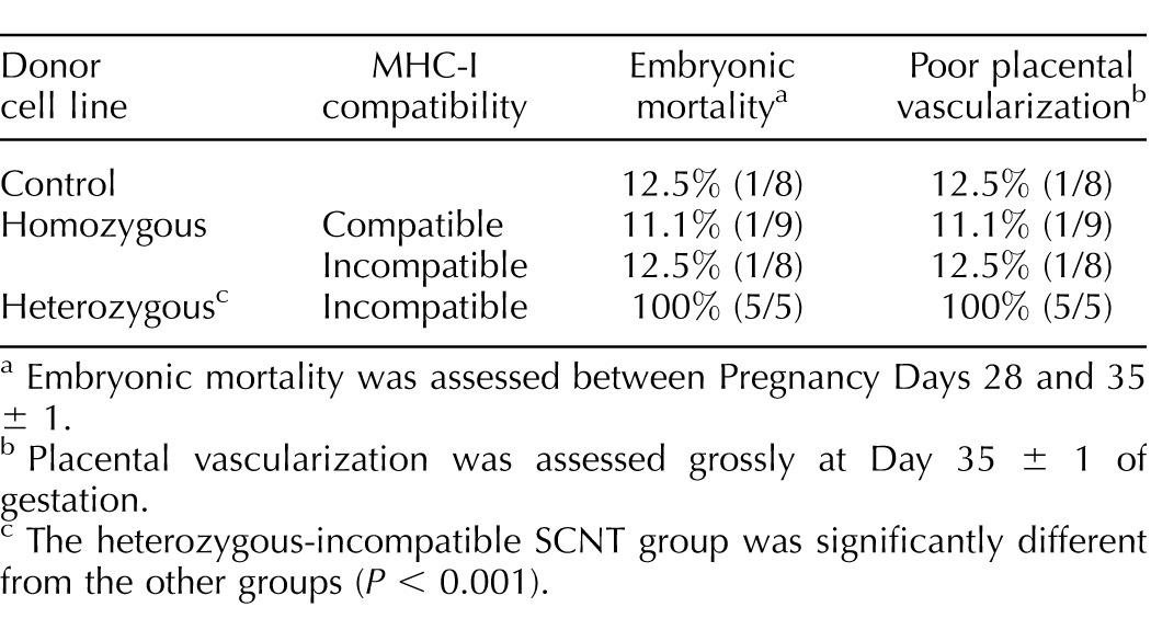
Embryonic mortality was assessed between Pregnancy Days 28 and 35 ± 1.
Placental vascularization was assessed grossly at Day 35 ± 1 of gestation.
The heterozygous-incompatible SCNT group was significantly different from the other groups (P < 0.001).
FIG. 1.
Crown-to-rump length (cm) and weight of embryos at Day 35 ± 1 of gestation. SCNT pregnancies were either MHC-I homozygous-compatible (n = 9), homozygous-incompatible (n = 8), or heterozygous-incompatible (n = 5). Age-matched control pregnancies (n = 8) were established by AI. There was no significant difference between any of the groups.
MHC-I Expression by Trophoblast Cells
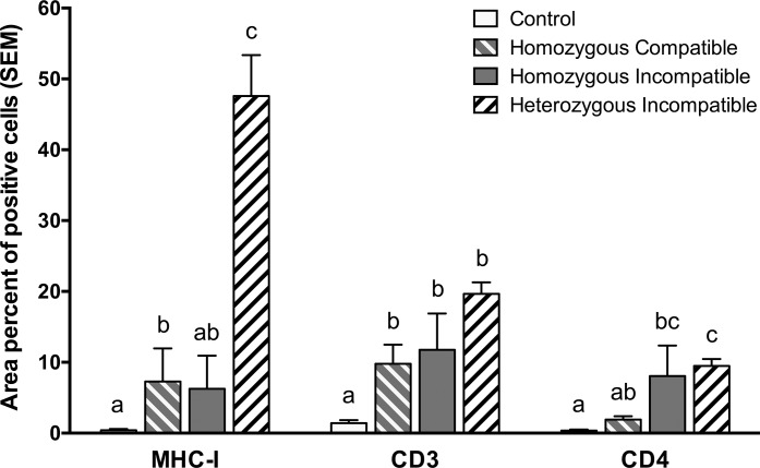
In the control animals, the trophoblast expression of MHC-I proteins was nearly undetectable, including in the placenta of the one dead embryo. Trophoblast cells from SCNT pregnancies expressed MHC-I proteins at different levels. The heterozygous-incompatible pregnancies expressed significantly higher levels of MHC-I proteins compared to the control, homozygous-compatible, and homozygous-incompatible pregnancies (P < 0.001, P = 0.006, and P = 0.01, respectively; Figs. 2 and 3). Although both the homozygous-compatible and -incompatible pregnancies expressed higher levels of MHC-I proteins than the control pregnancies, statistical significance was only achieved for the homozygous-compatible group (P = 0.035), not for the homozygous-incompatible group (P = 0.13; Fig. 2).
FIG. 2.
Percent area of MHC-I+ trophoblast cells in the placenta, and dispersed CD3+ and CD4+ T cells in the endometrium in control (n = 8), MHC-I homozygous-compatible SCNT (n = 9), MHC-I homozygous-incompatible SCNT (n = 8), and MHC-I heterozygous-incompatible SCNT (n = 5) pregnancies at Day 35 ± 1 of pregnancy. Age-matched control pregnancies were established by AI. Groups with different superscripts were significantly different (P < 0.05).
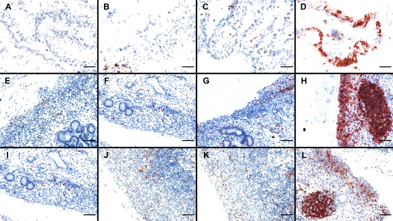
FIG. 3.
Immunohistochemical labeling of trophoblast for MHC-I (A–D), and endometrial tissue for CD3 (E–H) and CD4 (I–L) of cows at Day 35 ± 1 of pregnancy. A, E, and I) Pregnancies established by AI (control). B, F, and J) MHC-I homozygous-compatible SCNT pregnancies. C, G, and K) Homozygous-incompatible SCNT pregnancies. D, H, and L) Heterozygous-incompatible SCNT pregnancies. Bar = 50 μm.
Endometrial CD3+, CD4+, CD8+, and FOXP3+ Cells
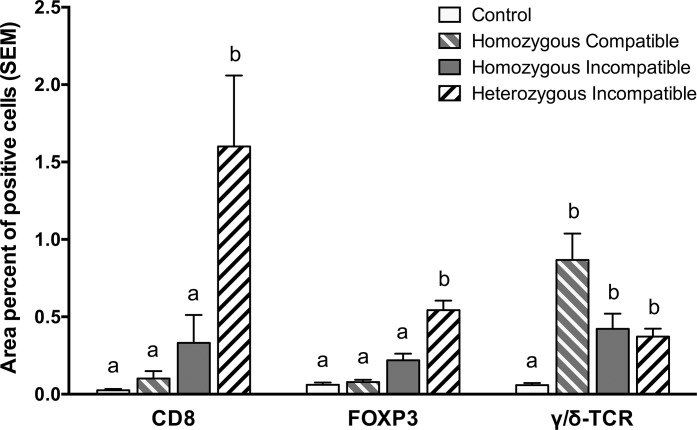
Endometrial lymphocyte infiltration and proliferation was assessed by the percent area of the superficial and deep endometrial stroma occupied by these cells. In the interplacentomal endometrium the presence of dispersed CD3+, CD4+, CD8+, and FOXP3+ cells in the superficial stroma followed the same trend as the trophoblast MHC-I expression (Figs. 2–4). All SCNT pregnancies had increased numbers of dispersed CD3+ cells compared to the control pregnancies (P < 0.001, P < 0.001, and P < 0.001 for MHC-I homozygous compatible, homozygous incompatible, and heterozygous incompatible, respectively). There were no significant differences between the SCNT pregnancies for the infiltration of CD3+ lymphocytes.
FIG. 4.
Percent area of dispersed CD8+, FOXP3+, and γ/δTCR+ lymphocytes in the endometrium of Day 35 ± 1 pregnancies established by SCNT, which were MHC-I homozygous compatible (n = 9), MHC-I homozygous incompatible (n = 8), or MHC-I heterozygous incompatible (n = 5). Age-matched control pregnancies were established by AI (n = 8). Groups with different superscripts were significantly different (P < 0.05).
Endometrial infiltration of CD4+ lymphocytes was significantly affected by embryonic viability (P = 0.002) as well as treatment (P < 0.001). This lymphocyte population was increased in live compared to dead fetuses in all treatment groups, which is the opposite of what would be predicted if the CD4+ cells were accumulating in response to the presence of a dead embryo. These cells were increased in SCNT homozygous- and heterozygous-incompatible pregnancies compared to the homozygous-compatible (P = 0.027 and P = 0.02, respectively) and control groups (P < 0.001 and P < 0.001, respectively; Figs. 2 and 3). The CD8+ and FOXP3+ cells were significantly increased in the MHC-I heterozygous-incompatible group compared to the control group (P = 0.01 for CD8+ cells and P < 0.001 for FOXP3+ cells; Fig. 4). Among the SCNT pregnancies, the MHC-I heterozygous-incompatible group was significantly increased compared with the homozygous-compatible and -incompatible pregnancies for CD8+ cells (P = 0.018 and P = 0.020, respectively) and FOXP3+ cells (P < 0.001 and P = 0.041, respectively; Fig. 4). Interestingly, while the CD3+, CD4+, CD8+, and FOXP3+ cells were disseminated throughout the superficial and glandular endometrial stroma in control pregnancies, in the SCNT pregnancies, the majority of these cells were located in the superficial stroma under the uterine epithelium. Lymphocytes were also found organized in large lymphoid nodules in the superficial endometrial stroma in the heterozygous-incompatible and, to a lesser extent, in the homozygous-compatible and -incompatible pregnancies (Fig. 3). These nodules were composed mainly of CD4+ cells, with the remaining cells consisting of CD8+, CR2+, and a very small percentage of γ/δTCR+ cells.
Endometrial γ/δTCR+ Cells
All SCNT pregnancies presented higher numbers of γ/δTCR+ cells than the control group (P < 0.001). However, the infiltration of these cells did not follow the same trend as the other T cell populations. Although there was not a statistical difference between SCNT pregnancies, the homozygous-compatible pregnancies had higher numbers of these cells than the homozygous- or heterozygous-incompatible pregnancies (Fig. 4).
Endometrial CR2+, MHC-II+, and NCR1+ Cells
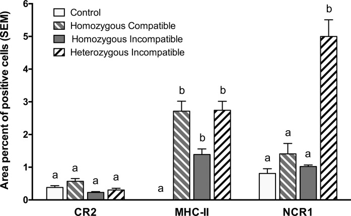
Figure 5 illustrates the area percent of dispersed CR2-, MHC-II-, and NCR1-positive cells in the different treatment groups. Very few CR2+ cells were found in the endometrium of pregnant cows at 35 days of pregnancy, and these cells were scattered throughout the glandular stroma. No significant difference was seen among treatment groups (P = 0.92). The MHC-II+ cells were significantly more abundant in the endometrium of cows carrying SCNT conceptuses compared with the control group (homozygous compatible, P = 0.002; homozygous incompatible, P = 0.011; heterozygous incompatible P = 0.003). These cells were mainly located immediately adjacent to the basement membrane of the uterine epithelium. The number of NCR1+ cells was dramatically increased in the heterozygous-incompatible pregnancies compared to the control, homozygous-compatible, and homozygous-incompatible groups (P = 0.009, P = 0.011, and P = 0.010, respectively).
FIG. 5.
Percent area of dispersed cells positive for CR2, MHC-II, and NCR1 in the Day 35 ± 1 pregnant endometrium. Pregnancies were established by either AI (control, n = 8) or SCNT. SCNT pregnancies were MHC-I homozygous compatible (n = 9), MHC-I homozygous incompatible (n = 8), or MHC-I heterozygous incompatible (n = 5). Groups with different superscripts were significantly different (P < 0.05).
Endometrial and Placental CD68+ Cells
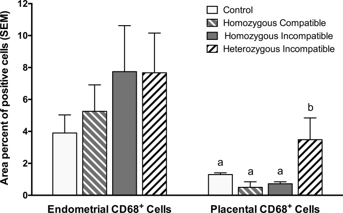
CD68 is a pan-macrophage marker. In all treatment groups, CD68+ cells were abundant in the endometrial stroma. The endometrial CD68+ cells were numerically increased in the MHC-I-incompatible treatment groups, but the increase was not statistically significant (P = 0.331; Fig. 6). As shown in Figure 6, increased numbers of macrophages were found in the placental tissue of MHC-I heterozygous-incompatible pregnancies compared with the control, homozygous-compatible, and homozygous-incompatible pregnancies (P = 0.02, P < 0.01, and P < 0.01, respectively). Two of the dead MHC-I heterozygous-incompatible conceptuses, the two with the smallest embryos, had particularly large numbers of CD68+ macrophages that comprised 3.9% and 11% of the area of the placenta, respectively. It is likely that these embryos died earlier than the other ones, and that more maternal macrophages infiltrated the placenta to clean up the cellular debris.
FIG. 6.
Percent area of CD68+ cells in the endometrium and placenta of pregnancies at Day 35 ± 1 of gestation. SCNT pregnancies were MHC-I homozygous compatible (n = 9), MHC-I homozygous incompatible (n = 8), or MHC-I heterozygous incompatible (n = 5). Age-matched control pregnancies were established by AI (n = 8). Groups with different superscripts were significantly different (P < 0.05).
Trophoblast MHC-I Expression, Lymphocyte Markers, and Embryonic Mortality
Logistic regression analyses revealed that the probability of embryonic death increased as the level of trophoblast MHC-I expression increased (P = 0.011) and as the number of CD3+ (P = 0.048) or CD4+ (P = 0.03) cells in the endometrium increased.
Cytokine Immunostaining
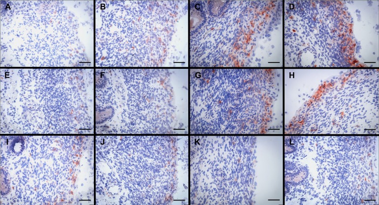
We were only able to conduct immunohistochemistry for TNF, IL12, and CSF2. Because cytokines are secreted proteins and are often dispersed in the tissue section, it was not possible to do a precise measurement of the area of positive cells. Therefore, cytokine protein expression was evaluated visually and scored on a scale from 0 to 5.
Immunostaining for TNF, IL12, and CSF2 is depicted in Figure 7. Immunostaining for TNF revealed that the MHC-I heterozygous- and homozygous-incompatible treatment groups had increased production of this cytokine compared to the homozygous-compatible and control groups. The mean TNF scores for the control, MHC-I homozygous-compatible, MHC-I homozygous-incompatible, and MHC-I heterozygous-incompatible pregnancies were 1.2, 2.1, 2.8, and 3.8, respectively. IL12 was only increased in the heterozygous-incompatible group compared to the other groups. Mean scores for IL12 staining in the control, homozygous-compatible, homozygous-incompatible, and heterozygous-incompatible pregnancies were 1.2, 1.0, 2.1, and 3.6, respectively. Interestingly, CSF2 was slightly increased in control compared to the SCNT pregnancies, with mean scores of 1.6 in the control, 0.9 in the homozygous-compatible, 1.2 in the homozygous-incompatible, and 1.3 in the heterozygous-incompatible pregnancies.
FIG. 7.
Immunohistochemical labeling of cells for TNF (A–D), IL12 (E–H), and CSF2 (I–L) in the endometrium of cows at Day 35 ± 1 of pregnancy. A, E, and I) Pregnancies established by AI (control). B, F, and J) MHC-I homozygous-compatible SCNT pregnancies. C, G, and K) Homozygous-incompatible SCNT pregnancies. D, H, and L) Heterozygous-incompatible SCNT pregnancies. Bar = 50 μm.
DISCUSSION
In cattle, trophoblast MHC-I expression is shut down during the first trimester of the pregnancy to avoid maternal immune recognition. As shown here and by others [15], trophoblast cells from SCNT conceptuses express significant amounts of MHC-I proteins during the first trimester of pregnancy compared with control pregnancies established by AI.
As a consequence, a pronounced endometrial lymphocyte infiltration composed mainly of CD3+ T lymphocytes was observed. The vast majority of both the dispersed lymphocytes and the lymphocytes in lymphoid nodules were CD4+ T helper (Th) cells (Figs. 3 and 4). Furthermore, because the lymphoid nodules were excluded when the leukocyte populations were quantified, the relative number of CD3 and CD4 lymphocytes shown in Figure 2 for the MHC-I-incompatible pregnancies, which tended to have more lymphoid nodules, are low estimates. The accumulation of CD4+ lymphocytes in the endometrium of MHC-I-incompatible SCNT pregnancies suggests that MHC-I proteins are processed and presented to the maternal immune system indirectly by MHC-II+ endometrial antigen-presenting cells. The indirect recognition of MHC-I proteins is also supported by the presence of significant numbers of MHC-II+ cells in the superficial stroma of the endometrium. It is likely that delivery of MHC-I proteins to the maternal immune system happens through the migration and fusion of MHC-I+ binucleate trophoblast cells with the maternal endometrial epithelium and eventual death of hybrid trinucleate cells [18–21].
Placental abnormalities have been suggested to be the main cause for the increased fetal demise of SCNT-produced calves [4, 9, 22, 23]. SCNT embryos and fetuses themselves present few macroscopic and histological abnormalities [23]. In the present study, there was complete correlation between the presence of grossly visible placental vasculature and embryonic mortality (Table 2 and Supplemental Fig. S1). It is likely that lack of visible blood vessels was due to embryonic circulatory failure and an absence of red blood cells in the placental vasculature. Lack of grossly visible placental blood vessels is not evidence for failure of placental vasculogenesis. Placental vascular development can only be assessed histologically, which was not done. Embryonic mortality between Days 28 and 35 of gestation was dramatically increased in the MHC-I heterozygous-incompatible group compared with other pregnancies. The fact that all of the embryos in this group were dead at the time of tissue collection is a potential confounding factor. Nevertheless, our data suggest that lymphocyte infiltration was the cause and not the consequence of fetal demise. The evidence for this conclusion is that the number of endometrial lymphocytes was not increased in the control pregnancy that had a dead fetus. This is also in accord with a previous study in which the only dead fetus observed in the control group did not trigger an increased endometrial lymphocytic infiltration [15]. Furthermore, the lack of a difference in fetal weight and crown-to-rump length suggests that SCNT and AI embryos were developmentally comparable and that embryonic death occurred immediately prior to sample collection.
Recent evidence indicates that T regulatory cells play an essential role in tolerance of the fetal allograft in mice and humans. In mice, the absence of T regulatory cells leads to implantation failure, due to a maternal immune response against fetal alloantigens [24, 25]. Expression of the transcription factor, FOXP3, is critical for the development and function of T regulatory cells. Deficient expression of FOXP3 is characterized by a lethal lymphoproliferative autoimmune disease in mice [26] and humans [27]. In the present study, we observed an increase in cells expressing FOXP3 in the heterozygous-incompatible group, which followed the same trend as the number of CD3+, CD4+, and CD8+ T cells present in the endometrium. This suggests that T regulatory cells respond to the same chemotactic signals as other T lymphocytes in the predominantly proinflammatory endometrial environment of MHC-I heterozygous-incompatible pregnancies. An alternative explanation is that FOXP3+ cells were specifically recruited to the endometrium as a way to control T cell responses in an attempt to prevent immune-mediated rejection of the conceptus.
As with the other T lymphocyte populations, γ/δTCR+ T cell infiltration was minimal in control pregnancies. Interestingly, the pattern of endometrial infiltration of these cells in the SCNT pregnancies was opposite to that of the other immune cell populations. Although the difference was not statistically significant, γ/δTCR+ T cells were more abundant in the endometrium of cows carrying MHC-I homozygous-compatible pregnancies than in the uterus of cows carrying MHC-I heterozygous- and homozygous-incompatible conceptuses. The role of γ/δTCR+ cells during pregnancy is still unclear. However, Hoek et al. [28] demonstrated that circulating bovine γ/δTCR+ cells could function as regulatory cells in culture systems. While in humans and rodents only 1%–10% of T lymphocytes are γ/δTCR+, in ruminants the percent of lymphocytes that are γ/δTCR+ can be quite large (20%–40%) [29]. A recent study showed that γ/δTCR+ cells are significantly increased both in peripheral blood and in the decidua of pregnant compared with nonpregnant women [30], and that these cells are highly biased toward IL10 and TGFB1 (previously known as TGF-β) production [30, 31]. Fan et al. [30] also found that these cells promoted trophoblast proliferation and invasion, and decreased trophoblast apoptosis through IL10 secretion. Gamma/deltaTCR+ cells accumulate in the endometrial epithelium of pregnant sheep as a result of unknown systemic signals [32, 33]. These cells may contribute to the growth of the conceptus and immune tolerance to fetal antigens. The increased mortality rate of the MHC-I heterozygous-incompatible embryos between 28 and 35 days of gestation may be compounded by the lower number of regulatory γ/δTCR+ cells in the endometrium providing a less-favorable immunological environment for embryonic survival.
Macrophages seem to play an essential role during early pregnancy, and the population of macrophages is expanded in the endometrium during pregnancy as early as Day 13 of gestation [34]. During attachment, apoptosis of endometrial epithelial cells and syncytial cells, hybrid cells resulting from fusion of endometrial epithelial cells and binucleate trophoblast cells, is critical for tissue remodeling and trophoblast invasion. The effective removal of apoptotic cells and cell debris by endometrial macrophages is a fundamental process during attachment because it promotes adequate tissue remodeling and prevents the release of paternal antigens. There was considerable variation in the number of CD68+ endometrial macrophages in all of the experimental groups, so the difference between the groups was not statistically significant (Fig. 6). Nevertheless, the number of endometrial macrophages in the MHC-I-incompatible pregnancies was increased in comparison to the control group. One explanation for the increased number of macrophages in the MHC-I heterozygous pregnancies is that these macrophages were attracted to the endometrium in response to the death of the conceptus. However, this explanation seems unlikely, since there were just as many macrophages in the MHC-I homozygous-incompatible pregnancies that had viable embryos. Another explanation for the increased numbers of endometrial macrophages in the MHC-I homozygous- and heterozygous-incompatible pregnancies is that endometrial macrophages were attracted or proliferated in conjunction with the increase in the number of CD4+ T lymphocytes (Fig. 2). This makes sense, since the primary role of CD4+ Th1 cells is to activate macrophages.
The signals that initiate the migration and differentiation of monocytes into macrophages in the pregnant endometrium still remain to be elucidated. Macrophages have a very high degree of plasticity, and differentiate according to their microenvironment. Macrophages are classified into two phenotypes: M1 and M2. The M1 phenotype promotes inflammation and cytotoxicity, while M2 macrophages are involved in immunosuppression and angiogenesis [35, 36]. In cattle pregnancies, at least a portion of the endometrial macrophages are M2 macrophages, which are involved in angiogenesis, immunoregulation, tissue remodeling, and apoptosis [37]. Trophoblast cells from healthy pregnancies seem to play a major role in stimulating endometrial macrophages to acquire an immunosuppressive, M2 phenotype. Exposure of macrophages to in vitro-cultured trophoblast debris induces the expression of immunosuppressive factors, such as IL10, IL6, IL1 receptor antagonist (IL1RN), and indoleamine 2,3-dioxygenase 1, and decreases the secretion of IL1B (previously known as IL1β), IL12 (previously known as IL12p70), and CXCL8 (previously known as IL8) [38]. Additionally, endometrial macrophages have been shown to secrete high levels of vascular endothelial growth factor A (previously known as VEGF), which suggests that they are involved with vascular remodeling [39].
We found that placental macrophages were increased in the MHC-I heterozygous-incompatible pregnancies compared to the control and other SCNT pregnancies. Since all of the MHC-I heterozygous embryos were dead at the time of tissue collection, the macrophages present in these placentae were probably maternal macrophages that were responding to the death of the embryo by invading the conceptus to phagocytize cellular debris.
The number of MHC-II+ cells was increased in the SCNT pregnancies compared with the control pregnancies. It would be reasonable to conclude that the majority of these MHC-II+ cells are macrophages; however, Oliveira and Hansen [40] have demonstrated that, in the interplacentomal endometrium of normal pregnancies, the vast majority of the MHC-II+ cells adjacent to the luminal epithelium do not express markers specific for macrophages, such as CD68 or CD14. Therefore, it is likely that a significant portion of the MHC-II+ cells that we observed in the superficial stroma were dendritic cells rather than macrophages. The CD68+ cells that were present could be mainly of the M2 phenotype, responsible for angiogenesis and immunoregulation, but not antigen presentation. While it is unclear if endometrial M2 macrophages are MHC-II negative, tumor-associated M2 macrophages have been shown to have significantly decreased MHC-II expression compared to M1 macrophages [41]. The MHC-II+ cell population adjacent to the endometrial glands was also mostly negative for CD68 and CD14, but was CR2+. Other data collected by our group suggest that these cells are likely dendritic cells (C.R. Thacker, H.M. Rutigliano, and C.J. Davies, unpublished results).
Natural killer (NK) cells recognize self-MHC-I proteins using receptors that deliver inhibitory signals that prevent destruction of the target cell. When there is absence or down-regulation of MHC-I, or the expression of nonself-MHC-I proteins, these inhibitory signals are not delivered and the target cell is killed either by the release of cytotoxic granzymes [42] or by induction of apoptosis [43]. In this study, the presence of increased numbers of NK cells in the MHC-I heterozygous-incompatible group suggests that these cells were recruited as a result of the proinflammatory environment in the endometrium. It is possible that NK cells interact with trophoblast cells that express allogeneic MHC-I proteins and induce their apoptosis.
Although at a lower magnitude, the endometrial lymphocyte infiltration in the contralateral uterine horn to the pregnancy followed the same trend as in the ipsilateral horn (Supplemental Fig. S2). This suggests the involvement of both local and systemic signals in lymphocyte accumulation. Majewski et al. [33], using a model where ovine pregnancies were confined to only one uterine horn, demonstrated that factors of maternal or conceptus origin act systemically, rather than locally, to regulate endometrial lymphocyte populations during pregnancy.
Unfortunately, we were not able to conduct immunohistochemistry for many of the cytokines in which we were interested, but the IL12 and TNF cytokine expression data presented here are in agreement with our lymphocyte population data, which suggests that there is a pronounced T cell and macrophage response mainly in the MHC-I heterozygous-incompatible pregnancies. IL12 is produced mainly by macrophages and dendritic cells. Its primary function is to stimulate the differentiation of naïve T cells into Th1 cells, which produce IFNG (previously known as IFNγ) and TNF. TNF is primarily secreted by M1 macrophages, but is also produced by CD4+ lymphocytes and NK cells; it is responsible for a variety of functions, including local inflammation, endothelial activation, apoptosis, and T and B cell proliferation. Our gene expression data, which will be presented in a subsequent aricle (H.M. Rutigliano et al., unpublished data), are consistent with what we have shown here, and demonstrates that, in the endometrium of MHC-I heterozygous-incompatible pregnancies, Th1 proinflammatory cytokines, such as IFNG, TNF, and IL12, are upregulated.
We have observed that MHC-I heterozygous SCNT conceptuses trigger an exacerbated endometrial immune response, which probably leads to fetal death. Davies et al. [13] showed that MHC-I homozygous conceptuses produced by SCNT had considerably higher survival rates from Pregnancy Days 28 to 90, 28 to 200, and 28 to term compared with MHC-I heterozygous conceptuses. The exacerbated immune response and decreased embryonic survival in the MHC-I heterozygous-incompatible pregnancies can be explained by the fact that trophoblast cells from these pregnancies express two MHC-I haplotypes that are antigenically different from the maternal haplotypes, while the homozygous-incompatible pregnancies express only one unmatched haplotype. The increased antigenicity of the MHC-I heterozygous conceptuses triggers an augmented immune response against fetal antigens in the endometrium, which is accompanied by increased levels of IFNG. It is known that IFNG increases MHC-I expression in somatic cells [44]. Therefore, a feedback loop may be formed with antigenic MHC-I proteins expressed by trophoblast cells of MHC-I-incompatible SCNT conceptuses promoting release of IFNG, which, in turn, promotes further expression of MHC-I by trophoblast cells.
To our knowledge, this is the first study to determine embryonic survival and uterine immune responses in MHC-I-compatible and -incompatible SCNT pregnancies. We have demonstrated that MHC-I compatibility between the fetus and the dam influences the maternal immune response to the conceptus. MHC-I-matched or -compatible SCNT pregnancies have lower endometrial lymphocytic infiltration and better embryonic survival rates than MHC-I-incompatible pregnancies.
Supplementary Material
ACKNOWLEDGMENT
We thank Dr. Qinggang Meng for skilled micromanipulation of embryos, and Dick Whittier for assisting with humane tissue collection. The authors are grateful to Dr. Koji Yoshinaga and the members of the National Institute of Child Health and Human Development Interdisciplinary Collaborative Team for Blastocyst Implantation Research for constructive discussion about this study during our team meetings.
Footnotes
This project was funded by the National Institute of Child Health and Human Development grant no. 1R01HD055502 from the National Institutes of Health to C.J.D. and K.L.W., and was also supported by the Utah Agricultural Experiment Station, Utah State University, and approved as journal paper number 8843. Presented in part at the 31st American Society for Reproductive Immunology Meeting, 19–22 May 2011, Salt Lake City, Utah.
REFERENCES
- Wilmut I, Schnieke AE, McWhir J, Kind AJ, Campbell KH. Viable offspring derived from fetal and adult mammalian cells. Nature. 1997;385:810–813. doi: 10.1038/385810a0. [DOI] [PubMed] [Google Scholar]
- Cibelli JB, Stice SL, Golueke PJ, Kane JJ, Jerry J, Blackwell C, Ponce de León FA, Robl JM. Cloned transgenic calves produced from nonquiescent fetal fibroblasts. Science. 1998;280:1256–1258. doi: 10.1126/science.280.5367.1256. [DOI] [PubMed] [Google Scholar]
- Wakayama T, Perry AC, Zuccotti M, Johnson KR, Yanagimachi R. Full-term development of mice from enucleated oocytes injected with cumulus cell nuclei. Nature. 1998;394:369–374. doi: 10.1038/28615. [DOI] [PubMed] [Google Scholar]
- Wells DN, Misica PM, Tervit HR. Production of cloned calves following nuclear transfer with cultured adult mural granulosa cells. Biol Reprod. 1999;60:996–1005. doi: 10.1095/biolreprod60.4.996. [DOI] [PubMed] [Google Scholar]
- Farin PW, Piedrahita JA, Farin CE. Errors in development of fetuses and placentas from in vitro-produced bovine embryos. Theriogenology. 2006;65:178–191. doi: 10.1016/j.theriogenology.2005.09.022. [DOI] [PubMed] [Google Scholar]
- Lonergan P, Khatir H, Piumi F, Rieger D, Humblot P, Boland MP. Effect of time interval from insemination to first cleavage on the developmental characteristics, sex ratio and pregnancy rate after transfer of bovine embryos. J Reprod Fertil. 1999;117:159–167. doi: 10.1530/jrf.0.1170159. [DOI] [PubMed] [Google Scholar]
- Hasler JF. In vitro culture of bovine embryos in Ménézo's B2 medium with or without coculture and serum: the normalcy of pregnancies and calves resulting from transferred embryos. Anim Reprod Sci. 2000;60–61:81–91. doi: 10.1016/s0378-4320(00)00086-5. [DOI] [PubMed] [Google Scholar]
- Edwards JL, Schrick FN, McCracken MD, Van Amstel SR, Hopkins FM, Welborn MG, Davies CJ. Cloning adult farm animals: a review of the possibilities and problems associated with somatic cell nuclear transfer. Am J Reprod Immunol. 2003;50:113–123. doi: 10.1034/j.1600-0897.2003.00064.x. [DOI] [PubMed] [Google Scholar]
- Hill JR, Burghardt RC, Jones K, Long CR, Looney CR, Shin T, Spencer TE, Thompson JA, Winger QA, Westhusin ME. Evidence for placental abnormality as the major cause of mortality in first-trimester somatic cell cloned bovine fetuses. Biol Reprod. 2000;63:1787–1794. doi: 10.1095/biolreprod63.6.1787. [DOI] [PubMed] [Google Scholar]
- Davies CJ, Fisher PJ, Schlafer DH. Temporal and regional regulation of major histocompatibility complex class I expression at the bovine uterine/placental interface. Placenta. 2000;21:194–202. doi: 10.1053/plac.1999.0475. [DOI] [PubMed] [Google Scholar]
- Davies CJ, Eldridge JA, Fisher PJ, Schlafer DH. Evidence for expression of both classical and non-classical major histocompatibility complex class I genes in bovine trophoblast cells. Am J Reprod Immunol. 2006;55:188–200. doi: 10.1111/j.1600-0897.2005.00364.x. [DOI] [PubMed] [Google Scholar]
- Joosten I, Sanders MF, Hensen EJ. Involvement of major histocompatibility complex class I compatibility between dam and calf in the aetiology of bovine retained placenta. Anim Genet. 1991;22:455–463. doi: 10.1111/j.1365-2052.1991.tb00717.x. [DOI] [PubMed] [Google Scholar]
- Davies CJ, Hill JR, Edwards JL, Schrick FN, Fisher PJ, Eldridge JA, Schlafer DH. Major histocompatibility antigen expression on the bovine placenta: its relationship to abnormal pregnancies and retained placenta. Anim Reprod Sci. 2004;82–83:267–280. doi: 10.1016/j.anireprosci.2004.05.016. [DOI] [PubMed] [Google Scholar]
- Benedictus L, Thomas AJ, Jorritsma R, Davies CJ, Koets AP. Two-way calf to dam major histocompatibility class I compatibility increases risk for retained placenta in cattle. Am J Reprod Immunol. 2012;67:224–230. doi: 10.1111/j.1600-0897.2011.01085.x. [DOI] [PubMed] [Google Scholar]
- Hill JR, Schlafer DH, Fisher PJ, Davies CJ. Abnormal expression of trophoblast major histocompatibility complex class I antigens in cloned bovine pregnancies is associated with a pronounced endometrial lymphocytic response. Biol Reprod. 2002;67:55–63. doi: 10.1095/biolreprod67.1.55. [DOI] [PubMed] [Google Scholar]
- Banham AH, Lyne L, Scase TJ, Blacklaws BA. Monoclonal antibodies raised to the human FOXP3 protein can be used effectively for detecting Foxp3+ T cells in other mammalian species. Vet Immunol Immunopathol. 2009;127:376–381. doi: 10.1016/j.vetimm.2008.10.328. [DOI] [PubMed] [Google Scholar]
- Aston KI, Li G-P, Hicks BA, Sessions BR, Pate BJ, Hammon DS, Bunch TD, White KL. The developmental competence of bovine nuclear transfer embryos derived from cow versus heifer cytoplasts. Anim Reprod Sci. 2006;95:234–243. doi: 10.1016/j.anireprosci.2005.10.011. [DOI] [PubMed] [Google Scholar]
- Wathes DC, Wooding FB. An electron microscopic study of implantation in the cow. Am J Anat. 1980;159:285–306. doi: 10.1002/aja.1001590305. [DOI] [PubMed] [Google Scholar]
- Wooding FB, Wathes DC. Binucleate cell migration in the bovine placentome. J Reprod Fertil. 1980;59:425–430. doi: 10.1530/jrf.0.0590425. [DOI] [PubMed] [Google Scholar]
- Wooding FB. The role of the binucleate cell in ruminant placental structure. J Reprod Fertil Suppl. 1982;31:31–39. [PubMed] [Google Scholar]
- Wooding FB. Current topic: the synepitheliochorial placenta of ruminants: binucleate cell fusions and hormone production. Placenta. 1992;13:101–113. doi: 10.1016/0143-4004(92)90025-o. [DOI] [PubMed] [Google Scholar]
- Stice SL, Strelchenko NS, Keefer CL, Matthews L. Pluripotent bovine embryonic cell lines direct embryonic development following nuclear transfer. Biol Reprod. 1996;54:100–110. doi: 10.1095/biolreprod54.1.100. [DOI] [PubMed] [Google Scholar]
- Hill JR, Roussel AJ, Cibelli JB, Edwards JF, Hooper NL, Miller MW, Thompson JA, Looney CR, Westhusin ME, Robl JM, Stice SL. Clinical and pathologic features of cloned transgenic calves and fetuses (13 case studies) Theriogenology. 1999;51:1451–1465. doi: 10.1016/s0093-691x(99)00089-8. [DOI] [PubMed] [Google Scholar]
- Aluvihare VR, Kallikourdis M, Betz AG. Regulatory T cells mediate maternal tolerance to the fetus. Nat Immunol. 2004;5:266–271. doi: 10.1038/ni1037. [DOI] [PubMed] [Google Scholar]
- Darrasse-Jèze G, Darasse-Jèze G, Klatzmann D, Charlotte F, Salomon BL, Cohen JL. CD4+CD25+ regulatory/suppressor T cells prevent allogeneic fetus rejection in mice. Immunol Lett. 2006;102:106–109. doi: 10.1016/j.imlet.2005.07.002. [DOI] [PubMed] [Google Scholar]
- Brunkow ME, Jeffery EW, Hjerrild KA, Paeper B, Clark LB, Yasayko SA, Wilkinson JE, Galas D, Ziegler SF, Ramsdell F. Disruption of a new forkhead/winged-helix protein, scurfin, results in the fatal lymphoproliferative disorder of the scurfy mouse. Nat Genet. 2001;27:68–73. doi: 10.1038/83784. [DOI] [PubMed] [Google Scholar]
- Bennett CL, Christie J, Ramsdell F, Brunkow ME, Ferguson PJ, Whitesell L, Kelly TE, Saulsbury FT, Chance PF, Ochs HD. The immune dysregulation, polyendocrinopathy, enteropathy, X-linked syndrome (IPEX) is caused by mutations of FOXP3. Nat Genet. 2001;27:20–21. doi: 10.1038/83713. [DOI] [PubMed] [Google Scholar]
- Hoek A, Rutten VP, Kool J, Arkesteijn GJ, Bouwstra RJ, Van Rhijn I, Koets AP. Subpopulations of bovine WC1(+) gammadelta T cells rather than CD4(+)CD25(high) Foxp3(+) T cells act as immune regulatory cells ex vivo Vet Res 2009. 40:06, 1–14. [DOI] [PMC free article] [PubMed] [Google Scholar]
- Mackay CR, Hein WR. A large proportion of bovine T cells express the gamma delta T cell receptor and show a distinct tissue distribution and surface phenotype. Int Immunol. 1989;1:540–545. doi: 10.1093/intimm/1.5.540. [DOI] [PubMed] [Google Scholar]
- Fan D-X, Duan J, Li M-Q, Xu B, Li D-J, Jin L-P. The decidual gamma-delta T cells up-regulate the biological functions of trophoblasts via IL-10 secretion in early human pregnancy. Clin Immunol. 2011;141:284–292. doi: 10.1016/j.clim.2011.07.008. [DOI] [PubMed] [Google Scholar]
- Nagaeva O, Jonsson L, Mincheva-Nilsson L. Dominant IL-10 and TGF-beta mRNA expression in gammadeltaT cells of human early pregnancy decidua suggests immunoregulatory potential. Am J Reprod Immunol. 2002;48:9–17. doi: 10.1034/j.1600-0897.2002.01131.x. [DOI] [PubMed] [Google Scholar]
- Lee CS, Meeusen E, Gogolin-Ewens K, Brandon MR. Quantitative and qualitative changes in the intraepithelial lymphocyte population in the uterus of nonpregnant and pregnant sheep. Am J Reprod Immunol. 1992;28:90–96. doi: 10.1111/j.1600-0897.1992.tb00766.x. [DOI] [PubMed] [Google Scholar]
- Majewski AC, Tekin S, Hansen PJ. Local versus systemic control of numbers of endometrial T cells during pregnancy in sheep. Immunology. 2001;102:317–322. doi: 10.1046/j.1365-2567.2001.01182.x. [DOI] [PMC free article] [PubMed] [Google Scholar]
- Mansouri-Attia N, Oliveira LJ, Forde N, Fahey AG, Browne JA, Roche JF, Sandra O, Reinaud P, Lonergan P, Fair T. Pivotal role for monocytes/macrophages and dendritic cells in maternal immune response to the developing embryo in cattle. Biol Reprod. 2012;87:123. doi: 10.1095/biolreprod.112.101121. [DOI] [PubMed] [Google Scholar]
- Mantovani A, Sozzani S, Locati M, Allavena P, Sica A. Macrophage polarization: tumor-associated macrophages as a paradigm for polarized M2 mononuclear phagocytes. Trends Immunol. 2002;23:549–555. doi: 10.1016/s1471-4906(02)02302-5. [DOI] [PubMed] [Google Scholar]
- Gordon S. Alternative activation of macrophages. Nat Rev Immunol. 2003;3:23–35. doi: 10.1038/nri978. [DOI] [PubMed] [Google Scholar]
- Oliveira LJ, McClellan S, Hansen PJ. Differentiation of the endometrial macrophage during pregnancy in the cow. PLoS One. 2010;5:e13213. doi: 10.1371/journal.pone.0013213. [DOI] [PMC free article] [PubMed] [Google Scholar]
- Abumaree MH, Chamley LW, Badri M, El-Muzaini MF. Trophoblast debris modulates the expression of immune proteins in macrophages: a key to maternal tolerance of the fetal allograft? J Reprod Immunol. 2012;94:131–141. doi: 10.1016/j.jri.2012.03.488. [DOI] [PubMed] [Google Scholar]
- Li C, Houser BL, Nicotra ML, Strominger JL. HLA-G homodimer-induced cytokine secretion through HLA-G receptors on human decidual macrophages and natural killer cells. Proc Natl Acad Sci U S A. 2009;106:5767–5772. doi: 10.1073/pnas.0901173106. [DOI] [PMC free article] [PubMed] [Google Scholar]
- Oliveira LJ, Hansen PJ. Phenotypic characterization of macrophages in the endometrium of the pregnant cow. Am J Reprod Immunol. 2009;62:418–426. doi: 10.1111/j.1600-0897.2009.00761.x. [DOI] [PubMed] [Google Scholar]
- Movahedi K, Laoui D, Gysemans C, Baeten M, Stangé G, Van den Bossche J, Mack M, Pipeleers D. In't Veld P, De Baetselier P, Van Ginderachter JA. Different tumor microenvironments contain functionally distinct subsets of macrophages derived from Ly6C(high) monocytes. Cancer Res. 2010;70:5728–5739. doi: 10.1158/0008-5472.CAN-09-4672. [DOI] [PubMed] [Google Scholar]
- Bryceson YT, March ME, Ljunggren H-G, Long EO. Synergy among receptors on resting NK cells for the activation of natural cytotoxicity and cytokine secretion. Blood. 2006;107:159–166. doi: 10.1182/blood-2005-04-1351. [DOI] [PMC free article] [PubMed] [Google Scholar]
- Screpanti V, Wallin RPA, Grandien A, Ljunggren H-G. Impact of FASL-induced apoptosis in the elimination of tumor cells by NK cells. Mol Immunol. 2005;42:495–499. doi: 10.1016/j.molimm.2004.07.033. [DOI] [PubMed] [Google Scholar]
- Rock KL, York IA, Saric T, Goldberg AL. Protein degradation and the generation of MHC class I-presented peptides. Adv Immunol. 2002;80:1–70. doi: 10.1016/s0065-2776(02)80012-8. [DOI] [PubMed] [Google Scholar]
Associated Data
This section collects any data citations, data availability statements, or supplementary materials included in this article.


