

Abstract
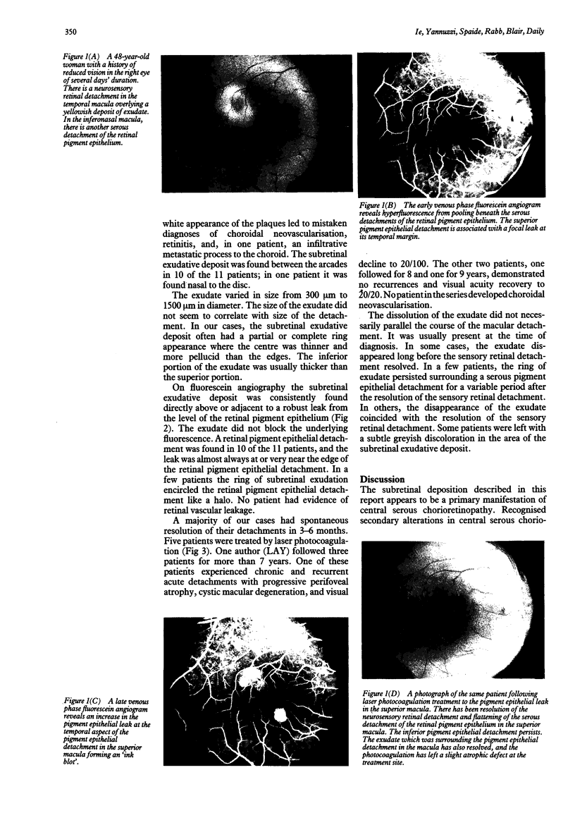
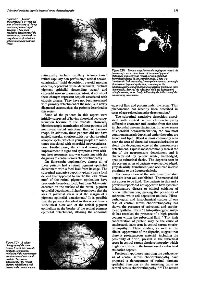
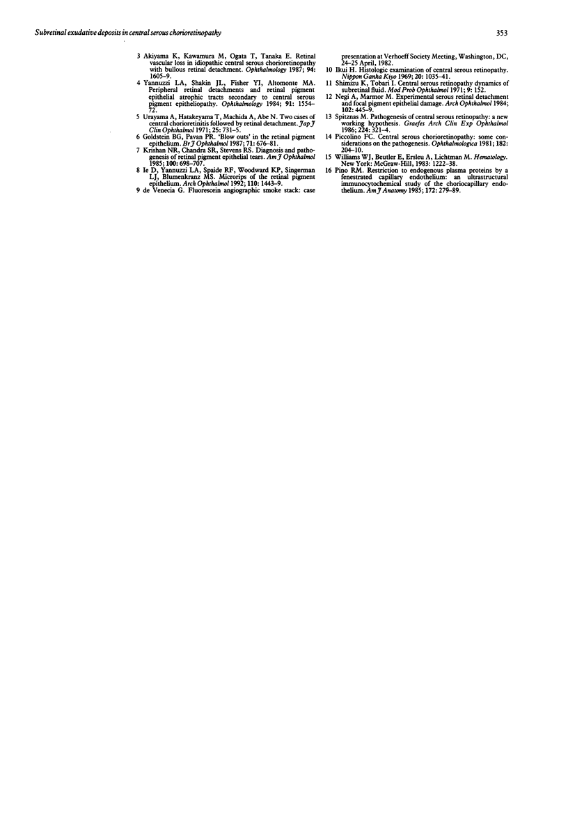
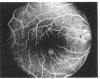
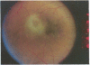
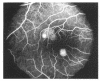
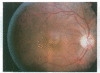
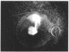
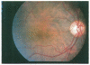
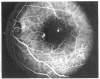
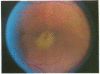
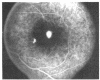
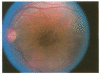
The presence of subretinal exudation in a patient with neurosensory detachment of the macula frequently suggests the diagnosis of choroidal neovascularisation. A retrospective chart review of newly diagnosed cases of central serous chorioretinopathy revealed 11 patients, seven men and four non-pregnant women, who had plaques of subretinal exudate, which presumably were fibrin. Each of these patients had a solitary plaque that ranged in size from 300 to 1500 microns in diameter. These patients had no signs or a clinical course suggestive of choroidal neovascularisation. In each case the subretinal plaque was overlying an exuberant leak in the retinal pigment epithelium. The exudate was generally present at the initial examination, and usually showed dissolution before or coincident with the resolution of the neurosensory detachment. After resolution of the central serous chorioretinopathy, patients were left with subtle alterations in the retinal pigment epithelium in the areas of the subretinal plaque. These findings are important for two reasons. Firstly, the presence of subretinal exudation does not necessarily rule out the diagnosis of central serous chorioretinopathy. Secondly, pathophysiological theories of central serous chorioretinopathy must explain how the plaques are deposited behind the retina.
Full text
PDF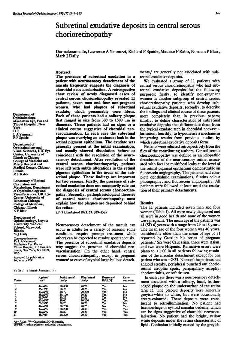
Images in this article
Selected References
These references are in PubMed. This may not be the complete list of references from this article.
- Akiyama K., Kawamura M., Ogata T., Tanaka E. Retinal vascular loss in idiopathic central serous chorioretinopathy with bullous retinal detachment. Ophthalmology. 1987 Dec;94(12):1605–1609. doi: 10.1016/s0161-6420(87)33243-9. [DOI] [PubMed] [Google Scholar]
- Gass J. D. Bullous retinal detachment. An unusual manifestation of idiopathic central serous choroidopathy. Am J Ophthalmol. 1973 May;75(5):810–821. doi: 10.1016/0002-9394(73)90887-8. [DOI] [PubMed] [Google Scholar]
- Gass J. D. Central serous chorioretinopathy and white subretinal exudation during pregnancy. Arch Ophthalmol. 1991 May;109(5):677–681. doi: 10.1001/archopht.1991.01080050091036. [DOI] [PubMed] [Google Scholar]
- Goldstein B. G., Pavan P. R. 'Blow-outs' in the retinal pigment epithelium. Br J Ophthalmol. 1987 Sep;71(9):676–681. doi: 10.1136/bjo.71.9.676. [DOI] [PMC free article] [PubMed] [Google Scholar]
- Ie D., Yannuzzi L. A., Spaide R. F., Woodward K. P., Singerman L. J., Blumenkranz M. S. Microrips of the retinal pigment epithelium. Arch Ophthalmol. 1992 Oct;110(10):1443–1449. doi: 10.1001/archopht.1992.01080220105030. [DOI] [PubMed] [Google Scholar]
- Ikui H. [Histological examination of central serous retinopathy]. Nihon Ganka Kiyo. 1969 Nov;20(11):1035–1043. [PubMed] [Google Scholar]
- Krishan N. R., Chandra S. R., Stevens T. S. Diagnosis and pathogenesis of retinal pigment epithelial tears. Am J Ophthalmol. 1985 Nov 15;100(5):698–707. doi: 10.1016/0002-9394(85)90626-9. [DOI] [PubMed] [Google Scholar]
- Negi A., Marmor M. F. Experimental serous retinal detachment and focal pigment epithelial damage. Arch Ophthalmol. 1984 Mar;102(3):445–449. doi: 10.1001/archopht.1984.01040030359038. [DOI] [PubMed] [Google Scholar]
- Piccolino F. C. Central serous chorioretinopathy: some considerations on the pathogenesis. Ophthalmologica. 1981;182(4):204–210. doi: 10.1159/000309115. [DOI] [PubMed] [Google Scholar]
- Spitznas M. Pathogenesis of central serous retinopathy: a new working hypothesis. Graefes Arch Clin Exp Ophthalmol. 1986;224(4):321–324. doi: 10.1007/BF02150023. [DOI] [PubMed] [Google Scholar]
- Yannuzzi L. A., Shakin J. L., Fisher Y. L., Altomonte M. A. Peripheral retinal detachments and retinal pigment epithelial atrophic tracts secondary to central serous pigment epitheliopathy. Ophthalmology. 1984 Dec;91(12):1554–1572. doi: 10.1016/s0161-6420(84)34117-3. [DOI] [PubMed] [Google Scholar]