Abstract
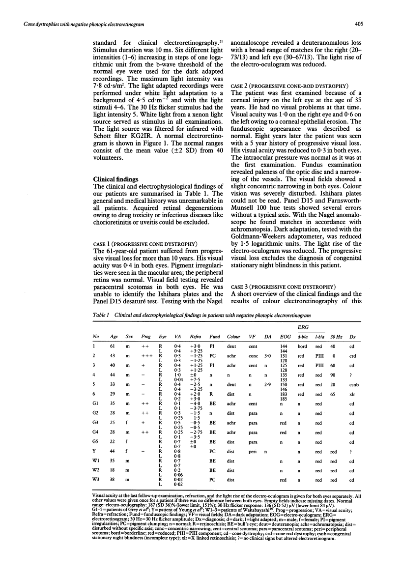
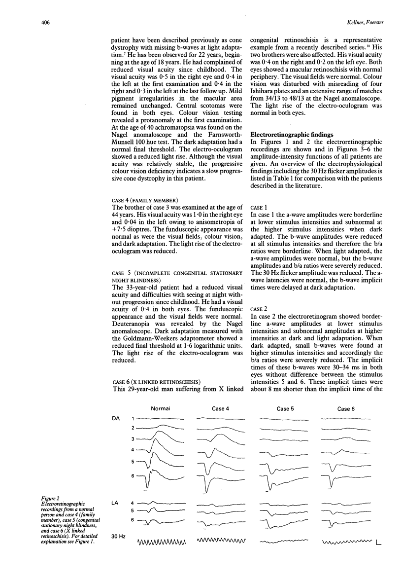
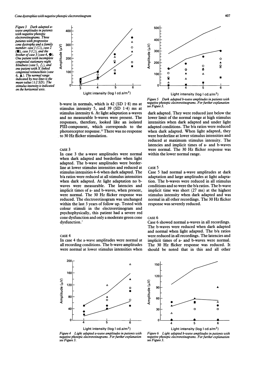
A scotopic electroretinogram with an a-wave amplitude larger than the b-wave amplitude traditionally is termed 'negative'. Six male patients with negative photopic electroretinograms were examined; three of them suffered from progressive cone dystrophy, in which negative electroretinograms are unusual. Another patient without symptoms was the brother of a patient with cone dystrophy. These patients are compared with others who characteristically have negative electroretinograms-one patient with incomplete congenital stationary night blindness and another with X linked congenital retinoschisis. Differential diagnosis between these unusual cases of cone dystrophies and X linked retinoschisis or congenital stationary night blindness was possible with funduscopy, adaptometry, and evaluation of progression, but not with the electroretinogram. Inner retinal defects may occur in cone dystrophies as indicated by the negative electroretinogram. The waveform variations between our patients may be due to different inner retinal defects. The findings in two brothers indicate that cone dystrophy and inner retinal defects may be inherited separately.
Full text
PDF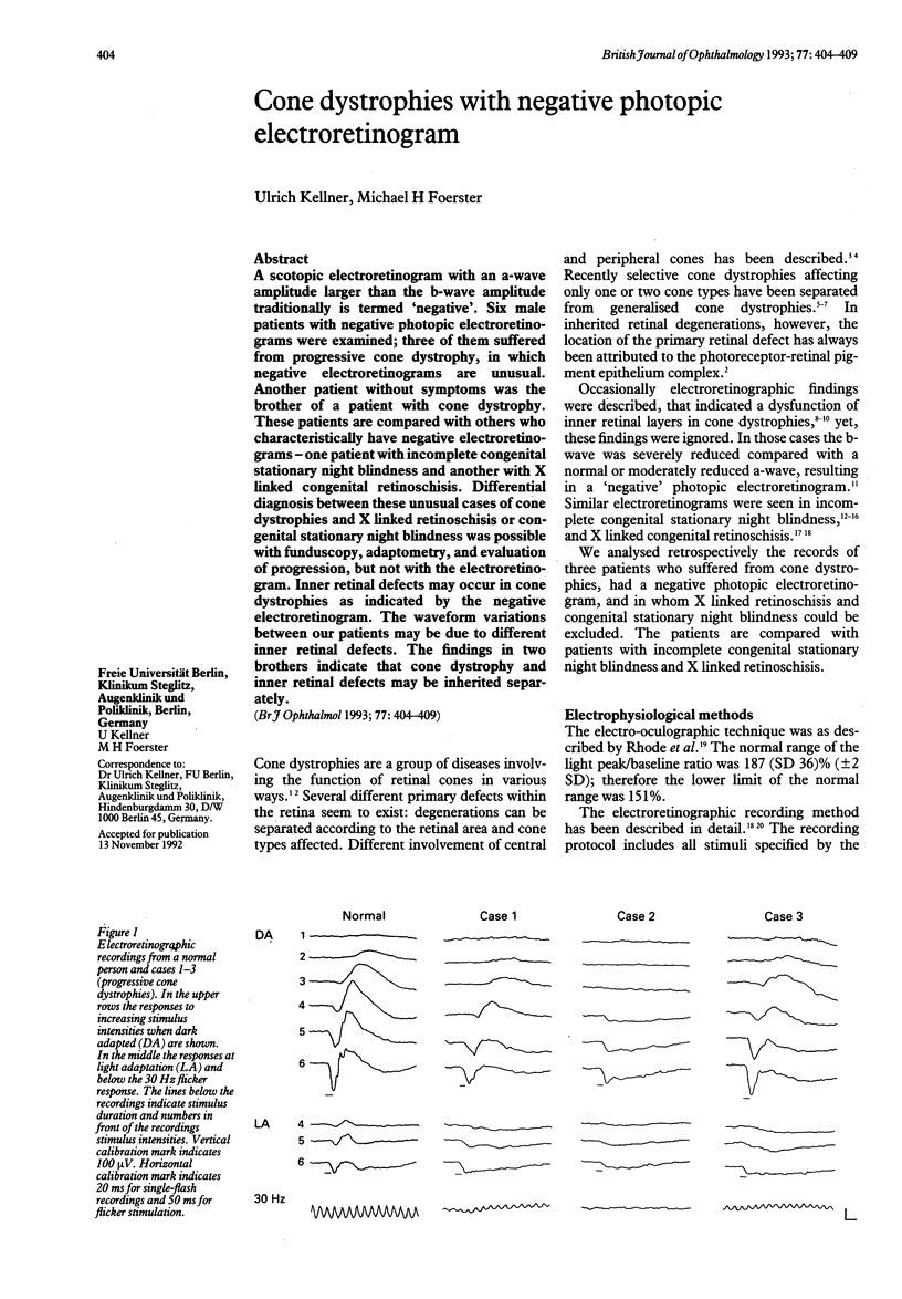
Selected References
These references are in PubMed. This may not be the complete list of references from this article.
- Bacon P., Spalton D. J., Smith S. E. Blindness from quinine toxicity. Br J Ophthalmol. 1988 Mar;72(3):219–224. doi: 10.1136/bjo.72.3.219. [DOI] [PMC free article] [PubMed] [Google Scholar]
- Condon G. P., Brownstein S., Wang N. S., Kearns J. A., Ewing C. C. Congenital hereditary (juvenile X-linked) retinoschisis. Histopathologic and ultrastructural findings in three eyes. Arch Ophthalmol. 1986 Apr;104(4):576–583. doi: 10.1001/archopht.1986.01050160132029. [DOI] [PubMed] [Google Scholar]
- Foerster M. H., Kellner U., Wessing A. Cone dystrophy and supernormal dark-adapted b-waves in the electroretinogram. Graefes Arch Clin Exp Ophthalmol. 1990;228(2):116–119. doi: 10.1007/BF00935718. [DOI] [PubMed] [Google Scholar]
- GOODMAN G., RIPPS H., SIEGEL I. M. CONE DYSFUNCTION SYNDROMES. Arch Ophthalmol. 1963 Aug;70:214–231. doi: 10.1001/archopht.1963.00960050216013. [DOI] [PubMed] [Google Scholar]
- Grey R. H., Blach R. K., Barnard W. M. Bull's eye maculopathy with early cone degeneration. Br J Ophthalmol. 1977 Nov;61(11):702–718. doi: 10.1136/bjo.61.11.702. [DOI] [PMC free article] [PubMed] [Google Scholar]
- Kellner U., Brümmer S., Foerster M. H., Wessing A. X-linked congenital retinoschisis. Graefes Arch Clin Exp Ophthalmol. 1990;228(5):432–437. doi: 10.1007/BF00927256. [DOI] [PubMed] [Google Scholar]
- Kellner U., Foerster M. H. Color electroretinography. A method for separation of dysfunctions of cones. Doc Ophthalmol. 1992;80(1):13–23. doi: 10.1007/BF00161227. [DOI] [PubMed] [Google Scholar]
- Kellner U., Foerster M. H. Netzhautdegeneration mit Blauzapfenhypersensitivität. Fortschr Ophthalmol. 1991;88(6):637–641. [PubMed] [Google Scholar]
- Miyake Y., Yagasaki K., Horiguchi M., Kawase Y., Kanda T. Congenital stationary night blindness with negative electroretinogram. A new classification. Arch Ophthalmol. 1986 Jul;104(7):1013–1020. doi: 10.1001/archopht.1986.01050190071042. [DOI] [PubMed] [Google Scholar]
- Pinckers A., Deutman A. F. Peripheral cone disease. Ophthalmologica. 1977;174(3):145–150. doi: 10.1159/000308592. [DOI] [PubMed] [Google Scholar]
- Reichel E., Bruce A. M., Sandberg M. A., Berson E. L. An electroretinographic and molecular genetic study of X-linked cone degeneration. Am J Ophthalmol. 1989 Nov 15;108(5):540–547. doi: 10.1016/0002-9394(89)90431-5. [DOI] [PubMed] [Google Scholar]
- Rohde N., Täumer R., Pernice D. Vorschlag eines verbesserten klinischen EOG-Testes. Ber Zusammenkunft Dtsch Ophthalmol Ges. 1977;74:747–750. [PubMed] [Google Scholar]
- SCHUBERT G., BORNSCHEIN H. Beitrag zur Analyse des menschlichen Elektroretinogramms. Ophthalmologica. 1952 Jun;123(6):396–413. doi: 10.1159/000301211. [DOI] [PubMed] [Google Scholar]
- Smith E. L., 3rd, Harwerth R. S., Crawford M. L., Duncan G. C. Contribution of the retinal ON channels to scotopic and photopic spectral sensitivity. Vis Neurosci. 1989 Sep;3(3):225–239. doi: 10.1017/s0952523800009986. [DOI] [PubMed] [Google Scholar]
- Standard for clinical electroretinography. International Standardization Committee. Arch Ophthalmol. 1989 Jun;107(6):816–819. doi: 10.1001/archopht.1989.01070010838024. [DOI] [PubMed] [Google Scholar]
- Weleber R. G., Pillers D. A., Powell B. R., Hanna C. E., Magenis R. E., Buist N. R. Aland Island eye disease (Forsius-Eriksson syndrome) associated with contiguous deletion syndrome at Xp21. Similarity to incomplete congenital stationary night blindness. Arch Ophthalmol. 1989 Aug;107(8):1170–1179. doi: 10.1001/archopht.1989.01070020236032. [DOI] [PubMed] [Google Scholar]
- Yagasaki K., Jacobson S. G. Cone-rod dystrophy. Phenotypic diversity by retinal function testing. Arch Ophthalmol. 1989 May;107(5):701–708. doi: 10.1001/archopht.1989.01070010719034. [DOI] [PubMed] [Google Scholar]
- Young R., Price J., Gorham N., Cowart M. Selective abnormality of the cone B-wave in a patient with retinal degeneration. Doc Ophthalmol. 1985 Aug 30;60(2):211–218. doi: 10.1007/BF00158037. [DOI] [PubMed] [Google Scholar]


