Abstract
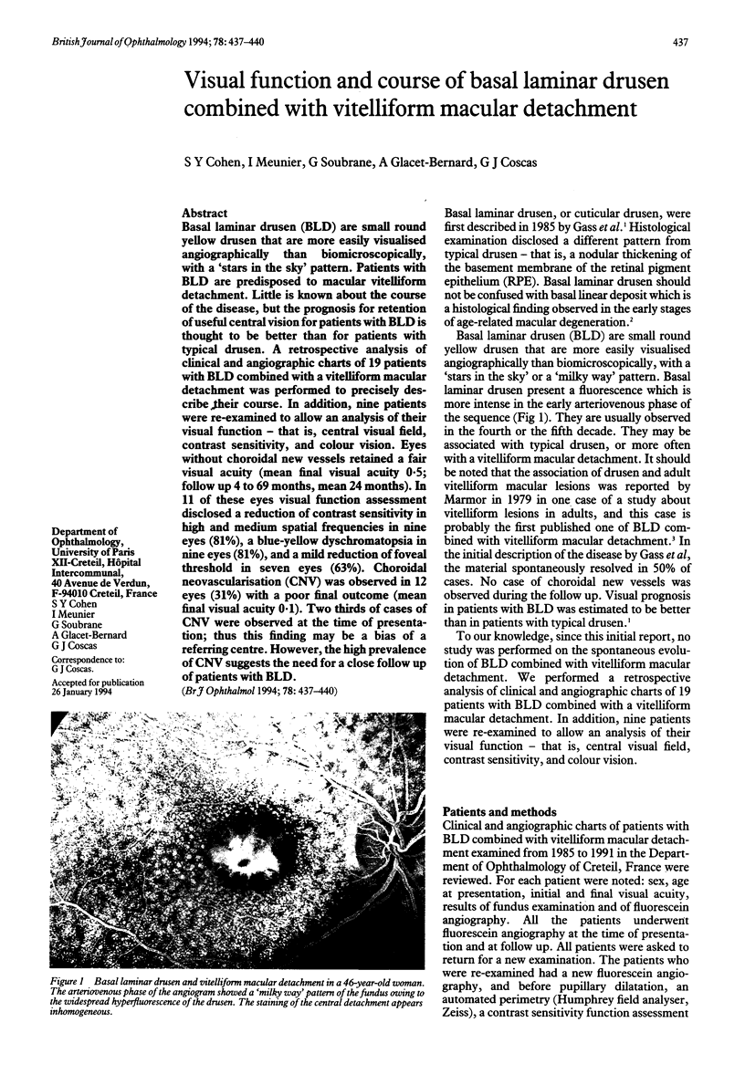
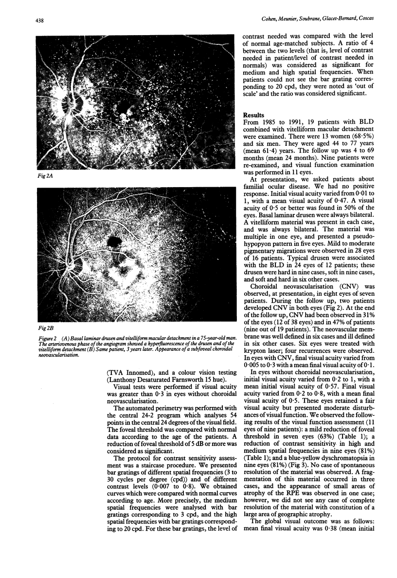
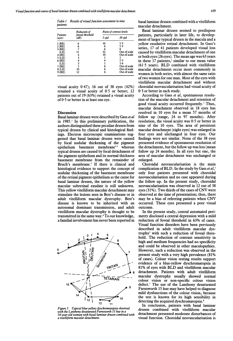
Basal laminar drusen (BLD) are small round yellow drusen that are more easily visualised angiographically than biomicroscopically, with a 'stars in the sky' pattern. Patients with BLD are predisposed to macular vitelliform detachment. Little is known about the course of the disease, but the prognosis for retention of useful central vision for patients with BLD is thought to be better than for patients with typical drusen. A retrospective analysis of clinical and angiographic charts of 19 patients with BLD combined with a vitelliform macular detachment was performed to precisely describe their course. In addition, nine patients were re-examined to allow an analysis of their visual function--that is, central visual field, contrast sensitivity, and colour vision. Eyes without choroidal new vessels retained a fair visual acuity (mean final visual acuity 0.5; follow up 4 to 69 months, mean 24 months). In 11 of these eyes visual function assessment disclosed a reduction of contrast sensitivity in high and medium spatial frequencies in nine eyes (81%), a blue-yellow dyschromatopsia in nine eyes (81%), and a mild reduction of foveal threshold in seven eyes (63%). Choroidal neovascularisation (CNV) was observed in 12 eyes (31%) with a poor final outcome (mean final visual acuity 0.1). Two thirds of cases of CNV were observed at the time of presentation; thus this finding may be a bias of a referring centre. However, the high prevalence of CNV suggests the need for a close follow up of patients with BLD.
Full text
PDF


Images in this article
Selected References
These references are in PubMed. This may not be the complete list of references from this article.
- Brecher R., Bird A. C. Adult vitelliform macular dystrophy. Eye (Lond) 1990;4(Pt 1):210–215. doi: 10.1038/eye.1990.28. [DOI] [PubMed] [Google Scholar]
- Gass J. D., Jallow S., Davis B. Adult vitelliform macular detachment occurring in patients with basal laminar drusen. Am J Ophthalmol. 1985 Apr 15;99(4):445–459. doi: 10.1016/0002-9394(85)90012-1. [DOI] [PubMed] [Google Scholar]
- Green W. R., Key S. N., 3rd Senile macular degeneration: a histopathologic study. Trans Am Ophthalmol Soc. 1977;75:180–254. [PMC free article] [PubMed] [Google Scholar]
- Kenyon K. R., Maumenee A. E., Ryan S. J., Whitmore P. V., Green W. R. Diffuse drusen and associated complications. Am J Ophthalmol. 1985 Jul 15;100(1):119–128. doi: 10.1016/s0002-9394(14)74993-1. [DOI] [PubMed] [Google Scholar]
- Lanthony P., Dubois-Poulsen A. Le Farnsworth--15 désaturé. Bull Soc Ophtalmol Fr. 1973 Sep-Oct;73(9-10):861–866. [PubMed] [Google Scholar]
- Marmor M. F. "Vitelliform" lesions in adults. Ann Ophthalmol. 1979 Nov;11(11):1705–1712. [PubMed] [Google Scholar]
- Sabates R., Pruett R. C., Hirose T. Pseudovitelliform macular degeneration. Retina. 1982;2(4):197–205. [PubMed] [Google Scholar]
- Sarks S. H. Ageing and degeneration in the macular region: a clinico-pathological study. Br J Ophthalmol. 1976 May;60(5):324–341. doi: 10.1136/bjo.60.5.324. [DOI] [PMC free article] [PubMed] [Google Scholar]


