Abstract
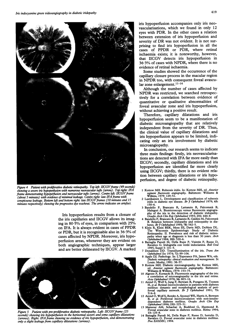
AIMS/BACKGROUND: Iris fluorescein angiography (IFA) is not commonly used in clinical practice, although its value has been demonstrated especially in cases of diabetic disease. IFA is able to show neovascular tufts in order to guide the laser treatment, and it is highly recommended in diabetic patients who need cataract surgery or vitrectomy. Nevertheless, IFA fails to demonstrate the iris vascular pattern in heavily pigmented iris and conspicuous leakage cases. The aim of the study was to evaluate the feasibility of iris indocyanine green videoangiography (IICGV) and to correlate its findings with those of IFA in diabetic iridopathy. METHODS: Thirty six patients affected in varying degrees by diabetic retinopathy underwent an ophthalmic examination including retinal fluorescein angiography, IFA, and IICGV. IICGV was performed using IMAGEnet System H1024. RESULTS: The results demonstrated that IICGV allows precise visualisation of the iris vascular pattern, also in cases of heavily pigmented iris. CONCLUSIONS: Three main findings seemed to be evident: firstly, iris neovascularisations are detected with IFA far more easily than IICGV; secondly, capillary dilatations and iris hypoperfusion are identified far more clearly using IICGV; thirdly, there is no evident relation between capillary dilatation or iris hypoperfusion, and degree of diabetic retinopathy.
Full text
PDF
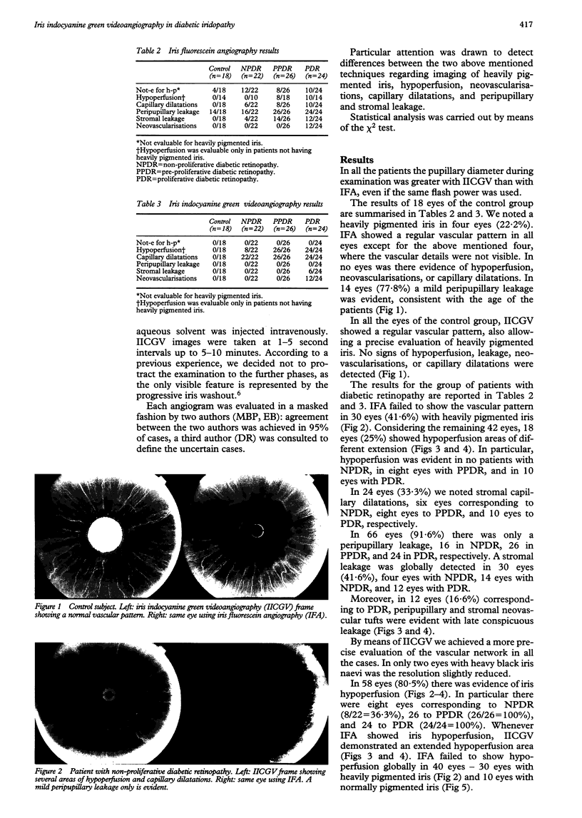
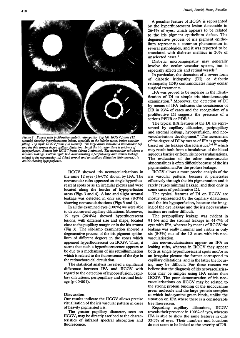
Images in this article
Selected References
These references are in PubMed. This may not be the complete list of references from this article.
- Algvere P., Kornacki B. Fluorescein angiography of the iris. A correlation of microangiopathy in the iris and retina. Acta Ophthalmol (Copenh) 1978 Oct;56(5):803–816. doi: 10.1111/j.1755-3768.1978.tb06645.x. [DOI] [PubMed] [Google Scholar]
- Arend O., Wolf S., Remky A., Sponsel W. E., Harris A., Bertram B., Reim M. Perifoveal microcirculation with non-insulin-dependent diabetes mellitus. Graefes Arch Clin Exp Ophthalmol. 1994 Apr;232(4):225–231. doi: 10.1007/BF00184010. [DOI] [PubMed] [Google Scholar]
- Bandello F., Brancato R., Lattanzio R., Falcomatà B., Malegori A. Biomicroscopy versus fluorescein angiography of the iris in the detection of diabetic iridopathy. Graefes Arch Clin Exp Ophthalmol. 1993 Aug;231(8):444–448. doi: 10.1007/BF02044229. [DOI] [PubMed] [Google Scholar]
- Bandello F., Brancato R., Lattanzio R., Galdini M., Falcomatà B. Relation between iridopathy and retinopathy in diabetes. Br J Ophthalmol. 1994 Jul;78(7):542–545. doi: 10.1136/bjo.78.7.542. [DOI] [PMC free article] [PubMed] [Google Scholar]
- Donaldson D. D. Transillumination of the iris. Trans Am Ophthalmol Soc. 1974;72:89–106. [PMC free article] [PubMed] [Google Scholar]
- Klein R., Klein B. E., Moss S. E., Davis M. D., DeMets D. L. The Wisconsin epidemiologic study of diabetic retinopathy. II. Prevalence and risk of diabetic retinopathy when age at diagnosis is less than 30 years. Arch Ophthalmol. 1984 Apr;102(4):520–526. doi: 10.1001/archopht.1984.01040030398010. [DOI] [PubMed] [Google Scholar]
- Laatikainen L. Development and classification of rubeosis iridis in diabetic eye disease. Br J Ophthalmol. 1979 Mar;63(3):150–156. doi: 10.1136/bjo.63.3.150. [DOI] [PMC free article] [PubMed] [Google Scholar]
- Mansour A. M., Schachat A., Bodiford G., Haymond R. Foveal avascular zone in diabetes mellitus. Retina. 1993;13(2):125–128. doi: 10.1097/00006982-199313020-00006. [DOI] [PubMed] [Google Scholar]


