Abstract
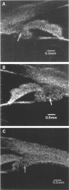
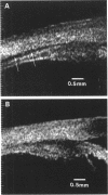
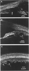
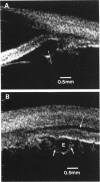
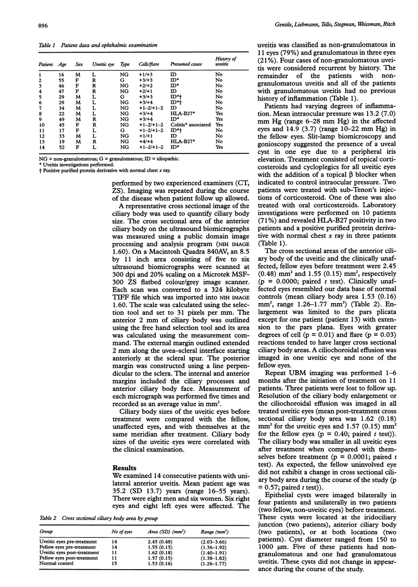
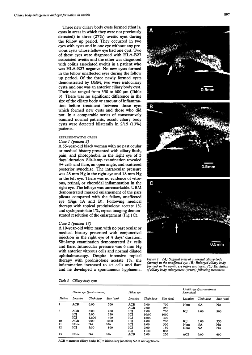
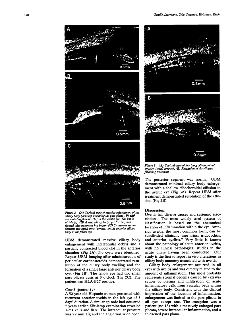
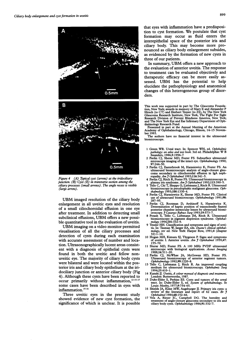
BACKGROUND: Acute anterior uveitis has diverse causes and systemic associations. Inflammation is predominantly localised to the iris and pars plicata. Little is known about the in vivo effects of uveitis on ciliary body anatomy. METHODS: Bilateral, high frequency, high resolution, ultrasound biomicroscopy was performed on consecutive patients with unilateral anterior uveitis to evaluate ciliary body anatomy. Imaging was repeated when possible during the clinical course. The cross sectional area of the anterior ciliary body was measured using image processing and analysis software. Measurements from the uveitic eyes were compared with the fellow eyes and the effect of treatment was evaluated. RESULTS: Fourteen patients were enrolled. Ultrasound biomicroscopy demonstrated a larger ciliary body cross sectional area in the uveitic eyes compared with the fellow, clinically uninvolved eyes (2.45 (SD 0.48) mm2 versus 1.55 (SD 0.15) mm2, (p = 0.0000; paired t test)). A ciliochoroidal effusion was present in one uveitic eye. Epithelial cysts were imaged bilaterally in four uveitic patients (29%) and unilaterally in unaffected eyes of two uveitic patients. Ciliary body cross sectional area decreased following steroid therapy (p = 0.0001; paired t test). New cysts were noted in three uveitic eyes during the follow up period and in none of the fellow, unaffected eyes. CONCLUSION: Ultrasound biomicroscopy offers a new approach to the evaluation of anterior uveitis. The response to treatment can be evaluated objectively and therapeutic efficacy can be more easily assessed. It has the potential to help elucidate the pathophysiology and anatomical changes of this heterogeneous group of disorders.
Full text
PDF
Images in this article
Selected References
These references are in PubMed. This may not be the complete list of references from this article.
- HOGAN M. J., KIMURA S. J., THYGESON P. Signs and symptoms of uveitis. I. Anterior uveitis. Am J Ophthalmol. 1959 May;47(5 Pt 2):155–170. doi: 10.1016/s0002-9394(14)78239-x. [DOI] [PubMed] [Google Scholar]
- Pavlin C. J., Easterbrook M., Harasiewicz K., Foster F. S. An ultrasound biomicroscopic analysis of angle-closure glaucoma secondary to ciliochoroidal effusion in IgA nephropathy. Am J Ophthalmol. 1993 Sep 15;116(3):341–345. doi: 10.1016/s0002-9394(14)71351-0. [DOI] [PubMed] [Google Scholar]
- Pavlin C. J., Harasiewicz K., Sherar M. D., Foster F. S. Clinical use of ultrasound biomicroscopy. Ophthalmology. 1991 Mar;98(3):287–295. doi: 10.1016/s0161-6420(91)32298-x. [DOI] [PubMed] [Google Scholar]
- Pavlin C. J., McWhae J. A., McGowan H. D., Foster F. S. Ultrasound biomicroscopy of anterior segment tumors. Ophthalmology. 1992 Aug;99(8):1220–1228. doi: 10.1016/s0161-6420(92)31820-2. [DOI] [PubMed] [Google Scholar]
- Pavlin C. J., Ritch R., Foster F. S. Ultrasound biomicroscopy in plateau iris syndrome. Am J Ophthalmol. 1992 Apr 15;113(4):390–395. doi: 10.1016/s0002-9394(14)76160-4. [DOI] [PubMed] [Google Scholar]
- Pavlin C. J., Rootman D., Arshinoff S., Harasiewicz K., Foster F. S. Determination of haptic position of transsclerally fixated posterior chamber intraocular lenses by ultrasound biomicroscopy. J Cataract Refract Surg. 1993 Sep;19(5):573–577. doi: 10.1016/s0886-3350(13)80002-8. [DOI] [PubMed] [Google Scholar]
- Pavlin C. J., Sherar M. D., Foster F. S. Subsurface ultrasound microscopic imaging of the intact eye. Ophthalmology. 1990 Feb;97(2):244–250. doi: 10.1016/s0161-6420(90)32598-8. [DOI] [PubMed] [Google Scholar]
- Potash S. D., Tello C., Liebmann J., Ritch R. Ultrasound biomicroscopy in pigment dispersion syndrome. Ophthalmology. 1994 Feb;101(2):332–339. doi: 10.1016/s0161-6420(94)31331-5. [DOI] [PubMed] [Google Scholar]
- Shields J. A., Kline M. W., Augsburger J. J. Primary iris cysts: a review of the literature and report of 62 cases. Br J Ophthalmol. 1984 Mar;68(3):152–166. doi: 10.1136/bjo.68.3.152. [DOI] [PMC free article] [PubMed] [Google Scholar]
- Tello C., Chi T., Shepps G., Liebmann J., Ritch R. Ultrasound biomicroscopy in pseudophakic malignant glaucoma. Ophthalmology. 1993 Sep;100(9):1330–1334. doi: 10.1016/s0161-6420(93)31479-x. [DOI] [PubMed] [Google Scholar]
- Tello C., Liebmann J. M., Ritch R. An improved coupling medium for ultrasound biomicroscopy. Ophthalmic Surg. 1994 Jun;25(6):410–411. [PubMed] [Google Scholar]
- Vela A., Rieser J. C., Campbell D. G. The heredity and treatment of angle-closure glaucoma secondary to iris and ciliary body cysts. Ophthalmology. 1984 Apr;91(4):332–337. doi: 10.1016/s0161-6420(84)34287-7. [DOI] [PubMed] [Google Scholar]


