Abstract
A new separation-free method for detection of single nucleotide polymorphisms (SNPs) is described. The method is based on the single base extension principle, fluorescently labeled dideoxy nucleotides and two-photon fluorescence excitation technology, known as ArcDia™ TPX technology. In this assay technique, template-directed single base extension is carried out for primers which have been immobilized on polymer microparticles. Depending on the sequence of the template DNA, the primers are extended either with a labeled or with a non-labeled nucleotide. The genotype of the sample is determined on the basis of two-photon excited fluorescence of individual microparticles. The effect of various assay condition parameters on the performance of the assay method is studied. The performance of the new assay method is demonstrated by genotyping the SNPs of human individuals using double-stranded PCR amplicons as samples. The results show that the new SNP assay method provides sensitivity and reliability comparable to the state-of-the-art SNaPshot™ assay method. Applicability of the new method in routine laboratory use is discussed with respect to alternative assay techniques.
INTRODUCTION
When the frequency of base variation in a certain locus of DNA within a population exceeds 1%, the locus is generally considered polymorphic (single nucleotide polymorphism, SNP) (1). SNPs are distributed relatively evenly in the human genome and occur in average once in 1000 bases (2,3). Most often an SNP occurs between two alternative bases, while polymorphism with three or four alternative bases exists less often in the human genome. Even though most of the human SNPs are located in the regions of the genome with no direct impact on the phenotype, the SNPs in the coding regions of genes cause most of the known inherited disorders, and these SNPs are routinely analysed for diagnostic purposes. It is likely that SNPs in the regulatory regions of genes influence the risk of developing polygenic diseases (4). The SNP data can be used for identification of individuals, for revealing disease-causing genes, and for pharmacogenetic analysis. Knowing the SNPs that have an impact on the drug metabolism of a patient, enables individualized design of therapy. The SNPs that are physically close to disease-predisposing alleles and inherited with them, can be used as markers in linkage disequilibrium (LD) mapping to identify genes predisposing individuals to multifactorial disorders (4,5).
Various techniques to detect SNPs have been developed during the last decade. SNP assay techniques can be categorized by their reaction principle, detection method and assay format (4,5). The mechanisms of allelic discrimination of sequence-specific SNP techniques are based either on allele-specific hybridization (6), enzymatic cleavage (7) or nucleotide incorporation (8). Methods based on nucleotide incorporation, such as single base extension (SBE) method, are particularly popular due to their sensitivity, accuracy and simple assay procedure (9). Most common detection methods for allelic discrimination products are based either on mass spectrometry (10), fluorometry (11), fluorescence resonance energy transfer (12), fluorescence polarization (13), chemiluminescence (14) or enzyme activity (15). Of the detection methods, fluorometry has become the most popular during the recent years. The chosen detection method is the main determinant for the assay format, which can be homogenous separation-free assay [such as TaqMan (16)], homogenous separation assay [such as SNaPshot™ (17)], solid-phase separation-free assay and solid-phase separation assay [such as oligonucleotide arrays (18)]. Separation-free assay format is desired, since it allows simple assay protocol, instrumentation and method automation.
The properties of an ideal SNP assay method are dependent on the application. For example, SNP assays in clinical diagnostics and in large-scale genotyping studies require different assay throughput and cost structure; thus, none of the assay methods currently available is ideal for all applications (5). Most of the work and resources in the field have been directed to development of technology allowing high-throughput applications; while simple and cost-effective assay technologies, including instrumentation and reagents, are still needed for routine clinical diagnostics and small-scale research applications. Such applications include, e.g. carrier screening, pharmacogenetics, and forensic and paternity analysis. Typically in these applications no more than few tens of SNPs are determined per subject.
Single base extension reaction principle has been extensively used for SNP assays in combination with different assay formats and detection techniques (10,11,13,17). This reaction principle, in the literature, is also called ‘minisequencing’ (19) or ‘template-directed dye-terminator incorporation’ (TDI) (20). SBE principle is based on extension of a primer that anneals on the target DNA next to the nucleotide position of interest. Polymerase is used to incorporate a labeled nucleotide into this position. When terminating dideoxy analogues of nucleotides (ddNTPs) are used, the polymerase cannot continue, but only a single nucleotide next to the primer is incorporated. Most of the published SBE methods are based on separation steps, and are thus not ideal for routine clinical use.
The objective of this work is to study applicability of the novel separation-free assay technique, ArcDia™ TPX, for SNP genotyping assays. This assay technology enables ultra-sensitive separation-free assays in microvolumes (21–23), and its potential as a general tool for bioaffinity assays has been recently demonstrated (24,25). The assay technology is based on the use of microparticles as solid-phase reaction carrier and detection of two-photon excited fluorescence from the surface of individual microparticles. In the present study, we have applied the ArcDia™ TPX assay platform in combination with the single base extension principle and fluorescently labeled ddNTPs. Accordingly, single base extension reaction takes place on the surface of primer coated microparticles. Depending on the sequence of the sample DNA, the primers are extended either with a labeled or with a non-labeled nucleotide, and the sample genotype is determined on the basis of two-photon excited fluorescence of individual microparticles.
MATERIALS AND METHODS
Sample preparation
PCR primers (forward primer: GGAGAGCTTCATAAAGCCACAGCA; reverse primer: AGACGTTCTTGTTCCGGCGC) were designed with the Primer Express™ software (Applied Biosystems, Foster City, CA). PCR amplification reactions included 100 ng of template DNA, 200 μM dNTP (MBI Fermentas, Vilnius, Lithuania), 400 nM primers (Eurogentec, Seraing, Belgium), 2 U Dynazyme EXT polymerase (Finnzymes, Espoo, Finland), and dimethyl sulfoxide 5% (Finnzymes), in 50 μl of Dynazyme detergent-free buffer (Finnzymes). The samples were cycled as follows: 5 min at 95°C, followed by 40 cycles of 45 s at 94°C, 45 s at 62°C and 45 s at 72°C, and final extension for 6 min at 73°C. The PCR products were treated with shrimp alkaline phosphatase (SAP 333 U/ml, UBS, Cleveland, OH) and Exonuclease I (133 U/ml, UBS) for 60 min at 37°C followed by 15 min denaturation at 90°C.
Coating of microparticles
Microparticles (ArcDia™ Biotin binding microspheres, 2.0 × 10⁷ p.c.s., Ø = 3.2 μm, ArcDia Ltd, Turku, Finland, cat no. D-101) were suspended in 200 μl of coating buffer (1 mM EDTA, 10 mM NaN3, 0.01% Tween-20, pH 8.0) containing variable amount (6.1 × 10⁶, 3.1 × 10⁶, 1.5 × 10⁶, 7.7 × 10⁵, 3.8 × 10⁵ or 1.9 × 10⁵ primers per microsphere) of biotinylated 35mer oligodeoxynucleotide primer [5′-biotin-(T)15AGTTTCCCCGACACCTCCAG-3′, Eurogentec, Seraing, Belgium]. The suspension was incubated overnight under continuous shaking (1300 r.p.m.; 23°C). Microspheres were washed with the coating buffer (4 × 500 μl) by sequential centrifugation (5000 g; 3 min)–aspiration–resuspension, and finally resuspended in 125 μl of the same buffer. The concentration of the microsphere suspension was determined with Multisizer 3 Coulter Counter (Beckman Coulter, Fullerton, CA), followed by dilution to concentration of 1.0 × 10⁵ p.c.s./μl.
Genotyping reaction conditions for TPX technique
PCR sample (8 μl) was added to genotyping reagent cocktail (17 μl in TopYield™ Strips (Nunc A/S, Roskilde, Denmark) resulting in a solution containing 27.5 mM of TRIS, 6.5 mM of MgCl2, 17.5 mM of KCl, 25 nM of dideoxynucleotide labeled with TAMRA (ddUTP or ddCTP, NEN™ Life Science Products, Inc., Boston, MA), 125 nM of three unlabeled ddNTPs (Invitrogen Life Technologies, Carlsbad, CA), 80 U/ml of Thermo Sequenace™ DNA polymerase (with Thermoplasma acidophilum Inorganic Pyrophosphatase, cat no. E79000Y, Amersham Biosciences, Piscataway, NJ), 4.0 × 10⁶ p.c.s./ml of microspheres with coating density of 3.1 × 10⁶ primers per microsphere. Samples were thermocycled with Eppendorf Mastercycler® gradient instrument (Eppendorf GmbH, Hamburg, Germany) using the following program: initial step 45 s at 95°C, followed by 20 cycles of 45 s at 95°C, 3 min at 58°C and 20 s at 72°C. After the cycles the samples were stored at +4°C. The samples were measured without separation steps with ArcDia™ TPX Plate reader (Model 5-003, Arctic Diagnostics Oy, Turku, Finland) using a measurement time of 60 s per well. The fluorescence data were filtered by omitting data points characterized with the optical trap duration time outside of the range 5–120 ms.
Optimization of assay conditions
SNP assays were optimized with respect to polymerase concentration, nucleotide concentration, annealing temperature and microparticle coating density. The sample used in the optimization studies had been previously typed as wild-type with SNaPshot™ technique. All optimization assays were run in three sample replicates. If not otherwise stated, the assay parameters for the optimization studies were as given in the previous chapter.
In the optimization of polymerase, the following concentrations were used: 195, 78, 31, 12, 5, 2 and 0 U/ml. Concentration of labeled nucleotide was 20 nM and annealing temperature was 60°C. The extension step at 72°C was not applied.
The optimization of nucleotides was carried out with the following concentration: 50, 33.3, 22.2, 14.8, 9.9, 6.6 and 4.4 nM. Annealing temperature was 60°C. The extension step at 72°C was not applied.
Optimization of the annealing temperature was carried out with the temperatures: 72.9, 71.0, 68.8, 66.2, 63.5, 60.9, 58.4 and 56.4°C. The extension step of 20 s at 72°C was used.
Microparticles of the following coating densities were studied: 6.1 × 10⁶, 3.1 × 10⁶, 1.5 × 10⁶, 7.7 × 10⁵, 3.8 × 10⁵ or 1.9 × 10⁵ primers per microsphere.
Genotyping with SNaPshot™ technique
Genotyping with SNaPshot™ technique was carried out according to the manufacturer's directions (Applied Biosystems, Foster City, CA). PCR products were diluted either 20- or 5-fold or used as such. The single base extension reaction contained 6 μl of ABIPRISM® SNaPshot™ Multiplex Kit (Applied Biosystems), 1 μM extension primer (GACT6)AGTTTCCCCGACACCTCCAG and 3 μl of PCR product in a total volume of 10 μl. The samples were cycled as follows: 25 cycles of 10 s at 96°C, 5 s at 50°C and 30 s at 60°C. The reaction products were treated with SAP and run with ABIPRISM® 3100 instrument (Applied Biosystems). The results were analyzed with GeneScan software (Applied Biosystems).
RESULTS
Assay principle
In this paper, we describe a new method for SNP genotyping. The method is based on ArcDia™ TPX detection technique and SBE reaction principle. This detection technique enables separation-free bioaffinity assay from microvolumes, including measurements of SBE reaction products of SNP assays. The separation-free assay format is based on the non-linear character of two-photon excitation, which results in generation of fluorescence only in the diffraction-limited focal volume of the laser illumination (26). In the SNP variant of this assay technique, microparticles are used to concentrate fluorescent dideoxy nucleotide molecules on their surface. When a such microparticle is brought into focus by optical forces of the illuminating near infra-red (IR) laser (1064 nm) (27), a scattering signal and a two-photon excited fluorescence signal are recorded in near IR and visible range of spectra, respectively. The intensity of fluorescence from the individual microparticle is proportional to the number of labeled nucleotides incorporated to the surface bound primers. Using microparticles 3 μm in diameter, upto 106 labeled nucleotides can be incorporated per particle. The optical configuration of the fluorometer and the physical phenomena related to the measurement process have been described in detail in previous publications (28,29).
In order to demonstrate applicability of the SNP assay technique, a method was developed for genotyping double-stranded PCR products of human samples. The assay was directed to SNP identified as GenBank rs2074170 A→G. This method was applied for 25 human samples, and the assay results were compared to those of commercial SNaPshot™ assay method. For each human sample two reactions were carried out—one reaction with labeled dideoxy uridine, and one with labelled dideoxy cytidine. Since the wild-type sample is characterized with adenine base, incorporation of dideoxy uridine is expected as default. Mutant samples with two alleles of the less frequent type, give positive results with labeled dideoxy guanosine, while heterozygote samples provide positive results with both the nucleotide reagents.
Optimization of the SNP assay method
The SNP assay method was first optimized with respect to various assay parameters, including polymerase concentration, nucleotide concentrations, annealing temperature and primer coating density. The optimization study was carried out with a sample, which had previously been genotyped as a wild-type using the SNaPshot™ assay technique. Thus, this sample produces a positive assay result with labeled uridine reagent. Later in this paper, the fluorescence signal that is obtained using the wild-type sample as template in combination with complementary uridine reagent is called a specific signal, while the signal obtained with the same template in combination with the cytidine reagent is called an unspecific signal. The ratio of these two signals (specific to unspecific signal) is considered as the main indicator of performance of a particular assay method, and denoted in this section as Signal ratio.
The allele discrimination efficiency of SBE reactions relies on the high accuracy of nucleotide incorporation by the DNA polymerase (8,19). The effect of concentration of this key component on the assay performance was studied and the result is shown in Figure 1. The figure shows that increase in polymerase concentration up to 78 U/ml is associated with continuous increase in both the specific and the unspecific signal. The slope of increase of the unspecific signal is, however, significantly smaller compared to that of the specific signal, and therefore, the Signal ratio shows a clear increase up to concentration of 78 U/ml. This polymerase activity corresponds to a typical concentration used in liquid-phase SBE reactions. The results of an assay carried out without polymerase showed that the unspecific binding of the labeled nucleotides on microparticles was very low. These results suggest that the polymerase enzyme is responsible for the unspecific signal obtained from the SNP assays.
Figure 1.
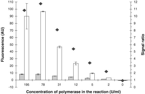
Effect of polymerase concentration on the SBE reaction. Grey bar represents unspecific signal, white bar represents specific signal and black diamond represents the ratio of specific signal to unspecific signal. The error bar represents error ± standard deviation.
Another key factor of the SNP assay method is the concentration of labeled nucleotides. In the separation-free assay format, this determines the level of background fluorescence, and thus, the lowest limit of the working range of the assay (21). On the other hand, nucleotide concentration can be the limiting factor of the enzyme catalysed nucleotide incorporation. Therefore, nucleotide concentration should be carefully optimized to obtain a sensitive and reliable genotyping assay. The result of the optimization study is shown in Figure 2. The figure indicates that the Signal ratio increases almost linearly from 4 to 33 nM, and the highest Signal ratio was achieved at nucleotide concentration of 50 nM. The increase of Signal ratio was, however, clearly levelling off at this nucleotide concentration, suggesting that a higher nucleotide concentration would not increase the assay performance.
Figure 2.
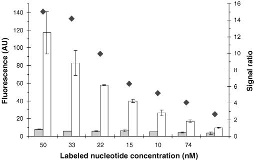
Effect of labeled nucleotide concentration on the SBE reaction. Grey bar represents unspecific signal, white bar represents specific signal and black diamond represents the ratio of specific signal to unspecific signal. The error bar represents error ± standard deviation.
The temperature profile of the primer extension reaction cycle was optimized by adjusting the annealing temperature. An unusually long annealing time was applied to compensate the reduced reaction kinetics of the solid-phase reaction. Also, an extra extension step of 20 s at 72 °C was used to compensate polymerase activity differences in different annealing temperatures. The highest Signal ratio was achieved at annealing temperature of 58.4°C (Figure 3). A small decrease in the unspecific signal was observed with increase in annealing temperature from 56 to 66°C, while above this temperature the unspecific signal remained constant.
Figure 3.
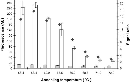
Effect of annealing temperature on the SBE reaction. Grey bar represents unspecific signal, white bar represents specific signal and black diamond represents the ratio of specific signal to unspecific signal. The error bar represents error ± standard deviation.
The result of the assay optimization study with respect to primer coating density is shown in Figure 4. The unspecific signal does not show any clear trend in this figure, and therefore, the specific signal was considered a better indicator of the assay performance than the Signal ratio. The assay shows a maximum specific signal at coating density of 3.1 × 10⁶ primers per particle. The specific signal increases strongly up to primer density of 1.5 × 10⁶ per particle, levels off and decreases slightly at the highest coating density. These results indicate that biotin binding capacity of the microparticles is higher than the optimal primer coating density.
Figure 4.
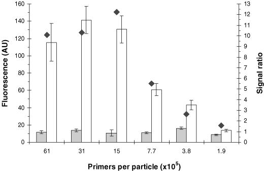
Effect of primer coating density on the SBE reaction. Grey bar represents unspecific signal, white bar represents specific signal and black diamond represents the ratio of specific signal to unspecific signal. The error bar represents error ± standard deviation.
Genotyping
The genotyping performance of the developed assay method was evaluated by genotyping 25 individuals of the Finnish population with respect to rs2074170 A→G. According to a recent study the frequency for this genetic marker among the Finnish population is 30% (30). The samples used in the study were double-stranded PCR amplicons, 807 bp in length. After treatment with SAP and EXO-1 enzymes, the samples were divided in two aliquots and analysed independently with the new assay method and a standard SNaPshot™ method. The SNaPshot™ platform was chosen as a reference because it is widely used and generally considered as one of the most versatile and efficient tool for genome research. The SNaPshot™ technique employs SBE reaction principle and separation of the assay products by capillary electrophoresis. In both SNaPshot™ and ArcDia™ TPX assay methods, SBE primers with the same specific sequence were used. The major difference between the two assay techniques relate to the SBE process, which takes place on solid phase and in homogeneous solution with ArcDia™ TPX and SNaPshot™ techniques, respectively.
The fluorescence signals of the SNP assays using two different labeled dideoxynucleotide reagents are shown graphically in Figure 5. In this figure, the fluorescence signal obtained with the uridine reagent is given on the X-axis, and the signal with cytidine reagent is given on the Y-axis. Wild-type samples with adenine bases are expected to provide signals on the X-axis and mutant samples with guanidine bases on the Y-axis. In Figure 5, three clearly distinguishable populations with 9, 10 and 3 cases can be recognized. The population that provides signal with both labeled dideoxy reagents (in the middle) represents heterozygote samples. In addition to the three main populations, three separate samples appear close to the origo. The same three samples gave extraordinarily low signal levels also with the reference SNaPshot™ method. The low signal levels of these samples are presumably due to insufficient primary PCR amplification.
Figure 5.
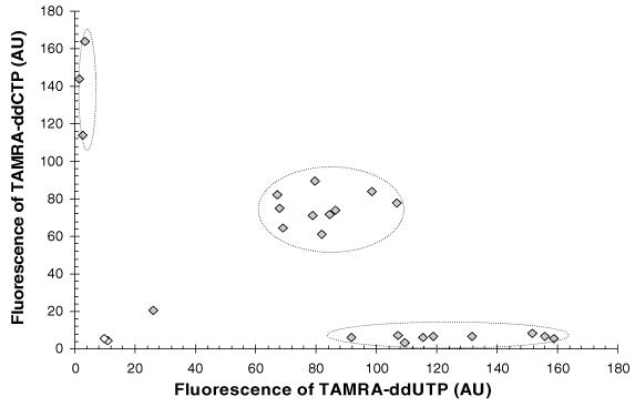
Scatter plot of the signals provided by the samples representing different genotypes. Vectors of the two white data points do not exceed the vector length threshold limit set for reliable genotyping.
Genotype assessment
The final genotype assessment of the samples was carried out by considering each data point as a vector (Figure 6), starting at the origo and ending up at the position of the original data point (Figure 5). In case the length of a vector exceeds the threshold value derived from the corresponding background fluorescence signals (equal to liquid fluorescence), the assay procedure including PCR, is considered successful. In this study the threshold value was set equal to twice the length of the corresponding background vector (see Figure 6). After passing this threshold criterion, the genotype assessment is continued by analysis of the vector angle. In case the vector angle of a data point falls within the range from 0 to π/6 radians (30°) the sample is judged as wild-type. A vector angle between π/3 and π/2 radians (60–90°) is an indication of a mutant, while vectors falling to the sector in the middle, i.e. angles between π/6 and π/3 radians (30–60°), are assigned heterozygotes in genome (see Figure 6). The advantage of this decision-making algorithm is the absolute character, meaning that standard samples are not required for reliable SNP genotyping.
Figure 6.
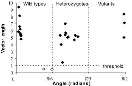
Vectors provided by the samples representing different genotypes. Vector lengths are normalized to threshold value (length of liquid fluorescence vectors). The two white data points do not exceed the threshold limit set for reliable genotyping. Vectors presented in the figure are background (liquid fluorescence) subtracted.
Using this genotype assessment algorithm, 23 samples out of the total number of 25 samples exceeded the threshold value and allowed reliable genotyping. Two of the three samples with extraordinary low fluorescence signal levels (see Figure 5), remained below the threshold level and were thus judged as examples of unsuccessful experimentation. One of the three samples, however, exceeded the threshold level and could thus be assigned as heterozygote. Using the SNaPshot™ reference technique with experimental conditions recommended by the manufacturer and diluted PCR samples, none of the three samples of low signals could be genotyped. However, with undiluted PCR samples, two of them could be assigned as heterozygotes, while one sample still remained untyped. The result of the genotyping assays are summarized in Table 1 for both assay methods. The table shows that the genotypes determined with the two alternative assay methods are in complete agreement. The frequency of the SNP among the genotyped population was calculated to be 37%.
Table 1. Shows genotypes determined with SNaPshot™ and TPX-method.
| Sample code | Genotype by TPX | Genotype by SNaPshot |
|---|---|---|
| 1 | Mutant | Mutant |
| 2 | Wild-type | Wild-type |
| 3 | Mutant | Mutant |
| 4 | Wild-type | Wild-type |
| 5 | Hetero | Hetero |
| 6 | Hetero | Hetero |
| 7 | Wild-type | Wild-type |
| 8 | Wild-type | Wild-type |
| 9 | Wild-type | Wild-type |
| 10 | Hetero | Hetero |
| 11 | Hetero | Heteroa |
| 12 | Hetero | Hetero |
| 13 | — | — |
| 14a | — | Hetero |
| 15 | Hetero | Hetero |
| 16 | Hetero | Hetero |
| 17 | Wild-type | Wild-type |
| 18 | Hetero | Hetero |
| 19 | Wild-type | Wild-type |
| 20 | Hetero | Hetero |
| 21 | Wild-type | Wild-type |
| 22 | Hetero | Hetero |
| 23 | Hetero | Hetero |
| 24 | Wild-type | Wild-type |
| 25 | Mutant | Mutant |
aIndicates that genotyping with the SNaPshot™ method was successful only when undiluted PCR samples were used.
Assay performance evaluation
The reliability of the new SNP assay method was evaluated by means of a recently introduced statistical tool called ‘screening window coefficient’ and abbreviated as ‘Z′-factor’ (31). The Z′-factor is a dimensionless, simple statistical characteristic which accounts for both the signal response and the signal variability of a screening assay. According to its definition, the Z′-factor exceeds 0.5 with an excellent assay and approaches unity with an ideal assay. When Z′-factor is applied as a measure of reliability of a SNP genotyping assay, three separate Z′-values are calculated, one Z′-value for each separation band. For the new SNP assay method the following Z′-values were calculated: 0.57 between wild-type and heterozygote populations, 0.67 between mutants and heterozygote populations, and 0.96 between wild-type and mutant populations. These values indicate a high reliability of SNP genotyping.
DISCUSSION
In this paper we have described a new technique for SNP genotyping, which is based on solid-phase SBE reaction and detection of two-photon excited fluorescence. The new technique is characterized by simple and separation-free assay procedure, and it was shown to enable reliable genotyping with comparable sensitivity to the SNaPshot™ technique. The limitations of the new technique are the need for sample preamplification and a rather long detection time per sample. To our knowledge this is the first reported technique where genotyping is carried out for double-stranded DNA templates using solid-phase SBE reaction principle.
Coating of microspheres with oligonucleotide primers
Covalent attachment of oligonucleotides on a solid surface has traditionally been carried out by means of carbodiimide chemistry. In this method, a surface containing carboxyl functional groups is activated with water-soluble carbodiimide, followed by substitution of the active intermediate with amino-modified oligonucleotide. This coupling method is simple and usually results in moderate to high absolute coating densities. The problem of the carbodiimide method relates to functionality of the resulting oligonucleotide coating, which often remains low mainly due to surface over-crowding. The reduced performance of oligonucleotide coatings prepared in this manner is a well-known phenomenon. This phenomenon has been accounted as a consequence of steric hindrance and electrostatic repulsion between complementary oligonucleotides and the coated oligonucleotide surface (32).
Despite careful optimization, microspheres coated covalently with the carbodiimide method never worked out satisfactorily for us. The experiments indicated that polymerase adsorbed on the surface of carbodiimide coated particles. This phenomenon could not be avoided by post-coating with globular proteins, since this led to reduced hybridization capacity and tendency to particle aggregation during thermocycling. Due to these problems, we decided to change the coating method and replaced the direct covalent coupling with biotin–avidin chemistry. This approach included the use of biotin binding proteins as the primary coating and secondary binding of biotinylated primers. The advantage of using this coating chemistry is the generic nature of the coating. In other words, once the coating process has been optimized for one primer, preparation of microspheres of another primer specificity is straightforward, thus, no extra optimization work is required. By means of the avidin–biotin chemistry, we were able to develop a generic microparticle reagent that allowed SNP genotyping assay with significantly improved performance compared to the standard carbodiimide chemistry. The results show that the avidin–biotin coating allows the use of biotinylated primers in high excess without remarkable loss in assay performance due to surface over-crowding. This property makes the coating procedure robust compared to standard covalent coupling methods.
Reaction conditions for single base extension
The SNP assay method was studied with respect to several parameters including the annealing temperature. The theoretical melting point of the primer-template hybrid used in the study was calculated to be 65°C (http://www.rnature.com/oligonucleotide.html). This temperature was chosen as the middle point of the studied temperature range. The results indicated, however, that the best Signal ratio was obtained at significantly lower annealing temperature (58°C). This result is understandable, since at lower temperatures higher degree of templates is hybridized. A high degree of hybridization does not, however, implicitly lead to higher signals due to the formation of unspecific hybrids and secondary structures that do the not lead to incorporation of labeled nucleotides. Also, the activity of polymerase is decreased at low temperatures.
The unspecific nucleotide incorporation was found to increase steadily below 66°C, while above this temperature it was found constant. Also, incorporation of nucleotides did not continue during prolonged standing of the cycled mixtures at room temperature. These two findings together suggest that a constant unspecific signal is independent of the thermocycling profile. The constant signal can be accounted for by affinity between DNA coated particles, polymerase and unincorporated nucleotides. This conclusion is also supported by the finding of a linear relationship between the unspecific signal and the polymerase concentration. The unspecific binding, however, is rather constant, and it does not jeopardize the reliability of the genotyping assay. Furthermore, it was found that the unspecific binding of labeled nucleotides can be decreased by including a washing step between the SBE reaction and the detection step (data not shown). This increased the calculated Signal ratios several fold, but did not significantly improve the assay reliability due to weakened signal precision. The extent of unspecific binding as well as the unspecific covalent incorporation of nucleotides are both critically dependent on the nature of polymerase. Throughout this study, ‘Thermo Sequenace™’ polymerase was used. This polymerase was chosen since it has been widely applied in SNP assay methods basing on SBE reaction principle (33–36). In this study, the genotyping assay condition was optimized with respect to concentration of the enzyme, but other polymerases were not tested. In order to further improve the Signal ratio of the new assay technique, alternative polymerase enzymes should be tested.
The results of the study show that SBE reactions can be carried out on solid phase by using reagent concentrations typically applied for SBE reactions in the liquid phase. This result is somewhat surprising, since the rate of reactions on solid phase is usually significantly reduced compared to that in homogeneous solution. This is due to steric hindrances and reduced molecular dynamics of the components bound on the solid phase, including vibrational, rotational and diffusion factors. The concentration of labeled nucleotides used in this study was 25 nM. This concentration is, in fact, exceptionally low compared to other SNP methods relying on SBE reaction principle. [For comparison with other methods see (33,37]. Other SNP assay methods that allow genotyping with equally low nucleotide concentrations are based on fluorescence polarization (13), fluorescence correlation and autocorrelation spectroscopy (38). Low consumption of expensive dideoxynucleotide reagents improves cost-efficiency of the new SNP assay technique.
The results of this study show also that the SBE reaction on the solid phase does not require higher polymerase concentrations than typically used for extension reactions in liquid phase. This result is of high value, since the enzyme is the most expensive component of the SNP assays constituting about 90% of the direct assay costs. Consequently, any change in the concentration of the polymerase is directly reflected, almost in the same proportion, to the cost of the SNP assay. This cost structure is real at the current price level of polymerases, which are held artificially high due to patent protection. Once those expire, it is evident that the cost structure of SNP assays will be dramatically changed.
Another strategy for decreasing the direct costs of SNP assays is the reduction of the assay volume. Exploitation of this strategy, however, has not been feasible with conventional detection techniques without significant compromises in the assay sensitivity. The detection technique applied in this study is unique in this respect. In contrast to the other detection techniques, ArcDia™ TPX technique does allow assay miniaturization (<1 μl) without compromising the assay performance (29). Such small volumes, however, are difficult to handle in the open atmosphere, and require special microfabricated closed assay chambers, preferably dispensing with microfluidics and operation with pre-packed dry-state reagents. Such microvolume assay platforms have aroused great interest among scientists and engineers during the last few years, and these have been widely claimed as the future platform of bioanalysis. In this study, however, SNP genotyping was still carried out in rather large assay volumes (25 μl) due to the lack of appropriate microcuvettes, which would allow thermocycling under hermetic assay conditions, and fluorescence detection through a transparent bottom window.
Multiplexing assay technique
Although ArcDia™ TPX technique allows multiplexed fluorescence reading (28), the SNP assays were carried out in this study in the single parameter mode. The samples were run in two parallel assay wells, one well for each of the two possible nucleotide bases. Alternatively, the same information could have been collected from a single assay well by running a dual parameter assay with two differently labeled dideoxynucleotides. Such a dual parameter assay strategy would be advantageous since it increases reliability of an SNP assay method. In such an assay, pipetting errors never lead to false data interpretation (false genotyping), but can always be recognized, flagged out and reanalysed. In fact, the increased reliability is the only clear advantage that we can find for multiplexed SNP assays. The advantage of running multiple, different SNPs in a single well, instead, is questionable. Such multiplexed assays do save assay wells but are associated with decrease in sensitivity due to unspecific nucleotide incorporation, and reduced genotype call rate due to sample dilution. In conclusion, multiplexing of SNP assays is advantageous whenever this is performed with respect to nucleotide bases, but identification of different SNPs is better when carried out in parallel assay wells.
When large amounts of SNPs are genotyped, like in linkage disequilibrium mapping, a high-throughput assay with minimal reagent cost is essential (37). However, when only a few or few tens of SNPs are studied like in clinical diagnostics or prognostics, high-throughput techniques become inefficient and costly. The SNP genotyping techniques which rely on monitoring of real-time PCR are in turn simple and cost-effective when single SNPs are studied, but suffer from the lack of multiplexing power. Currently a great deal of SNP assays is carried out in research laboratories, where large numbers of SNPs are run from limited number of subjects. In the future, the assaying trend will change. The collected genetic information will lead to an increasing number of clinical applications, where limited number of SNPs are analysed from a large number of individual patients. In order to cover the need for the emerging clinical applications, new assay platforms and detection technologies must be developed allowing simple, reliable and cost-effective SNP assays.
The major advantage of the new separation-free technique introduced in this paper rises from its compatibility with microvolume assay platforms. In combination with these, the new technique allows design of ready-to-use, pre-packed diagnostics product, which includes several parallel chambers for multiparameter SNP assays. The physical size of such assay chip could be a square inch, and the production cost, including the chip and the reagents is ∼1 USD. When combined with an inexpensive two-photon fluorescence reader, the microchip assay platforms would enable sensitive, separation-free and cost-effective genotyping assays with low to medium sample throughput. In conclusion, the presented SNP technique enables miniaturized and multiplexed SNP assays and thus seems to be rather competent for clinical applications, where a set of SNPs are analysed from individual patient samples.
REFERENCES
- 1.Li W.-H., Grauer,D. (1991) Fundamentals of Molecular Evolution. Sinauer Associates, Sunderland, MA. [Google Scholar]
- 2.Sachidanandam R., Weissman,D., Schmidt,S.C., Koko,J.M., Stein,L.D., Marth,G., Sherry,S., Mullikai,J.C., Mortimore,B.J., Willey,D.L. et al. (2001) A map of human genome sequence variation containing 1.42 million single nucleotide polymorphisms. Nature, 409, 928–933. [DOI] [PubMed] [Google Scholar]
- 3.Venter J.C., Adams,M.D., Myers,E.W., Li,P.W., Mural,R.J., Sutton,G.G., Smith,H.O., Yandell,M., Evans,C.A., Holt,R.A. et al. (2001) The sequence of the human genome. Science, 291, 1304–1351. [DOI] [PubMed] [Google Scholar]
- 4.Syvänen A.-C. (2001) Accessing genetic variation: genotyping single nucleotide polymorphisms. Nature Rev. Genet., 2, 930–942. [DOI] [PubMed] [Google Scholar]
- 5.Kwok P.Y. (2001) Methods for genotyping single nucleotide polymorphisms. Annu. Rev. Genomics Hum. Genet., 2, 235–258. [DOI] [PubMed] [Google Scholar]
- 6.Conner B.J., Reyes,A.A., Morin,C., Itakura,K., Teplitz,R.L. and Wallace,R.B. (1983) Detection of sickle cell beta S-globin allele by hybridization with synthetic oligonucleotides. Proc. Natl Acad. Sci. USA, 80, 278–282. [DOI] [PMC free article] [PubMed] [Google Scholar]
- 7.Lyamichev V., Mast,A.L., Hall,J.G., Prudent,J.R., Kaiser,M.W., Takova,T., Kwiatkowski,R.W., Sander,T.J., de Arruda,M., Arco,D.A., Neri,B.P. and Brow,M.A. (1999) Polymorphism identification and quantitative detection of genomic DNA by invasive cleavage of oligonucleotide probes. Nat. Biotechnol., 17, 292–296. [DOI] [PubMed] [Google Scholar]
- 8.Syvänen A.C., Aalto-Setälä,K., Harju,L., Kontula,K. and Soderlund,H. (1990) A primer-guided nucleotide incorporation assay in the genotyping of apolipoprotein E. Genomics, 8, 684–692. [DOI] [PubMed] [Google Scholar]
- 9.Chen X. (2003) Fluorescence polarization for single nucleotide polymorphism genotyping. Comb. Chem. High Throughput Screen., 6, 213–223. [DOI] [PubMed] [Google Scholar]
- 10.Ross P., Hall,L., Smirnov,I. and Haff,L. (1998) High level multiplex genotyping by MALDI-TOF mass spectrometry. Nat. Biotechnol., 16, 1347–1351. [DOI] [PubMed] [Google Scholar]
- 11.Tully G., Sullivan,K.M., Nixon,P., Stones,R.E. and Gill,P. (1996) Rapid detection of mitochondrial sequence polymorphisms using multiplex solid-phase fluorescent minisequencing. Genomics, 34, 107–113. [DOI] [PubMed] [Google Scholar]
- 12.Tyagi S., Bratu,D.P. and Kramer,F.R. (1998) Multicolor molecular beacons for allele discrimination. Nat. Biotechnol., 16, 49–53. [DOI] [PubMed] [Google Scholar]
- 13.Chen X., Levine,L. and Kwok,P.Y. (1999) Fluorescence polarization in homogeneous nucleic acid analysis. Genome Res., 9, 492–498. [PMC free article] [PubMed] [Google Scholar]
- 14.Nyren P., Pettersson,B. and Uhlen,M. (1993) Solid phase DNA minisequencing by an enzymatic luminometric inorganic pyrophosphate detection assay. Anal. Biochem., 208, 171–175. [DOI] [PubMed] [Google Scholar]
- 15.Nikiforov T.T., Rendle,R.B., Goelet,P., Rogers,Y.H., Kotewicz,M.L., Anderson,S., Trainor,G.L. and Knapp,M.R. (1994) Genetic Bit Analysis: a solid phase method for typing single nucleotide polymorphisms. Nucleic Acids Res., 22, 4167–4175. [DOI] [PMC free article] [PubMed] [Google Scholar]
- 16.Livak K.J. (1999) Allelic discrimination using fluorogenic probes and the 5′ nuclease assay. Genet. Anal., 14, 143–149. [DOI] [PubMed] [Google Scholar]
- 17.Turner D., Choudhury,F., Reynard,M., Railton,D. and Navarrete,C. (2002) Typing of multiple single nucleotide polymorphisms in cytokine and receptor genes using SNaPshot. Hum. Immunol., 63, 508–513. [DOI] [PubMed] [Google Scholar]
- 18.Hacia J.G., Sun,B., Hunt,N., Edgemon,K., Mosbrook,D., Robbins,C., Fodor,S.P., Tagle,D.A. and Collins,F.S. (1998) Strategies for mutational analysis of the large multiexon ATM gene using high-density oligonucleotide arrays. Genome Res., 8, 1245–1258. [DOI] [PubMed] [Google Scholar]
- 19.Pastinen T., Kurg,A., Metspalu,A., Peltonen,L. and Syvänen,A.-C. (1997) Minisequencing: a specific tool for DNA analysis and diagnostics on oligonucleotide arrays. Genome Res., 7, 606–614. [DOI] [PubMed] [Google Scholar]
- 20.Chen X. and Kwok P.Y. (1997) Template-directed dye-terminator incorporation (TDI) assay: a homogeneous DNA diagnostic method based on fluorescence resonance energy transfer. Nucleic Acids Res., 25, 347–53. [DOI] [PMC free article] [PubMed] [Google Scholar]
- 21.Hänninen P.E., Soini,A., Meltola,N.J., Soini,J.T., Soukka,J. and Soini,E. (2000) A new microvolume technique for bioaffinity assays using two-photon excitation. Nat. Biotechnol., 18, 548–550. [DOI] [PubMed] [Google Scholar]
- 22.Soini J.T., Soukka,J.M., Meltola,N.J., Soini,A.E., Soini,E. and Hänninen,P.E. (2000) Ultra sensitive bioaffinity assay for micro volumes. Single Molecules, 1, 203–206. [Google Scholar]
- 23.Koskinen J.O., Vaarno,J., Meltola,N.J., Soini,J.T., Hänninen,P.E., Luotola, J, Waris,M.E., Soini,A.E. (2004) Fluorescent nanopartilces as labels for immunometric assay of C-reactive protein using two-photon excitation assay technology. Anal. Biochem., 328, 210–218. [DOI] [PubMed] [Google Scholar]
- 24.Waris M.E., Meltola,N.J., Soini,J.T., Soini,E., Peltola,O.J. and Hänninen,P.E. (2002) Two-photon excitation fluorometric measurement of homogeneous microparticle immunoassay for C-reactive protein. Anal. Biochem., 309, 67–74. [DOI] [PubMed] [Google Scholar]
- 25.Hänninen P.E., Waris,M.E., Kettunen,M. and Soini,E. (2003) Reaction kinetics of a two-photon excitation microparticle based immunoassay—from modelling to practise. Biophys. Chem., 105, 23–28. [DOI] [PubMed] [Google Scholar]
- 26.Lakowicz J.R.(ed.), (1997) Non-linear and Two-Photon-Induced Fluorescence. Topics in Fluorescence Spectroscopy. Plenum Press, NY, Vol. 5. [Google Scholar]
- 27.Ashkin A., Dziedzic,J.M., Bjorkholm,J.E. and Chu,S. (1986) Observation of a single-beam gradient force optical trap for dielectric particles. Opt. Lett., 11, 288–288. [DOI] [PubMed] [Google Scholar]
- 28.Soini J.T., Soukka,J.M., Soini,E. and Hänninen,P.E. (2002) Two-photon excitation microfluorometer for multiplexed single-step bioaffinity assays. Rev. Sci. Instrum., 73, 2680–2685. [Google Scholar]
- 29.Soini J.T. (2002) Development of instrumentation for single-step, multiplexed, microvolume bioaffinity assays. Academic Dissertation, Turun Yliopisto, Turku, Finland. [Google Scholar]
- 30.Ylikoski E., Kinos,R., Sirkkanen,N., Pykäläinen,M., Savolainen,J., Laitinen,L.A., Kere,J., Laitinen,T. and Lahesmaa,R. (2004) Association study of 15 novel single nucleotide polymorphisms of the T-bet locus among Finnish asthma families. Clin. Exp. Allergy, 34, 1049–1055. [DOI] [PubMed] [Google Scholar]
- 31.Zhang J.H., Chung,T.D. and Oldenburg,K.R. (1999) A simple statistical parameter for use in evaluation and validation of high throughput screening assays. J. Biomol. Screen., 4, 67–73. [DOI] [PubMed] [Google Scholar]
- 32.Southern E., Mir,K. and Shchepinov,M. (1999) Molecular interactions on microarrays. Nat. Genet., 21, 5–9. [DOI] [PubMed] [Google Scholar]
- 33.Hirschhorn J.N., Sklar,P., Lindblad-Toh,K., Lim,Y.-M., Ruiz-Gutierrez,M., Bolk,S., Langhorst,B., Schaffner,S., Winchester,E. and Lander,E.S. (2000) SBE-TAGS: an array-based method for efficient single-nucleotide polymorphism genotyping. Proc. Natl Acat. Sci. USA, 97, 12164–12169. [DOI] [PMC free article] [PubMed] [Google Scholar]
- 34.Fan J.B., Chen,X., Halushka,M.K., Berno,A., Huang,X., Ryder,T., Lipshutz,R.J., Lockhart,D.J. and Chakravarti,A. (2000) Parallel genotyping of human SNPs using generic high-density oligonucleotide tag arrays. Genome Res., 10, 853–60. [DOI] [PMC free article] [PubMed] [Google Scholar]
- 35.Cai H., White,P.S., Torney,D., Deshpande,A., Wang,Z., Keller,R.A., Marrone,B. and Nolan,J.P. (2000) Flow cytometry-based minisequencing: a new platform for high-throughput single-nucleotide polymorphism scoring. Genomics, 66, 135–43. [DOI] [PubMed] [Google Scholar]
- 36.Lindblad-Toh K., Winchester,E., Daly,M.J., Wang,D.G., Hirschhorn,J.N., Laviolette,J.P., Ardlie,K., Reich,D.E., Robinson,E., Sklar,P. et al. (2000) Large-scale discovery and genotyping of single-nucleotide polymorphisms in the mouse. Nature Genet., 24, 381–6. [DOI] [PubMed] [Google Scholar]
- 37.Chen J., Iannone,M.A., Li,M.-S., Taylor,J.D., Rivers,P., Nelsen,A.J., Slentz-Kesler,K.A., Roses,A. and Weiner,M.P. (2000) A microsphere-based assay for multiplexed single nucleotide polymorphism analysis using single base chain extension. Genome Res., 10, 549–557. [DOI] [PMC free article] [PubMed] [Google Scholar]
- 38.Twist C.R., Winson,M.K., Rowland,J.J. and Kell,D.B. (2004) Single-nucleotide polymorphism detection using nanomolar nucleotides and single-molecule fluorescence. Anal. Biochem., 327, 35–44. [DOI] [PubMed] [Google Scholar]


