Abstract
Background
Ebola virus (EBOV) is a Category A pathogen that is a member of Filoviridae family that causes hemorrhagic fever in humans and non-human primates. Unpredictable and devastating outbreaks of disease have recently occurred in Africa and current immunoprophylaxis and therapies are limited. The main limitation of working with pathogens like EBOV is the need for costly containment. To potentiate further and wider opportunity for EBOV prophylactics and therapies development, innovative approaches are necessary.
Methods
In the present study, an antigen delivery platform based on a recombinant bovine herpesvirus 4 (BoHV-4), delivering a synthetic EBOV glycoprotein (GP) gene sequence, BoHV-4-syEBOVgD106ΔTK, was generated.
Results
EBOV GP was abundantly expressed by BoHV-4-syEBOVgD106ΔTK transduced cells without decreasing viral replication. BoHV-4-syEBOVgD106ΔTK immunized goats produced high titers of anti-EBOV GP antibodies and conferred a long lasting (up to 6 months), detectable antibody response. Furthermore, no evidence of BoHV-4-syEBOVgD106ΔTK viremia and secondary localization was detected in any of the immunized animals.
Conclusions
The BoHV-4-based vector approach described here, represents: an alternative antigen delivery system for vaccination and a proof of principle study for anti-EBOV antibodies generation in goats for potential immunotherapy applications.
Electronic supplementary material
The online version of this article (doi:10.1186/s12967-016-1084-5) contains supplementary material, which is available to authorized users.
Keywords: Ebola virus, Bovine herpesvirus 4, Vaccine platform, Viral vector, Recombineering
Background
Ebola virus (EBOV) is a pathogen responsible of outbreaks of human hemorrhagic fever in African countries, including the last epidemic, which ended with more than 11,300 deaths in Guinea, Liberia and Sierra Leone (updated since September 20th; http://apps.who.int/ebola/ebola-situation-reports). EBOV infection is characterized by systemic viral replication, host immunity hyper responsiveness along with a cytokine storm and disseminated intravascular coagulation similar to septic shock [1]. EBOV belongs to the Filoviridae family which includes two genera, Ebolavirus and Marburgvirus. The genus Ebolavirus includes three species pathogenic in humans, Zaire ebolavirus [case fatality report (CFR) 70–90%]; Sudan ebolavirus (CFR ~50%) and Bundibugyo ebolavirus (CFR ~25%) [2].
Vaccine production and availability is widely dependent on commercial factors. Indeed, it is not a mere coincidence if vaccines dedicated to important diseases of undeveloped countries are less prevalent on the market than those for diseases of developed countries. An important exception could be represented by EBOV vaccines. Although the disease has been known by the scientific community since 1976, an effective, commercially available vaccine is still lacking. The recent EBOLA outbreak, which began in December 2013, affected both people in isolated rural areas and in large cities. The outbreak reached global dimensions and EBOV-infected patients have been hospitalized not only in Africa but also in USA and Europe. This phenomenon captured the attention of the global scientific community. However, research activity in this field is hampered by the need of costly facilities which is the most important issue in dealing with infectious pathogens for which there are few available vaccines and no effective treatment.
So far a dozen vaccines proved effective protection in non-human primates from lethal EBOV infection and several ones are in advanced trial phases. Most of these vaccine approaches are viral vector-based, where the immune-dominant full length membrane glycoprotein (GP) open reading frame is delivered by a recombinant viral vector. Platforms based on recombinant adenovirus serotype 5 (rAd5) vectors [3], combined DNA/rAd5 vectors [4], combined rAd serotype 26 and 35 vectors, recombinant chimpanzee adenovirus serotype 3 (rChAd3) vectors [3], alphavirus replicons based on recombinant human parainfluenza virus 3 (rHPIV3) [5], rabies virus [6], and recombinant vesicular stomatitis virus (sVSV) [7], have been exploited with successful results [8]. Vectorialized viruses are not only mere delivery systems but also a sort of adjuvant which strongly induce an active immunity. There are several types of viral vectors, derived from different classes of viruses and each of them possess particular characteristics. It is therefore difficult to predict which virus will best achieve the vaccine-vector goal. It must be kept in mind that a specific viral-vector could be suitable for the immunization toward a specific pathogen, but not toward others. Consequently, it would be of great interest to explore new vaccine-vector agents based on different viruses. Bovine herpesvirus 4 (BoHV-4)-is a relatively new viral vector derived from bovine gammaherpesvirus. Recombinant BoHV-4s cloned as bacterial artificial chromosome (BAC), delivering ORFs coding for immune-dominant antigens from different pathogens, were shown to successfully elicit a functional immune-response in mice [9, 10], rats [11], rabbits [12], sheep [13], swine [14] and goats [15]. In the present paper, a BoHV-4-based vector platform was generated exploiting a synthetic gene approach; a recombinant BoHV-4 delivering EBOV GP ORF expression cassette was constructed and goats were successfully immunized.
Methods
Cell lines
Bovine embryo kidney [(BEK) were from Dr. M. Ferrari, Istituto Zooprofilattico Sperimentale, Brescia, Italy; (BS CL-94)], BEK expressing cre recombinase (BEK cre) [16] and human embryo kidney 293T [(HEK 293T) ATCC: CRL-11268] cell lines were cultured in complete growth medium Eagle’s minimal essential medium (EMEM, LONZA) containing 10% fetal bovine serum (FBS), 2 mM of l-glutamine (SIGMA), 100 IU/mL of penicillin (SIGMA), 100 μg/mL of streptomycin (SIGMA) and 2.5 μg/mL of Amphotericin B (SIGMA) and incubated at 37 °C, 5% CO2.
Constructs
Synthetic Zaire Ebola virus Mayinga glycoprotein (GP) ORF, tagged at the carboxy-terminal with gD106 peptide (syEBOVgD106) was excised from pUC57sy EBOVgD106 (EUROFINS, GENOMICS) with NheI and SmaI enzymes and the 2153 bp fragment was inserted inside NheI/SmaI cut pINT2EGFPTK shuttle vector [17] to generate pINT2CMVsyEBOVgD106. EBOV secreted fragment (EBOVsec), without the trans-membrane domain, was obtained by amplification from pINT2CMVsyEBOVgD106 with NheI EBOGP sense (5′- ggggctagcccaccatgggcgtg-3′) and SalI EBOGP antisense (5′-ggggtcgacctggcgccagccggtccaccagtt 3′) primers. The generated 1967 bp NheI-EBOsec-SalI was inserted inside NheI/SalI digested pIgkE2gD106 to obtain the pCMVEBOsecgD106 construct.
Transient transfection
Confluent HEK 293T cells were seeded into six well plates (3 × 10⁵ cells/well) and incubated at 37 °C with 5% CO2. When the cells were sub-confluent, the culture medium was removed and the cells were transfected with pINT2CMVsyEBOVgD106 using polyethylenimine (Pei) transfection reagent (POLYSCIENCES, INC.). Briefly, 3 μg of DNA were mixed with 7.5 μg PEI (1 mg/mL) (ratio 1:2.5 DNA-Pei) in 200 μL of Dulbecco’s modified essential medium (DMEM) high glucose (EUROCLONE) without serum. After 15 min at RT, 800 μL of medium without serum were added and the transfection solution was transferred to the cells and left for 6 h at 37 °C with 5% CO2, in a humidified incubator. The transfection mixture was then replaced with fresh medium (EMEM, with 10% FBS, 50 IU/mL of penicillin, 50 μg/mL of streptomycin and 2.5 μg/mL of Amphotericin B) and incubated for 24 h at 37 °C with 5% CO2.
Viruses and viral replication
BoHV-4-syEBOVgD106ΔTK and BoHV-4-A were propagated by infecting confluent monolayers of BEK cells at a multiplicity of infection (MOI) of 0.5 tissue culture infectious doses 50 (TCID50) per cell and maintained in medium with only 2% FBS for 2 h. The medium was then removed and replaced with fresh EMEM containing 10% FBS. When the cytopathic effect (CPE) affected the majority of the cell monolayer (~72 h post infection), the virus was prepared by freezing and thawing cells three times and pelleting the virions through a 30% sucrose cushion, as described previously [18]. Virus pellets were then resuspended in cold EMEM without FBS. TCID50 were determined with BEK cells by limiting dilution.
Semi-reducing western immunoblotting
Protein cell extracts were obtained from a six-well confluent plate of HEK 293T transfected with pINT2CMVsyEBOVgD106 and from 25-cm2 confluent flasks of BEK infected with BoHV-4- syEBOVgD106ΔTK by adding 100 μL of cell extraction buffer (50 mM Tris–HCl, 150 mM NaCl, and 1% NP-40; pH 8). A 10% SDS-PAGE gel electrophoresis was used to analyze cell extracts containing 50 μg of total protein, after protein transfer in nylon membranes by electroblotting, the membranes were incubated with primary bovine anti BoHV-1 glycoprotein D monoclonal antibody (clone 1B8-F11; VRMD, Inc., Pullman, WA, USA), diluted 1:15.000, and then with a secondary antibody probed with horseradish peroxidase-labelled anti-mouse immunoglobulin (SIGMA), diluted 1:10.000, to be visualized by enhanced chemiluminescence (ECL KIT; PIERCE). Cell supernatant obtained from HEK 293T transfected with pCMVEBOsecgD106 was collected at different time points (16, 24, 40, 48, 50, 60 and 70 h after transfection) and analyzed as above.
BAC recombineering and selection
Recombineering was performed as previously described [19] with some modifications. After recombineering, only those colonies that were kanamycin negative and chloramphenicol positive were kept and grown overnight in 5 mL of LB containing 12.5 mg/mL of chloramphenicol. BAC DNA was purified and analyzed through HindIII restriction enzyme digestion. DNA was separated by electrophoresis in a 1% agarose gel, stained with ethidium bromide, and visualized through UV light. Original detailed protocols for recombineering can also be found at the recombineering website (http://recombineering.ncifcrf.gov).
Southern blotting
DNA from 1% agarose gel was capillary transferred to a positively charged nylon membrane (ROCHE), and cross-linked by UV irradiation by standard procedures [16].
The membrane was pre-hybridized in 50 mL of hybridization solution (7% SDS, 0.5 M phosphate, pH 7.2) for 1 h at 65 °C in a rotating hybridization oven (TECHNA INSTRUMENTS). The 1967 bp amplicon for EBO digoxigenin-labeled probe was generated by PCR with NheI EBOGP sense (5′-ggggctagcccaccatgggcgtg-3′) and SalI-EBOGP antisense (5′-ggggtcgacctggcgccagccggtccaccagtt 3′) primers, as previously described [12].
Cell culture electroporation and recombinant virus reconstitution
BEK or BEK cre cells were maintained as a monolayer with complete DMEM growth medium with 10% FBS, 2 mM l-glutamine, 100 IU/mL penicillin and 10 mg/mL streptomycin. When cells were sub-confluent (70–90%) they were split to a fresh culture flask (i.e., every 3–5 days) and were incubated at 37 °C in a humidified atmosphere of 95% air–5% CO2. BAC DNA (5 μg) was electroporated in 600 μL DMEM without serum (EQUIBIO APPARATUS, 270 V, 960 mF, 4-mm gap cuvettes) into BEK and BEK cre cells from a confluent 25-cm2 flask. Electroporated cells were returned to the flask, after 24 h the medium was replaced with fresh medium, and cells were split 1:2 when they reached confluence at 2 days post-electroporation. Cells were left to grow until the appearance of CPE. Recombinant viruses were propagated by infecting confluent monolayers of BEK cells at a M.O.I. of 0.5 TCID50/cell and maintaining them in EMEM with 10% FBS for 2 h.
Viral growth curves
BEK cells were infected with BoHV-4-A and BoHV-4syEBOVgD106ΔTK at a M.O.I. of 0.1 TCID50/cell and incubated at 37 °C for 4 h. Infected cells were washed with serum-free EMEM and then overlaid with EMEM containing 10% FBS, 2 mM l-glutamine, 100 IU/mL penicillin, 100 mg/mL streptomycin and 2.5-mg/mL Amphotericin B. The supernatants of infected cultures were harvested after 24, 48, 72 and 96 h, and the amount of infectious virus was determined by limiting dilution on BEK cells.
Samples collection and ELISA procedure
Blood samples were processed for ELISA test. Briefly, microplates (MICROLON HIGH BINDING) were coated overnight at 4 °C with 50 ng/well EBOsecgD106 protein supernatant obtained from 175-cm2 sub-confluent HEK 293T cells, transfected with pCMVEBOsecgD106 (Additional file 1: Figure S1) and diluted in 0.1 M carbonate/bicarbonate buffer pH 9.6. After blocking with 1% bovine serum albumin (BSA), serum samples at different dilutions (1/10, 1/100, 1/1000 and 1/10,000) were incubated for 1 h at room temperature. After three washing steps, 50 μL of donkey anti-goat IgG-HRP (SANTA CRUZ BIOTECNOLOGY, Germany) diluted 1:1.000 was added to each well and the plate was incubated as above. Following the final washing step, the reaction was developed with 3,3′,5,5′-tetramethylbenzidine (TMB), stopped with 0.2 M H2SO4 and read at 450 nm.
Results
In silico design of an EBOLA virus tagged ORF
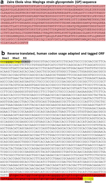
The Zaire Ebola virus Mayinga strain glycoprotein (GP) sequence, identical to the Gabon-94 strain and highly conserved with the new isolates (accession number: KJ660346.2, KJ660347, KJ660348), was used as a template to obtain its nucleotide sequence by reverse translation, which was human codon usage adapted and tagged at the carboxy-terminal with gD106 tag peptide [10, 20] (Fig. 1a, b). Furthermore, a Kozak’s sequence (to improve translation) and restriction enzyme sites (to facilitate the sub-cloning in suitable vector) were added to the synthetic ORF antigen (Fig. 1b), which we termed “syEBOVgD106”.
Fig. 1.

In silico design of EBOV GP synthetic ORF. a Zaire EBOV Mayinga strain GP sequence and b deduced nucleotide sequence provided of a tag peptide (gD 106; highlighted in red) a Kozak’ s sequence (KS; highlighted in grey) and restriction enzyme sites (SmaI and NheI; highlighted in yellow) for subcloning
Generation and expression of syEBOVgD106 antigen expression cassette
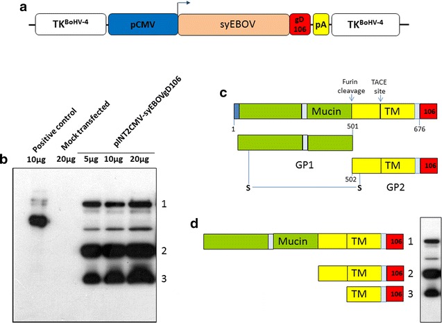
syEBOVgD106 synthetic ORF was cut out from its plasmid back-bone and sub-cloned in pINT2EGFP [17] shuttle vector containing a CMV-EGFP expression cassette, where EGFP ORF was substituted with syEBOVgD106 ORF. The resulting construct, pINT2CMV-syEBOVgD106, has two BoHV-4 TK region sequences, flanking the CMV-syEBOVgD106 expression cassette under the transcriptional control of the CMV early promoter and the bovine growth hormone polyadenylation signal sequence (Fig. 2a). The pINT2CMV-syEBOVgD106 construct was validated in terms of protein expression by transient transfection assay in HEK 293T cells and by western blotting using a specific monoclonal antibody against gD106 tag peptide. As expected, syEBOVgD106 glycoprotein was very well expressed in the pINT2CMV-syEBOVgD106 transfected HEK 293T cells (Fig. 2b) and showed the predicted banding pattern (Fig. 2c, d) generated by the cellular metalloprotease TACE [TNFα-converting enzyme, a member of the ADAM (a disintegrin and metalloproteinase proteinase family)] and Furin [21, 22].
Fig. 2.

Expression of syEBOVgD106. a Diagram (not on scale) of pINT2CMV-syEBOVgD106 targeting vector delivering the tagged syEBOVgD106 ORF (syEBOV, orange; gD106, red), under the control of the CMV promoter (CMV, blue) and the bovine growth hormone polyadenylation signal (PA, yellow). CMV-syEBOVgD106 expression cassette is flanked by BoHV-4 TK homologous sequences (white). b Western immunoblotting of pINT2CMV-syEBOVgD106 transfected HEK 293T cells extracts. Lanes were loaded with different amounts of total protein cell extract (5, 10 and 20 μg); cells transfected with pEGFPC-1 served as negative controls (Mock). The peculiar immune-blotting banding pattern is the result of the syEBOVgD106 protein processing by furin and TACE proteases (c), as revealed by the anti gD106 tag monoclonal antibody directed against three predicted and detected peptides (1 uncleaved; 2 only cleaved by Furin protease; 3 cleaved by Furin and TACE proteases) (d)
Vectorization of syEBOVgD106 expression cassette in BoHV-4-based vector
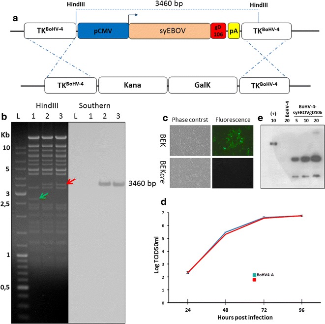
pINT2CMV-syEBOVgD106 shuttle plasmid construct was employed to generate pBAC-BoHV-4-syEBOVgD106ΔTK by heat-inducible homologous recombination in SW102 E. coli containing pBAC-BoHV-4-A-KanaGalKΔTK (Fig. 3a). KanaGalK negative selected colonies were amplified in liquid media and their respective BAC analyzed by HindIII restriction enzyme and successively by southern hybridization (Fig. 3b) using a specific, non-isotopic labeled probe directed against syEBOVgD106 ORF. pBAC-BoHV-4-syEBOVgD106ΔTK stability was assessed by host bacterial serial passages (over 20) and absence of aberrant recombination was detected by restriction enzyme digestion (data not shown).
Fig. 3.

BoHV-4-syEBOVgD106ΔTK constructs and characterization. a Diagram (not to scale) illustrating the re-targeting event (i.e., replacement of the Kana/GalK cassette with the CMV-syEBOVgD106 cassette) obtained by heat-inducible homologous recombination in SW102 E. coli cells containing pBAC-BoHV-4-A-TK-KanaGalK-TK. b Representative, 2-deoxy-galactose resistant colonies, tested by HindIII restriction enzyme analysis and southern blotting performed with a specific probe for syEBOVgD106 ORF. The 2650 bp band (indicated by a green arrow) corresponding to the non-retargeted pBAC-BoHV-4-A-TK-KanaGalK-TK control (lane 1) is replaced by 3460 bp band (indicated by a red arrow) in pBAC-BoHV-4-syEBOVgD106ΔTK (lanes 2 and 3). Phase contrast and fluorescent microscopy images of the plaques formed by viable, reconstituted recombinant BoHV-4-syEBOVgD106ΔTK (c) after electroporation of the corresponding BAC DNA clones into BEK or BEKcre cells (magnification, ×10). d Replication rate of BoHV-4-syEBOVgD106ΔTK grown in BEK cells and compared with that of the parental BoHV-4-A isolate. The data are the mean ± standard error of triplicate measurements (P > 0.05 for all time-points; Student’s t test). e Immunoblotting analyses conducted on extracts from cells infected with BoHV-4-syEBOVgD106ΔTK (numbers indicate the micrograms of total protein loaded). BoHV-4-A infected cells served as negative controls
Infectious BoHV-4-syEBOVgD106ΔTK viral particles were obtained by transfecting, through electroporation, BEK cells or BEK cells expressing cre recombinase (BEKcre) [16] to deplete the BAC cassette from pBAC-BoHV-4-syEBOVgD106ΔTK. In both cases, viable BoHV-4-syEBOVgD106 producing CPE and plaques was generated, although BoHV-4-syEBOVgD106ΔTK produced in BEKcre lost GFP expression due to the removal of GFP expression cassette associated to the BAC plasmid back-bone (Fig. 3c). Next, the growth characteristics of BoHV-4-syEBOVgD106ΔTK were compared with that one of the BoHV-4-A parental virus and no differences between them were observed (Fig. 3d). Furthermore, BoHV-4-syEBOVgD106ΔTK infected cells expressed syEBOVgD106 glycoprotein (Fig. 3e). Since the EBOV GP is a typical, type 1 integral membrane glycoprotein, its ability to be incorporated on the BoHV-4 envelope was investigated. A virus preparation from BoHV-4-A-CMV-IgK-gE2gD-TM was purified and analyzed by Western immunoblotting (Additional file 2: Figure S2). As expected, EBOV GP signal was detected only in BoHV-4-syEBOVgD106ΔTK virions, but not in the wild-type BoHV-4 virions, thus indicating the incorporation of syEBOVgD106 into BoHV-4 virus particles.
Goat immunization and humoral immune-response analysis
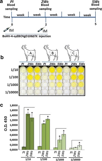
For testing the ability of BoHV-4-syEBOVgD106ΔTK to induce a humoral immune response in a large animal model, a pilot immunization study was performed in goats. In agreement with the current legislation on animal experimentation, which suggests to minimize the number of animals employed, three adult goats, after collection of the pre-immune serum, were inoculated subcutaneously with 1 mL of 106 TCID50 of BoHV-4-syEBOVgD106ΔTK. A second inoculation with an identical dose of BoHV-4-syEBOVgD106ΔTK was done 2 weeks apart from the first inoculation. Blood samples were collected at two, just before the second inoculum, and 5 weeks from the first inoculum (Fig. 4a). Serum samples were analyzed by ELISA and all three goats developed a very good antibody response, with high titers (Fig. 4b, c). As previously described for BoHV-4-based vaccine vector inoculation in goats [15], in the present study, a single inoculation of BoHV-4-syEBOVgD106ΔTK was able to elicit a humoral immune response 2 weeks post inoculation. Moreover, the antibody titer did not decrease when evaluated 6 months later (data not shown), showing long lasting immunization.
Fig. 4.

Kinetics of the humoral immune responses of goats immunized with BoHV-4-syEBOVgD106ΔTK. a Diagram showing the goats immunization scheme and blood sample collection. b Dilutes sera (1/10, 1/100, 1/1000 and 1/10,000) collected at 2 and 5 weeks (2 Ws; 5 Ws) from three (A, B and C) BoHV-4-syEBOVgD106ΔTK inoculated goats, were compared with pre-immune sera (PI) for anti-GP antibodies by ELISA. c Antibodies detected were expressed as the optical density at 450 nm and each value represents the mean response of the three goats sera, ± the standard error of the mean, at the same dilution and collected at the same time. Response differences between pre- and post-immune sera were measured by Student’s t test (*P ≤ 0.005)
Discussion
In this work, the potential utility of BoHV-4 as a safe, potent, large-capacity gene delivery vector for EBOV antigen was shown. A workflow strategy to construct a BoHV-4-based vector was generated and it was able to deliver an immune-dominant antigen derived from a BSL4 pathogen, thus avoiding all the economical and safety requirements necessary for the manipulation of this kind of biological agent. Furthermore, it was able to elicit a strong humoral immune-response. The results were obtained through a synthetic gene approach, currently based on solid-phase DNA synthesis, which allows the complete synthesis of a double-stranded DNA molecule with no apparent limits in nucleotide sequence or size.
As a model pathogen, EBOV was the primary choice. Firstly because a severe Ebola outbreak was taking place in African countries at the beginning of this project and at that time no approved prophylactic or therapeutic protocols were available. Secondly, EBOV it is classified as a Category A priority pathogen by NIAID and a Category A agent of bioterrorism by the CDC.
The EBOV genome is a single negative-strand RNA molecule encoding seven structural proteins, among which EBOV GP is a type I transmembrane glycoprotein of 676 amino acids in length and its transcript is made by an unusual transcriptional editing [23]. Full length GP has been shown to be responsible for eliciting a protective humoral immune response in infected individuals [8]. The cleavage of surface GP, by cellular metalloprotease, tumor necrosis factor α-converting enzyme (TACE), generates shed GP [21], which contributes to the host protective immune response [22]. Firstly, in silico customized full length GP ORF was successfully expressed in eukaryotic cells with a suitable expression vector and then integrated in BAC BoHV-4 genome through homologous recombination. BoHV-4-syEBOVgD106ΔTK replicated in cell culture at the same extent as the parental virus, thus no detrimental effect induced by the topological location of the foreign DNA in the BoHV-4 genome was observed. BoHV-4 is a Gammaherpesvirus belonging to the Rhadinovirus genera. Although BoHV-4 natural host is cattle, the virus has been isolated from other ruminants, including zebu (Bos indicus), American bison (Bison bison), African buffalo (Syncerus caffer), and sheep. Like other Herpesviruses, BoHV-4 is able to establish persistent infection in cells of the monocyte/macrophage lineage [24, 25] and in a bovine macrophage cell line (BoMAC) [26]. Furthermore, BoHV-4 experimental inoculation in rabbit demonstrates how spleen, as well as macrophages, are the main site of viral persistence [27]. Due to the lack of a direct correlation between BoHV-4 infection and specific lesions or pathology, BoHV-4 is not considered a primary pathogen and its genome was cloned as bacterial artificial chromosome (BAC), in order to be exploited as a gene delivery vector for immunization purposes and oncolysis. BoHV-4-based vector delivering antigens have been employed to immunize mice [9], rats [11], rabbits [12], sheep [13], swine [14], cows (paper in preparation) and goats [15] without any associated clinical signs or pathology.
Although goats are not susceptible to EBOV infection, BoHV-4-syEBOVgD106ΔTK immunization was performed in adults goats as they have previously been shown to be an appropriate large animal model for BoHV-4-based vector immunization [15]. Further, goats could be exploited as a source of antibodies production for antibody-based therapeutic in post-exposure treatment of EBOV disease. A pair of newly published studies [28, 29], demonstrated the efficacy of an ovine polyclonal antibody therapy against EBOV disease when tested in the stringent guinea pig model of EBOV disease.
The sub-cutaneous route of BoHV-4-based vector administration was shown to facilitate antigen production and vector replication takes place only at the site of inoculation [15], without spreading to the rest of the animal body. No BoHV-4-syEBOVgD106ΔTK viremia was detected in inoculated animals, although all BoHV-4-syEBOVgD106ΔTK inoculated animals were successfully immunized and high titers of EBOV GP antibodies were detected. The ability of BoHV-4-syEBOVgD106ΔTK to efficiently transduce goat skin cells, which consecutively expressed large amounts of GP, explains the consistent titer of antibodies produced and detected by the ELISA assay.
Whether the antibodies induced by BoHV-4-syEBOVgD106ΔTK in goats serum, correlates with a potential protection following a passive transfer in a suitable stringent model of EBOV disease remains unknown. Based on previous studies, which demonstrated the ability of BoHV-4 based vectors to efficiently protect against the Category A agent Monkeypox virus [10], it can be assumed that the use of BoHV-4 vector based platform represents an effective tool to test unknown antigens and vectors for class A pathogens.
Conclusion
In this study, the possibility to generate a BoHV-4-based vector delivering an immune-dominant antigen coming from EBOV and proven to be able to generate high titer of antibodies in inoculated goats was shown.
Authors’ contributions
DG conceived the experiments. AR, SJ, GT, ST, VF, FM and DG performed the experiments. SC and GK contributed with reagents. GD and DPK analysed the data. GD wrote the paper. All authors read and approved the final manuscript.
Acknowledgements
We would like to tank Dr. Laura Kramer for the English editing of the manuscript.
Competing interests
The authors declare that they have no competing interests.
Availability of data and materials
Available under request.
Ethics approval and consent to participate
Adult goats were used for the in vivo immunization study. Animal experimentation was conducted under the approval of the Ethical Committee of the University of Parma and the Italian Ministry of Health and it was carried out at the University of Parma, which follows the guidelines of the National Guide for Care and Use of Experimental Animals (n. 3/2014). The Animal Care and Use Program at Parma University are fully accredited by the European Accreditation of Laboratory Animal Care.
Funding
This work was supported by Italian Ministry of University and Scientific Research (Italian National Grant MIUR, PRIN 2010-2011).
Abbreviations
- EBOV
Ebola virus
- BoHV-4
bovine herpesvirus 4
- GP
glycoprotein
- ELISA
enzyme linked immunosorbent assay
- ORF
open reading frame
- CFR
case fatality report
- rVSV
recombinant vesicular stomatitis virus
- ZEBOVΔG
Zaire EBOV glycoprotein
- rAd5
recombinant adenovirus serotype 5
- rChAd3
recombinant chimpanzee adenovirus serotype 3
- rHPIV3
recombinant human parainfluenza virus 3
- BAC
bacterial artificial chromosome
- BSL
biosafety level
- BEK
bovine embryo kidney
- HEK 293T
human embryo kidney 293T
- EMEM
Eagle’s minimal essential medium
- FBS
fetal bovine serum
- EBOsec
EBOV secreted fragment
- PEI
polyethylenimine
- DMEM
Dulbecco’s modified essential medium
- MOI
multiplicity of infection
- TCID50
tissue culture infectious doses 50
- CPE
cytopathic effect
- SDS-PAGE
sodium dodecyl sulfate-polyacrylamide gel electrophoresis
- BoHV-1
bovine herpesvirus 1
- HRP
horseradish peroxidase
- LB
Luria–Bertani
- BSA
bovine serum albumin
- TMB
tetramethylbenzidine
- TK
thymidine kinase
- TACE
tumor necrosis factor α-converting enzyme
- PI
pre-immune
- BoMAC
bovine macrophage cell line
- FDA
Food and Drug Administration
- PA
polyadenylation
Additional files
Additional file 1: Figure S1. Overall strategy employed for cloning and expression of EBOV GP as a soluble secreted form. A Diagram (not to scale) showing the structure of gD106 tagged (red) EBOV GP. The EBOsecgD106 has been obtained by eliminating the transmembrane domain (TM). EBOsecgD106 peptide has been produced in serum free medium of HEK 293T cells transfected with pCMVEBOsecgD106. B A time course to optimize EBOsecgD106 expression at different time post transfection as analyzed by western immunoblotting was used. Twenty-four hours was considered as the best time post transfection to collect the cell sovranatant.
Additional file 2: Figure S2. Incorporation of EBOV GP into recombinant BoHV-4 particles. Extracts of purified viruses (BoHV-4-A and BoHV-4-syEBOVgD106) analyzed by Western immunoblotting.
Contributor Information
Alfonso Rosamilia, Email: alfonso.rosamilia@studenti.unipr.it.
Sarah Jacca, Email: sarah.jacca@studenti.unipr.it.
Giulia Tebaldi, Email: giulia.tebaldi1@studenti.unipr.it.
Silvia Tiberti, Email: silvia.tiberti@studenti.unipr.it.
Valentina Franceschi, Email: valentina.franceschi@unipr.it.
Francesca Macchi, Email: francesca.macchi@studenti.unipr.it.
Sandro Cavirani, Email: sandro.cavirani@unipr.it.
Gary Kobinger, Email: gary.kobinger@phac-aspc.gc.ca.
Donald P. Knowles, Email: don.knowles@ars.usda.gov
Gaetano Donofrio, Phone: 00390521902677, Email: gaetano.donofrio@unipr.it.
References
- 1.de La Vega MA, Wong G, Kobinger GP, Qiu X. The multiple roles of sGP in Ebola pathogenesis. Viral Immunol. 2015;28:3–9. doi: 10.1089/vim.2014.0068. [DOI] [PMC free article] [PubMed] [Google Scholar]
- 2.Martines RB, Ng DL, Greer PW, Rollin PE, Zaki SR. Tissue and cellular tropism, pathology and pathogenesis of Ebola and Marburg viruses. J Pathol. 2015;235:153–174. doi: 10.1002/path.4456. [DOI] [PubMed] [Google Scholar]
- 3.Stanley DA, Honko AN, Asiedu C, Trefry JC, Lau-Kilby AW, Johnson JC, Hensley L, Ammendola V, Abbate A, Grazioli F, et al. Chimpanzee adenovirus vaccine generates acute and durable protective immunity against ebolavirus challenge. Nat Med. 2014;20:1126–1129. doi: 10.1038/nm.3702. [DOI] [PubMed] [Google Scholar]
- 4.Ledgerwood JE, Costner P, Desai N, Holman L, Enama ME, Yamshchikov G, Mulangu S, Hu Z, Andrews CA, Sheets RA, et al. A replication defective recombinant Ad5 vaccine expressing Ebola virus GP is safe and immunogenic in healthy adults. Vaccine. 2010;29:304–313. doi: 10.1016/j.vaccine.2010.10.037. [DOI] [PubMed] [Google Scholar]
- 5.Meyer M, Garron T, Lubaki NM, Mire CE, Fenton KA, Klages C, Olinger GG, Geisbert TW, Collins PL, Bukreyev A. Aerosolized Ebola vaccine protects primates and elicits lung-resident T cell responses. J Clin Investig. 2015;125:3241–3255. doi: 10.1172/JCI81532. [DOI] [PMC free article] [PubMed] [Google Scholar]
- 6.Johnson RF, Kurup D, Hagen KR, Fisher C, Keshwara R, Papaneri A, Perry DL, Cooper K, Jahrling PB, Wang JT, et al. An inactivated rabies virus-based Ebola vaccine, FILORAB1, adjuvanted with glucopyranosyl lipid A in stable emulsion confers complete protection in nonhuman primate challenge models. J Infect Dis. 2016;214:S342–S354. doi: 10.1093/infdis/jiw231. [DOI] [PMC free article] [PubMed] [Google Scholar]
- 7.Henao-Restrepo AM, Longini IM, Egger M, Dean NE, Edmunds WJ, Camacho A, Carroll MW, Doumbia M, Draguez B, Duraffour S, et al. Efficacy and effectiveness of an rVSV-vectored vaccine expressing Ebola surface glycoprotein: interim results from the Guinea ring vaccination cluster-randomised trial. Lancet. 2015;386:857–866. doi: 10.1016/S0140-6736(15)61117-5. [DOI] [PubMed] [Google Scholar]
- 8.Mire CE, Matassov D, Geisbert JB, Latham TE, Agans KN, Xu R, Ota-Setlik A, Egan MA, Fenton KA, Clarke DK, et al. Single-dose attenuated vesiculovax vaccines protect primates against Ebola Makona virus. Nature. 2015;520:688–691. doi: 10.1038/nature14428. [DOI] [PMC free article] [PubMed] [Google Scholar]
- 9.Franceschi V, Capocefalo A, Calvo-Pinilla E, Redaelli M, Mucignat-Caretta C, Mertens P, Ortego J, Donofrio G. Immunization of knock-out alpha/beta interferon receptor mice against lethal bluetongue infection with a BoHV-4-based vector expressing BTV-8 VP2 antigen. Vaccine. 2011;29:3074–3082. doi: 10.1016/j.vaccine.2011.01.075. [DOI] [PubMed] [Google Scholar]
- 10.Franceschi V, Parker S, Jacca S, Crump RW, Doronin K, Hembrador E, Pompilio D, Tebaldi G, Estep RD, Wong SW, et al. BoHV-4-based vector single heterologous antigen delivery protects STAT1(−/−) mice from monkeypoxvirus lethal challenge. PLoS Negl Trop Dis. 2015;9:e0003850. doi: 10.1371/journal.pntd.0003850. [DOI] [PMC free article] [PubMed] [Google Scholar]
- 11.Donofrio G, Martignani E, Poli E, Lange C, Martini FM, Cavirani S, Cabassi CS, Taddei S, Flammini CF. Bovine herpesvirus 4 based vector interaction with liver cells in vitro and in vivo. J Virol Methods. 2006;136:126–136. doi: 10.1016/j.jviromet.2006.04.008. [DOI] [PubMed] [Google Scholar]
- 12.Donofrio G, Franceschi V, Capocefalo A, Taddei S, Sartori C, Bonomini S, Cavirani S, Cabassi CS, Flammini CF. Cellular targeting of engineered heterologous antigens is a determinant factor for bovine herpesvirus 4-based vaccine vector development. Clin Vaccine Immunol. 2009;16:1675–1686. doi: 10.1128/CVI.00224-09. [DOI] [PMC free article] [PubMed] [Google Scholar]
- 13.Donofrio G, Sartori C, Ravanetti L, Cavirani S, Gillet L, Vanderplasschen A, Taddei S, Flammini CF. Establishment of a bovine herpesvirus 4 based vector expressing a secreted form of the bovine viral diarrhoea virus structural glycoprotein E2 for immunization purposes. BMC Biotechnol. 2007;7:68. doi: 10.1186/1472-6750-7-68. [DOI] [PMC free article] [PubMed] [Google Scholar]
- 14.Donofrio G, Taddei S, Franceschi V, Capocefalo A, Cavirani S, Martinelli N, Ottonello S, Ferrari M. Swine adipose stromal cells loaded with recombinant bovine herpesvirus 4 virions expressing a foreign antigen induce potent humoral immune responses in pigs. Vaccine. 2011;29:867–872. doi: 10.1016/j.vaccine.2010.11.048. [DOI] [PubMed] [Google Scholar]
- 15.Donofrio G, Franceschi V, Lovero A, Capocefalo A, Camero M, Losurdo M, Cavirani S, Marinaro M, Grandolfo E, Buonavoglia C, Tempesta M. Clinical protection of goats against CpHV-1 induced genital disease with a BoHV-4-based vector expressing CpHV-1 gD. PLoS ONE. 2013;8:e52758. doi: 10.1371/journal.pone.0052758. [DOI] [PMC free article] [PubMed] [Google Scholar]
- 16.Donofrio G, Sartori C, Franceschi V, Capocefalo A, Cavirani S, Taddei S, Flammini CF. Double immunization strategy with a BoHV-4-vectorialized secreted chimeric peptide BVDV-E2/BoHV-1-gD. Vaccine. 2008;26:6031–6042. doi: 10.1016/j.vaccine.2008.09.023. [DOI] [PubMed] [Google Scholar]
- 17.Donofrio G, Cavirani S, Simone T, van Santen VL. Potential of bovine herpesvirus 4 as a gene delivery vector. J Virol Methods. 2002;101:49–61. doi: 10.1016/S0166-0934(01)00419-0. [DOI] [PubMed] [Google Scholar]
- 18.Donofrio G, Cavaggioni A, Bondi M, Cavirani S, Flammini CF, Mucignat-Caretta C. Outcome of bovine herpesvirus 4 infection following direct viral injection in the lateral ventricle of the mouse brain. Microbes Infect. 2006;8:898–904. doi: 10.1016/j.micinf.2005.10.016. [DOI] [PubMed] [Google Scholar]
- 19.Warming S, Costantino N, Court DL, Jenkins NA, Copeland NG. Simple and highly efficient BAC recombineering using galK selection. Nucleic Acids Res. 2005;33:e36. doi: 10.1093/nar/gni035. [DOI] [PMC free article] [PubMed] [Google Scholar]
- 20.Capocefalo A, Franceschi V, Mertens PP, Castillo-Olivares J, Cavirani S, Di Lonardo E, Leni Z, Donofrio G. Expression and secretion of Bluetongue virus serotype 8 (BTV-8)VP2 outer capsid protein by mammalian cells. J Virol Methods. 2010;169:420–424. doi: 10.1016/j.jviromet.2010.08.002. [DOI] [PubMed] [Google Scholar]
- 21.Escudero-Perez B, Volchkova VA, Dolnik O, Lawrence P, Volchkov VE. Shed GP of Ebola virus triggers immune activation and increased vascular permeability. PLoS Pathog. 2014;10:e1004509. doi: 10.1371/journal.ppat.1004509. [DOI] [PMC free article] [PubMed] [Google Scholar]
- 22.Dolnik O, Volchkova VA, Escudero-Perez B, Lawrence P, Klenk HD, Volchkov VE. Shedding of Ebola virus surface glycoprotein is a mechanism of self-regulation of cellular cytotoxicity and has a direct effect on virus infectivity. J Infect Dis. 2015;212(Suppl 2):S322–S328. doi: 10.1093/infdis/jiv268. [DOI] [PubMed] [Google Scholar]
- 23.Volchkov VE, Becker S, Volchkova VA, Ternovoj VA, Kotov AN, Netesov SV, Klenk HD. GP mRNA of Ebola virus is edited by the Ebola virus polymerase and by T7 and vaccinia virus polymerases. Virology. 1995;214:421–430. doi: 10.1006/viro.1995.0052. [DOI] [PubMed] [Google Scholar]
- 24.Osorio FA, Reed DE, Rock DL. Experimental infection of rabbits with bovine herpesvirus-4: acute and persistent infection. Vet Microbiol. 1982;7:503–513. doi: 10.1016/0378-1135(82)90045-1. [DOI] [PubMed] [Google Scholar]
- 25.Osorio FA, Rock DL, Reed DE. Studies on the pathogenesis of a bovine cytomegalo-like virus in an experimental host. J Gen Virol. 1985;66(Pt 9):1941–1951. doi: 10.1099/0022-1317-66-9-1941. [DOI] [PubMed] [Google Scholar]
- 26.Donofrio G, van Santen VL. A bovine macrophage cell line supports bovine herpesvirus-4 persistent infection. J Gen Virol. 2001;82:1181–1185. doi: 10.1099/0022-1317-82-5-1181. [DOI] [PubMed] [Google Scholar]
- 27.Lopez OJ, Galeota JA, Osorio FA. Bovine herpesvirus type-4 (BHV-4) persistently infects cells of the marginal zone of spleen in cattle. Microb Pathog. 1996;21:47–58. doi: 10.1006/mpat.1996.0041. [DOI] [PubMed] [Google Scholar]
- 28.Dowall SD, Bosworth A, Rayner E, Taylor I, Landon J, Cameron I, Coxon R, Al Abdulla I, Graham VA, Hall G, et al. Post-exposure treatment of Ebola virus disease in guinea pigs using EBOTAb, an ovine antibody-based therapeutic. Sci Rep. 2016;6:30497. doi: 10.1038/srep30497. [DOI] [PMC free article] [PubMed] [Google Scholar]
- 29.Dowall SD, Callan J, Zeltina A, Al-Abdulla I, Strecker T, Fehling SK, Krahling V, Bosworth A, Rayner E, Taylor I, et al. Development of a cost-effective ovine polyclonal antibody-based product, EBOTAb, to treat Ebola virus infection. J Infect Dis. 2016;213:1124–1133. doi: 10.1093/infdis/jiv565. [DOI] [PMC free article] [PubMed] [Google Scholar]
Associated Data
This section collects any data citations, data availability statements, or supplementary materials included in this article.
Data Availability Statement
Available under request.


