Abstract
The opportunistic fungal pathogen Candida albicans is both a benign gut commensal and a frequently fatal systemic pathogen. The interaction of C. albicans with the host's innate immune system is the primary factor in this balance; defects in innate immunity predispose the patient to disseminated candidiasis. Because of the central importance of phagocytic cells in defense against fungal infections, we have investigated the response of C. albicans to phagocytosis by mammalian macrophages using genomic transcript profiling. This analysis reveals a dramatic reprogramming of transcription in C. albicans that occurs in two successive steps. In the early phase cells shift to a starvation mode, including gluconeogenic growth, activation of fatty acid degradation, and downregulation of translation. In a later phase, as hyphal growth enables C. albicans to escape from the macrophage, cells quickly resume glycolytic growth. In addition, there is a substantial nonmetabolic response imbedded in the early phase, including machinery for DNA damage repair, oxidative stress responses, peptide uptake systems, and arginine biosynthesis. Further, a surprising percentage of the genes that respond specifically to macrophage contact have no known homologs, suggesting that the organism has undergone substantial evolutionary adaptations to the commensal or pathogen lifestyle. This transcriptional reprogramming is almost wholly absent in the related, but nonpathogenic, yeast Saccharomyces cerevisiae, suggesting that these large-scale and coordinated changes contribute significantly to the ability of this organism to survive and cause disease in vivo.
The polymorphic yeast Candida albicans, like many pathogens, has both a benign and pathogenic association with its host. Candida is part of the commensal flora: healthy humans are estimated to have carriage rates of >50% in the gastrointestinal tract and between 10 and 20% in the oral cavity, anorectal tract, and vagina (29). In its pathogenic state, it causes a wide variety of diseases, including common mucosal manifestations such as oropharyngeal thrush and vaginitis. In hospitalized patients, disseminated candidiasis is the fourth most common infection (outpacing all gram-negative species) and carries a mortality rate in excess of 30% (17, 24, 41). Since there are limited treatment options (18), development of the next generation of antifungal agents will require a further understanding of the biology of infection.
Analysis of the response of Candida to macrophages provides a window into the alterations necessary for the organism to survive its first encounter with the immune system. Once inside the macrophage, the yeast form of Candida differentiates into the filamentous hyphal form, which can pierce the macrophage, allowing it to resume proliferation. This internalization and escape can be observed in an in vitro system in which C. albicans and cultured macrophages are mixed. It is clear, however, that the morphogenetic change is only part of the response to phagocytosis. One method for analyzing this encounter more completely is to identify the alterations in transcription as Candida is undergoing phagocytosis.
Until recently comprehensive, transcription studies were limited because the genome sequence of C. albicans was not available. However, whole-genome microarrays were available to analyze the phagocytosis of the related but nonpathogenic yeast, Saccharomyces cerevisiae. These studies identified upregulation of the glyoxylate cycle when S. cerevisiae was internalized. Based on these findings, the glyoxylate cycle was also found to be induced in C. albicans and to play a significant role in virulence in a mouse model (23). A role for the glyoxylate cycle in microbial virulence has now been reported in many organisms including Mycobacterium tuberculosis (25), and the plant pathogens Leptosphaera maculans (fungal [14]), Magnaporthe grisea (fungal [40]), and Rhodococcus fascians (bacterial [39]). Isocitrate lyase, one of the key enzymes of the glyoxylate cycle, is upregulated in Cryptococcus neoformans (fungal) isolated from cerebrospinal fluid, but it is not required for virulence in this model (33). These studies have helped to demonstrate the potential of genomic analysis of host-pathogen interactions.
The recent completion of the C. albicans genome sequence (http://sequence-www.stanford.edu/group/candida/index.html; http://candida.bri.nrc.ca/candida) enables genomic analysis directly in C. albicans, which promises to be a valuable tool in the study of this asexual diploid yeast. Genomic studies of morphogenesis (26, 27) and responses to stress (9), blood (10), and neutrophils (32) have begun to elucidate the complex transcriptional programs that C. albicans uses to survive in vivo. Here we identify the global transcriptional response of C. albicans upon internalization by mammalian macrophages. Phagocytosis stimulates an immediate transcriptional response. The early pattern is characterized by a dramatic upregulation of the gluconeogenesis/glyoxylate pathways and downregulation of the genes encoding the translation apparatus. The nonfilamentous Δcph1 Δefg1 mutant (21), which is internalized but cannot escape, remains frozen in the early pattern. We have also identified a specific nonmetabolic response distinct from filamentation embedded in the early pattern that responds to stresses presented by macrophage contact. This early transcription profile switches to a later profile as the cells reprogram to escape the macrophage and resume rapid growth. This response is absent in the nonpathogenic S. cerevisiae, underscoring the pathogen/nonpathogen dichotomy and revealing a highly coordinated system in C. albicans for immune evasion.
MATERIALS AND METHODS
Strains, media, and growth conditions.
The C. albicans wild-type strain SC5314 was used for most experiments (11). The Δcph1 Δefg1 (HLC54) and Δtup1 (BCa2-9) strains have been described (4, 22). Candida strains were propagated in standard media, including YPD (1% yeast extract, 2% peptone, 2% dextrose) and YNB (0.17% yeast nitrogen base without amino acids, 0.5% ammonium sulfate, 2% dextrose) as described previously (35), except for the media used in the starvation experiments (described below). Murine macrophage line J774A (ATCC TIB-67) was grown in RPMI plus 10% fetal bovine serum (FBS) at 37°C in 5% CO2 in normal air.
Coculture conditions.
On day −1, macrophages were collected, washed and counted with a hemocytometer. A total of 4 × 10⁷ cells were plated in RPMI plus 10% FBS in 750-ml tissue culture flasks and grown overnight in 5% CO2 at 37°C. C. albicans strains were grown overnight at 37°C in YPD with 4% glucose to maintain cells in the yeast form. In the morning these cells were diluted 1:50 and grown for ∼4 h. Cultures were collected by centrifugation, washed in phosphate-buffered saline and counted. Repeated cell counts of the overnight macrophage cultures indicated a small increase to ca. 5 × 10⁷ cells per flask. We added 108 C. albicans cells to these flasks (a 2:1 C. albicans/macrophage ratio) and incubated for 1 to 6 h. For control cultures, 2 × 10⁸ C. albicans cells were inoculated to tissue culture flasks in 50 ml of RPMI plus 10% serum. Cells were collected by scraping the flasks with rubber scrapers in ice-cold water (to promote macrophage lysis) and transferred to 50-ml conical tubes. Tubes were centrifuged at 3,000 rpm at 4°C, washed twice with water, transferred to 1.5-ml microfuge tubes,and frozen on dry ice.
Nutrient starvation conditions.
SC5314 was grown overnight in YNB with 2% glucose and 0.5% ammonium sulfate at 37°C. Cultures were diluted into the same medium to an optical density at 600 nm (OD600) of ∼0.5 and grown for two doublings (to an OD600 of ∼2, ca. 4 h). Cells were collected by centrifugation and washed once with water. These cells were used to inoculate fresh medium at an OD600 of ∼0.5. For each starvation condition, the medium was 1.7 g of yeast nitrogen base/liter, which contains no usable nitrogen or carbon source. To this medium, we added 2% glucose (no nitrogen), 0.5% ammonium sulfate (no carbon), 2% potassium acetate, and 0.5% ammonium sulfate (acetate), or nothing (no carbon and no nitrogen). We also grew cells in YNB plus 2% glucose and 0.5% ammonium sulfate (YNB complete) as a rich medium control. Cultures were grown for 1 to 3 h (corresponding to the times at which C. albicans is inside the phagosome in the macrophage experiments) at 37°C and collected by filtration. Filters were frozen on dry ice-ethanol in 50-ml conical tubes. RNA was prepared directly from the collected cells on the filters as described below.
RNA isolation, microarray hybridization, and data analysis.
RNA was prepared from the frozen pellets or filters by the hot acidic phenol protocol (1). For the macrophage-ingested cells and the medium-alone controls, RNA was amplified via a cDNA approach by using a kit from Arcturus (Mountain View, Calif.). RNA was not amplified from the starvation experiments. Microarray labeling and hybridization were done via established protocols (http://microarrays.org). All experimental conditions were hybridized against a complex reference mixture of mRNAs prepared from ∼20 conditions (M. Lorenz, D. Inglis, A. Johnson, and G. Fink, unpublished data). The same pool of reference RNA was used for all hybridizations and was always labeled with Cy5. Experimental conditions were labeled with Cy3. Because of the constant reference condition, dye-swap experiments were not necessary.
Arrays were scanned with an Axon 4000B scanner, and data were extracted by using the Axon GenePix program. The data from each array were normalized to a constant value to equalize fluorescence intensity across slides. Flagged spots from the GenePix program due to high background intensity or below-background spot intensities were omitted from further analysis.
For each condition, we did at least two biological replicates, each hybridized in duplicate, for a minimum of four arrays per condition. Replicate data for each gene were averaged; genes with dissimilar replicates were discarded. All analyses were done by using ratios of the experimental (usually plus macrophages) condition over the control (medium alone) condition at the same time point.
Cluster analysis.
Hierarchical clustering (8) was used to identify patterns of regulation in the various datasets. In initial experiments, looking at carbon metabolism and ribosome function, patterns were easily evident and clustering was used only as a tool for data visualization. In later experiments (see Fig. 5 and 6 and Table 4), clustering was used for gene identification. For any given gene to be included in the analyses, there had to be reliable data for at least 90% of the conditions and a minimum variance (2- to 2.5-log2 units or 4- to 5.6-fold, depending on the experiment) between the lowest and highest expression values across all conditions to eliminate genes whose expression did not change substantially. We used three datasets: the 6-h phagocytosis time courses of the wild-type (SC5314) and Δcph1 Δefg1 (HLC54) strains and the 3-h time course of the wild-type strain under carbon starvation (in medium lacking carbon, lacking both carbon and nitrogen, or containing acetate as a carbon source [see above]), a total of 19 conditions. Clustering of these data identified genes coregulated by starvation and phagocytosis (see Fig. 5) and differentially regulated genes. Because of the different effects of the Δcph1 Δefg1 mutations, the 227 differentially regulated genes were found in multiple clusters that, for the purposes of visualization, were combined and reclustered to produce Fig. 6 and supplementary Fig. 3. (The complete, normalized data set and supplementary figures are available for download at http://www.lorenzlab.org).
FIG. 5.
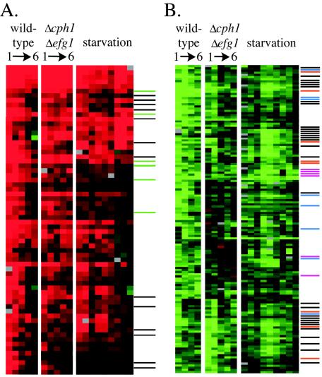
Similarities between starvation and phagocytosis. Data from the macrophage experiments in wild-type and Δcph1 Δefg1 strains were clustered with the starvation conditions (no nitrogen and no carbon, no carbon, or acetate as a carbon source, with a 1-, 2-, and 3-h time point for each condition). (A) Cluster of genes upregulated by macrophages and by starvation. Genes related to carbon metabolism are indicated by a black line; nutrient transporters are indicated by a green line. Of the 66 genes in this cluster, 19 fit one of these categories. (B) Downregulated cluster. Ribosomal proteins are indicated by black lines, tRNA synthases are indicated in blue, translational factors are indicated in orange, and glycolytic genes are indicated in purple. Of the 145 genes, 52 fit one of these categories.
FIG. 6.
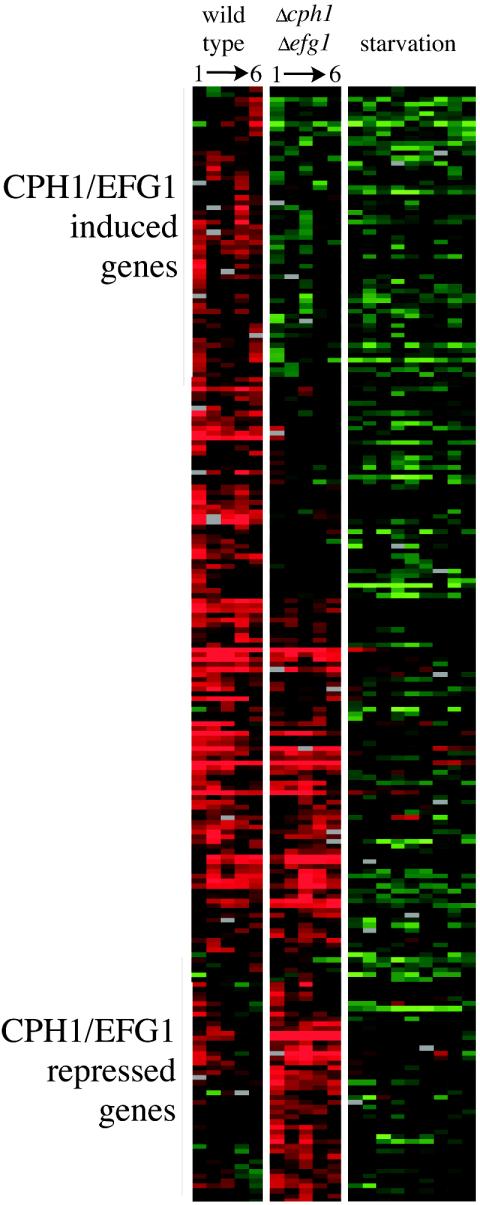
Macrophage-signature expression cluster. Data from the wild-type and Δcph1 Δefg1 strains with macrophages and the wild-type strain in various starvation conditions were clustered. This cluster contains 227 genes that are upregulated by macrophage contact but not by starvation. All 19 conditions shown were clustered together; the separation of the three experiments is for clarity only.
TABLE 4.
Genes upregulated by macrophage contacta
| Category | Gene(s) | ORF19 | Function |
|---|---|---|---|
| DNA damage repair | APN1 | 7428 | AP endonuclease (apurinic/apyrimidinic DNA lyase) |
| CDC45 | 1988 | Required for initiation of DNA replication | |
| HPR5 | 5970 | DNA helicase involved in repair | |
| POL2 | 2365 | DNA Pol e, involved in replication and repair | |
| POL30 | 4614 | Proliferating cell nuclear antigen, required for replication and repair | |
| RAD57 | 2174 | Required for recombination and recombinatorial repair | |
| RDH54 | 5367 | Required for recombination and recombinatorial repair | |
| Cytochrome activity | COQ3 | 3400 | Protein involved in biosynthesis of coenzyme Q |
| COQ4 | 3008 | Protein involved in biosynthesis of coenzyme Q | |
| CYC3 | 1957 | Holocytochrome c synthase | |
| FESUR1 | 6794 | Ubiquinone reductase | |
| QCR2 | 2644 | Ubiquinol cytochrome c reductase | |
| RIB3 | 5228 | DBP synthase, riboflavin biosynthesis | |
| Oxidative stress | CCP1 | 7868 | Cytochrome c peroxidase, destroys toxic radicals |
| MXR1 | 8675 | Methinone sulfoxide reductase | |
| GPX3 | 85 | Glutathione peroxidase | |
| GRE3 | 4317 | Xylose reductase | |
| AYR1 | 6167 | Putative oxidoreductase | |
| YPL088-1 | 8893 | Putative oxidoreductase | |
| YPL088-4 | 4477 | Putative oxidoreductase | |
| YBL064 | 5180 | Putative 1-Cys peroxidase | |
| YHB1 | 3707 | NO-responsive flavohemoglobin | |
| IDP1 | 5211 | Isocitrate dehydrogenase | |
| Metal homeostasis | CIP1 | 7761 | Cadmium induced protein |
| FRE3/CFL1 | 6139 | Ferric reductase | |
| FRE7 | 7077 | Ferric reductase | |
| CTR1 | 3646 | Copper transporter | |
| ZRT2 | 1585 | Low-affinity zinc transporter | |
| YFH1 | 1413 | Human frataxin homolog, iron homeostasis |
The table lists a portion of the 227 gene macrophage-induced cluster.
FIG. 3.
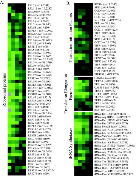
Widespread repression of translation by phagocytosis. Hierarchical clustering of expression data for genes related to translation during the entire 6-h time course. (A) All 54 ribosomal proteins included on our array. (B) All of the translation initiation factors (n = 23), translation elongation factors (n = 11), and cytoplasmic tRNA synthetases (n = 20) found on our array. Gene names are from S. cerevisiae homologs. Red represents induced genes, green represents repressed genes, and black indicates no change.
Microarrays.
Microarrays were based on open reading frames (ORFs) found in assemblies four to six of the C. albicans genome sequence (http://sequence-www.stanford.edu/group/candida/index.html). PCR products for ∼7,600 ORFs were spotted; about half are present in duplicate for a total of 10,831 spots per array. We have recently been able to map these 7,600 putative ORFs to ∼6,600 actual ORFs identified in the most recent genome assembly. The lower number is due, for the most part, to sequencing errors or incomplete assemblies in early releases that split single ORFs into multiple fragments. Arrays were spotted by using an Omnigrid robot (Genemachines, San Mateo, Calif.). This array has been described (2, 32, 38).
RESULTS
Macrophage-C. albicans coculture system.
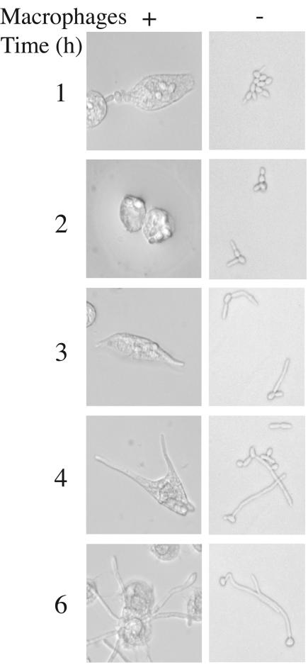
The transcriptional analysis of phagocytosis involved coincubation of J774A murine macrophage-like cells, together with C. albicans yeast cells in tissue culture medium (RPMI plus 10% FBS) at 37°C. At a Candida/macrophage ratio of 1:2, virtually all (>98%) of C. albicans cells are associated with macrophages (either attached or internalized) within 1 h of coculture (Fig. 1). The polarized growth of these cells eventually ruptures the macrophage, destroying the phagocyte and releasing the C. albicans cell. We determined transcript profiles of phagocytosed cells at various points during this interaction (at 1, 2, 3, 4, and 6 h postinoculation) and compared these profiles to cells grown in the same media for the same times in the absence of macrophages (see Materials and Methods).
FIG. 1.
In vitro C. albicans-macrophage interaction system. C. albicans strain SC5314 was incubated in RPMI plus 10% FBS at 37°C with or without macrophage line J774A. Germ tube formation, which occurs with roughly the same kinetics with or without macrophages, allows escape from the macrophage during the 6-h experiment.
Switch to a nutrient-poor growth mode.
The molecular composition of the phagosome, in terms of usable nutrients, is essentially unknown, but it is unlikely to be a rich environment; in particular, we would not expect it to contain substantial quantities of glucose or other sugars. Our previous work, demonstrating induction of the glyoxylate cycle and a virulence role for this metabolic pathway, was consistent with this assumption (23), as are the transcript profiles from phagocytosed C. albicans cells.
Early response to internalization reflects glucose deprivation.
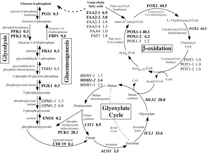
The initial transcriptional response to the internal milieu of the macrophage is a switch from glycolysis to gluconeogenesis (Fig. 2). In addition to being a preferred energy source, glucose is the precursor of other macromolecules, notably nucleotides via the pentose phosphate pathway, and must be synthesized if not available. The overall profiles show both the switch to gluconeogenic growth and a likely flow of carbon from fatty acids through the glyoxylate cycle to glucose.
FIG. 2.
Complete induction of alternative carbon metabolism upon phagocytosis. The pathways of β-oxidation, the glyoxylate cycle, gluconeogenesis, and glycolysis are shown, with the C. albicans gene names. Induction ratios (i.e., with macrophages/without macrophages) at 1 h are given, with genes in boldface regulated at least threefold. The dashed arrow shows the net reaction of these pathways: the conversion of fatty acids to glucose.
The switch from glycolysis to gluconeogenesis in phagocytosed cells can be seen most clearly in the two steps at which these pathways differ (see Fig. 2); both of which are key rate-limiting regulatory steps. In the first, there is an ∼30-fold expression difference between the enzymes that interconvert fructose-6-phosphate and fructose-1,6-bisphosphate. The gluconeogenic enzyme FBP1 is induced 9.6-fold above control cells, while the glycolytic PFK1 and PFK2 are each repressed ∼3.3-fold. Even more striking is the control of genes affecting the level of phosphoenolpyruvate, which is converted to citrate by CDC19 (10-fold repressed), whereas it is synthesized from oxaloacetate by PCK1 (induced 28.1-fold). Thus, upon internalization, there is nearly a 300-fold expression difference between these competing enzymes. Shared steps, in contrast, are mildly downregulated (averaging 2.5-fold repression), a finding consistent with a general reduction of cellular metabolism, as will be seen below.
In poor nutrient conditions, generating oxaloacetate from simple carbon compounds requires the glyoxylate cycle. We had previously shown that the key enzymes isocitrate lyase and malate synthase (ICL1 and MLS1) are strongly induced in phagocytosed populations of both C. albicans and S. cerevisiae (23). The microarray data confirm this in C. albicans, showing 33.6- and 28.0-fold induction of these enzymes. Three other enzymes participate in both the glyoxylate and tricarboxylic acid cycles: aconitase (ACO1) and citrate synthase (CIT1) are upregulated 3.5- and 8.9-fold, respectively. There are three isoforms of the third enzyme, malate dehydrogenase (MDH1), induced 1.3-, 2.5-, and 3.4-fold. Thus, all glyoxylate steps are induced to some degree (averaging 9.2-fold). Nonglyoxylate tricarboxylic acid cycle steps are not induced (ratio of 0.8; see supplementary Fig. 1A). Thus, the transcript profiles show how the gluconeogenic precursor oxaloacetate is generated, i.e., via the glyoxylate cycle.
Our data strongly suggest that the acetyl coenzyme A (acetyl-CoA) that drives the glyoxylate cycle is the product of β-oxidation of fatty acids. Every step of this pathway (except the three isoforms of POT1; Fig. 2) is significantly upregulated. Thus, transcript profiles show a clear flow of carbon from fatty acids to acetyl-CoA to glucose via these three biochemical pathways.
In addition, numerous other proteins related to the glyoxylate shunt are induced (see supplementary Fig. 1B), including transport proteins, peroxisomal biogenesis genes, and acetyl-CoA synthetase. The dramatic clarity of this pathway (virtually every enzyme required for the conversion of fatty acids to glucose is induced substantially), coupled with the distinct difference between gluconeogenesis and glycolysis (supplementary Fig. 1A and C), is striking and suggests a profoundly important role for the utilization of alternative carbon sources in vivo. Our previous data suggested this, but these genome-wide studies provide direct evidence for the suggestion (23) that phagocytosis induces a reprogramming of metabolism to produce glucose.
Other nutrient acquisition processes are also upregulated in the early response.
A large number of transport proteins, including multiple oligopeptide transporters (Table 1), are also induced after phagocytosis. Whereas S. cerevisiae has two proven (OPT1 and OPT2) and one putative (YGL114w) oligopeptide transporters, C. albicans has 10 homologs of these genes (all have BLASTp e-values to OPT1 or OPT2 of <10−48). ScPTR2 is a peptide transporter and has three C. albicans homologs (e-values of ≤10−43). This multiplicity in itself suggests that C. albicans makes use of small peptides, presumably as nutrients, at various points of its life cycle. In particular, the import of peptides may be important during macrophage contact, an idea supported by the macrophage-specific induction of several secreted peptidases (see below). The average induction for these 13 genes inside the phagosome is 4.2-fold at the 1 h time point, with 7 of 13 genes induced >4-fold. Clearly, not all of these genes are induced, but that may indicate regulatory specialization as a result of the duplication of these genes, permitting the cell to respond to different conditions.
TABLE 1.
Family of oligopeptide transporters induced by phagocytosisa
| ORF | Closest yeast hit | BLASTp e-value | Expression ratio at:
|
|
|---|---|---|---|---|
| 1 h (avg = 4.2) | 2 h (avg = 4.1) | |||
| 19.2292 | OPT2 | 10−170 | 33.5 | 80.9 |
| 19.11233 | OPT2 | <10−200 | 14.1 | 24.1 |
| 19.3718 | OPT2 | 10−73 | 19.9 | 20.5 |
| 19.3746 | OPT2 | <10−200 | 5.7 | 7.8 |
| 19.2584 | OPT1 | 10−48 | 4.9 | 6.7 |
| 19.6937 | PTR2 | 10−180 | 5.0 | 4.8 |
| 19.3749 | OPT2 | 10−163 | 4.9 | 2.9 |
| 19.2602 | OPT1 | 10−81 | 3.6 | 2.7 |
| 19.5673 | OPT2 | 10−61 | 2.6 | 1.9 |
| 19.5770 | YGL114w | <10−200 | 1.3 | 1.2 |
| 19.4655 | OPT2 | <10−200 | 1.0 | 1.2 |
| 19.2583 | PTR2 | 10−43 | 1.3 | 0.7 |
| 19.5121 | OPT2 | <10−200 | 1.3 | 0.5 |
Thirteen putative oligopeptide permeases in C. albicans are listed (by the systematic assembly 19 designation) with the closest S. cerevisiae homolog, BLAST score, and expression ratios at 1 and 2 h.
Other than peptides, C. albicans induces other permeases for carbon compounds, including four hexose transporter homologs (3.7- to 7.5-fold), a maltose transporter (6.0-fold), broad-specificity amino acid permeases (the general amino acid permease GAP1, 29.8-fold; the dicarboxylic amino acid permease DIP5, 3.4-fold; and the basic amino acid permease CAN1, 8.1-fold). Transporters for myo-inositol (ITR1, 4.7-fold), polyamine (UGA4, 4.5-fold), potassium (HAK1, 6.9-fold), and nicotinic acid (TNA1, 8.5-fold) are also induced. Taken together, it is clear that a primary component of the phagocytosis response is a reorganization of uptake and assimilation functions to utilize the altered spectrum of nutrients available in the phagosome.
The early response is coupled with wholesale repression of the translation machinery.
Concomitant with the induction of gluconeogenesis, there is a dramatic downregulation of components of the translation apparatus. Our array contains clear homologs for 54 of the 78 predicted S. cerevisiae ribosomal proteins (30). The remaining 24 genes were either not present in earlier genome assemblies, are missing in C. albicans (many of the S. cerevisiae ribosomal proteins are duplicated), or failed in our PCR amplification for array construction. Within 1 h of phagocytosis, these 54 genes are repressed an average of fivefold compared to cells grown in medium alone (with macrophage/without macrophage ratio of 0.21). Only 11 of these genes are repressed by <3-fold (data not shown). The expression of these genes can be seen throughout the interaction time course in Fig. 3A. In contrast, in phagocytosed S. cerevisiae cells the expression ratio for ribosomal protein genes is 0.9, essentially representing no change in expression (M. C. Lorenz and G. R. Fink, unpublished data). The speed and magnitude of this repression is striking and suggests that C. albicans (but not S. cerevisiae) immediately recognizes some signal inherent in macrophage contact and actively downregulates ribosome function.
In addition to the decrease in ribosomal protein mRNA abundance, other aspects of translation are similarly repressed. Dozens of genes encoding translation factors, ribosome biogenesis activities, tRNA synthases, and RNA polymerase I and III subunits are downregulated at least fourfold compared to cells grown in medium alone. Figure 3B shows the expression of C. albicans homologs (when a clear homolog could be found) of every known S. cerevisiae transcription initiation and elongation factor, and all cytoplasmic tRNA synthetases, a total of 54 genes.
The coordination of repression of these genes is remarkable. Of the 108 genes shown in Fig. 3, 45 are repressed at least fourfold (data not shown). When we include other genes involved in rRNA processing and ribosome biogenesis, this number rises to 56 genes related to translation downregulated more than fourfold. The response of C. albicans to macrophage contact is much more comprehensive than that for S. cerevisiae (Table 2), with >500 genes regulated >4-fold in C. albicans versus ∼50 in S. cerevisiae. This is likely an important differentiation between pathogens and nonpathogens.
TABLE 2.
Magnitude of S. cerevisiae and C. albicans responses to phagocytosis
| Organism | No. of genesa:
|
|
|---|---|---|
| Induced | Repressed | |
| C. albicans | 339 | 206 |
| S. cerevisiae | 31 | 22 |
Number of genes up- or downregulated at least fourfold in phagocytosed cells (at 1 h).
In contrast to the regulation of the cytoplasmic translation machinery, mitochondrial translation appears to be essentially unaffected by phagocytosis. The expression ratio for ∼50 mitochondrial ribosome components is 0.8 at 1 h (data not shown). This is consistent with an important role for mitochondrial activity during the stress of phagocytosis. Both the glyoxylate cycle and β-oxidation (which, as shown above, are significantly induced under these conditions), require mitochondrial activities for segments of the pathways. In addition, the neutralization of oxidizing agents requires the mitochondria (see below).
Late response: a remodeling of the early transcriptional profile.
The initially intense response to phagocytosis (induction of alternate carbon metabolism [see supplementary Fig. 1] and repression of translation [Fig. 3]) is modulated as the cells begin to escape the macrophage. Thus, a late phase of this interaction consists of C. albicans resuming a fast, glycolytic, growth mode upon release. Table 3 shows a comparison of early-phase (1 h) and late-phase (6 h) expression ratios for the genes involved in the nutrient-regulated processes described above. By 6 h, both carbon metabolism and the translational machinery show essentially the same expression level as in cells that have never seen macrophages. Supplementary Fig. 2 tracks the expression of every gene regulated (up or down) by >4-fold at the early (1 h) time point. The similarities in magnitude and timing of the induced and repressed genes are striking. By 6 h, both induced and repressed gene sets have returned to near control expression (averaging 1.5-fold induction and 1.4-fold repression).
TABLE 3.
Transcript changes upon escape from macrophages
| Process | No. of genesa | Expression levelb at:
|
Ratio (6 h/1 h) | |
|---|---|---|---|---|
| 1 h | 6 h | |||
| Ribosomal proteins | 54 | 0.21 | 0.81 | 3.9 |
| Glyoxylate cycle | 4c | 13.1 | 1.39 | 0.11 |
| Gluconeogenesisd | 2 | 12.7 | 0.61 | 0.05 |
| Glycolysisd | 3 | 0.20 | 1.10 | 5.4 |
| Other translatione | 55 | 0.40 | 0.82 | 2.1 |
That is, the number of genes included for each process.
That is, the macrophage population/medium at each time.
The three isoforms of malate dehydrogenase were omitted.
Excludes enzymes for reversible reactions shared by both pathways.
That is, translation initiation and elongation factors and tRNA synthetases.
Relationship to the morphogenetic switch.
These transcriptional changes are coincident with the switch from yeast form to hyphal form, which raises the question as to whether the patterns are unique to phagocytosis or are the consequence of the morphogenesis. Our data indicate that morphogenesis is independent of the transcriptional responses discussed above. As can be seen in Fig. 1, the control population filaments at essentially the same rate as cells in contact with macrophages. The tissue culture medium in which both populations are grown has neutral pH (∼7.4), contains 10% serum, and is at 37°C, all conditions that promote vigorous hyphal differentiation. Genes associated with the yeast to hyphal switch, such as ECE1 and HWP1 (3, 36), are significantly induced in filamentous cells, regardless of the presence of macrophages (data not shown). Because of this parallel morphogenesis in both the macrophage-grown cells and the control cells, hyphal marker genes are not among the genes induced specifically by macrophage contact. The alteration in carbon metabolism and the reduction in the expression of genes involved in translation occurred only in the cells exposed to macrophages. This analysis confirms our initial hypothesis that morphogenesis represents only one segment of the response to phagocytosis and that there is an underlying filamentation-independent metabolic component.
Identifying nonnutritional responses.
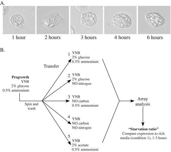
The data provide compelling evidence of a massive metabolic shift in phagocytosed cells, suggesting that nutritional stress—the need to find accessible stores of usable nutrients—is a prominent part of the early response to phagocytosis. With time the early pattern disappears and is replaced by the late pattern. It is clear, however, that there are many nonmetabolic genes regulated by phagocytosis as well. Assessing their significance was difficult, however, given the massive metabolic shift, so we designed further experiments (Fig. 4A) to validate these additional responses. First, we used a nonfilamentous mutant that alters the nature of the Candida-macrophage interaction. In addition, we used a series of starvation conditions in vitro to identify transcriptional responses that occur in the absence of the other cues resulting from macrophage contact.
FIG. 4.
Schematic for additional array experiments. (A) Time course of the interaction between the Δcph1 Δefg1 strain (HLC54) and macrophages. Because this strain is nonfilamentous, it cannot escape the macrophage. (B) Schematic for the in vitro starvation experiments. Cells pregrown in minimal medium with abundant carbon and nitrogen were shifted to medium lacking either nitrogen or carbon (or both). A ratio was calculated for each gene in each condition compared to condition 1 (medium with carbon and nitrogen).
A nonfilamentous mutant displays only the early response.
The Δcph1 Δefg1 strain fails to filament under most hypha-inducing conditions (22) because it lacks transcription factors responsive to both MAP kinase signaling (CPH1) and cyclic AMP signaling (EFG1), both pathways known to regulate filamentation (12, 16, 20, 21, 37). Because of this, it is unable to escape the macrophage after phagocytosis (Fig. 4A). Even though the mutant cells are “trapped” inside the macrophage, they are not killed. In fact, the Δcph1 Δefg1 cells undergo approximately one doubling while inside the macrophage during the 6-h experiment, as determined by using a standard XTT dye conversion assay (data not shown).
To analyze the response of cells that could not escape the macrophage, we collected transcript profiles from the Δcph1 Δefg1 strain HLC54 during a 1- to 6-h time course with macrophages. Transcript profiles were compared to the same strain grown in medium alone for the same times. These data were clustered with those from the wild-type data set. The analysis (Fig. 5 and data not shown) showed that gene regulation is broadly similar between the two experiments, particularly for metabolic genes, except that expression changes tend to persist in the Δcph1 Δefg1 mutant trapped within the macrophage. Maintenance of the early transcription pattern by cells unable to escape from the macrophage substantiates that this pattern is generated by the macrophage environment and draws a clear distinction between the early and late program.
Similarly, we have used a constitutively filamentous strain to demonstrate that the transcriptional profile that we attribute to macrophage phagocytosis is indeed due to ingestion and not to other factors, such as undetected changes in the medium. TUP1, a global repressor of transcription, is a negative regulator of filamentous growth and Δtup1 strains are locked in the pseudohyphal form (4). Due to the large size of these chains of elongated cells, they are phagocytosed poorly by macrophages. In this experiment, we see very few changes between the with-macrophage and without-macrophage conditions (data not shown). Although the Δtup1 mutation has pleiotropic effects, the limited transcriptional response in this experiment does suggest that exposure exposure to the internal macrophage environment is indeed necessary for the changes we observe.
Identifying a starvation profile in vitro.
The profiles described above for macrophage-ingested cells are very similar to those observed for S. cerevisiae cells deprived of nutrients in vitro (7). Repression of translation and activation of alternate nutrient metabolism are all hallmarks of this response. To identify specific responses in C. albicans to macrophage contact, we filtered out transcript changes due to starvation by assaying transcript profiles in several nutrient-poor conditions.
We shifted cells growing in minimal medium, with ample glucose and a preferred nitrogen source (YNB; 2% glucose and 0.5% ammonium sulfate), to medium lacking carbon, nitrogen, or both and collected cells at 1, 2, and 3 h after the transfer (diagrammed in Fig. 4B). Because of the induction of the glyoxylate cycle seen above, we also included a condition in which acetate was the sole carbon source. We created a ratio of the expression of each gene in the starvation conditions compared to cells shifted to the original YNB medium (2% glucose and 0.5% ammonium sulfate) for the same period of time (e.g., a ratio of the no carbon condition at 1 h to the rich YNB condition at 1 h). These data, to be discussed more fully below, clearly delineated similarities and, importantly, differences between transcript profiles in phagocytosed versus starved cells.
Macrophage signature expression profile.
We combined the data from phagocytosed wild-type and mutant cells with the starvation profiles and used hierarchical clustering to identify similarities and differences between starvation, the early pattern of gene expression in the wild-type strain, and persistent patterns in the mutant. Similarities, shown in Fig. 5, mostly include alternate carbon metabolism (in the induced genes) and translation activities (repressed genes). It was clear from these data that the similarities between phagocytosed cells and starvation were only to cells deprived of a carbon source. Nitrogen-depleted cells show a significantly different pattern of gene expression, with little to no overlap with ingested cells (data not shown). For this reason, the nitrogen starvation data set was excluded from further analysis.
Other clusters highlighted differences between the starvation and ingestion profiles. A cluster of 227 such genes is shown in Fig. 6 (an annotated version of this figure, with accompanying gene names is available as supplementary Fig. 3). Most of these genes are upregulated; we found relatively few macrophage-repressed genes that were not coregulated by nutrient limitation. Including the Δcph1 Δefg1 data in this analysis also allowed us to identify aspects of this response that may be regulated by one or both of these transcription factors. The top of Fig. 6 shows several genes that are induced in phagocytosed wild-type cells but not in Δcph1 Δefg1; these genes are likely to be positively regulated by these transcription factors. At the bottom are genes induced more strongly in the mutant strain and are possibly repressed by CPH1 or EFG1.
Arginine biosynthesis.
Our control conditions were very successful in eliminating metabolic genes from the macrophage induced profile. One glaring exception is arginine biosynthesis; every enzyme but one (ARG2) is strongly induced, averaging 5.6-fold upregulation at 1 h for these nine genes. Arginine genes are the only amino acid metabolic genes regulated in this manner. Since the base tissue culture medium in which both the macrophage samples and the controls are grown is rich in all amino acids, including arginine, the specific induction of this pathway is unlikely to result from arginine starvation prior to entry into the macrophage. It is not clear why arginine metabolism would be specifically regulated in this manner.
Transporters.
Of the 227 genes, 17 encode transporters for various compounds. Five of these are oligopeptide transporters, again suggesting a role for oligopeptide import in this environment. Further, this cluster includes homologs of aminopeptidase I (ScLAP4), carboxypeptidase Y (ScPRC1), a dipeptidyl aminopeptidase (ScDAP2), protease B (ScPRB1), and a weak homolog of aspartyl proteases (not one of the SAP genes). Thus, it seems clear that proteolysis and utilization of small peptides is important in surviving phagocytosis.
In addition, there are transporters for sulfite, phosphorous, choline, oxidocarboxylate, and allantoate. Several metal transporters are also induced; these will be discussed below.
Stress responses.
The phagocytosing macrophages would be expected to release reactive oxygen and nitrogen compounds as part of the antimicrobial burst (6). The cultured J774A cells used in our model have a relatively weak oxidative burst; it is sufficient to kill S. cerevisiae but not C. albicans. Similarly, J774 cells kill E. coli (via DNA damage) but not pathogenic Salmonella enterica serovar Typhimurium strains (34). There are a number of genes, listed in Table 4, that help to detoxify these radicals, including several peroxidases and reductases. Further, many aspects of the cytochrome electron transport system, which is critical for detoxification, are also induced (Table 4).
Iron homeostasis is critical in cells since iron is an essential cofactor for cytochrome activity and is an electron acceptor for several antioxidant proteins, like oxidoreductases and superoxide dismutase, making the uptake and storage of iron a critical aspect of the oxidative stress response. Iron uptake is particularly important within the host, where iron is scarce. Many pathogens have secreted siderophores and/or high-affinity uptake systems to acquire iron. C. albicans is no exception, and these systems are required for aspects of virulence (13, 31). We found two homologs of transmembrane ferric reductases, important in iron uptake, in this cluster. Metal ion homeostasis is interdependent, tying iron availability to that of other metals, like copper and zinc, and transporters for both of these are upregulated, as is the cadmium-induced protein CIP1.
The outcome of oxidative stress is DNA and protein damage. One heat shock protein (HSP78) is induced, as are multiple aspects of the DNA recombination-repair system (Table 4). In particular, genes that mediate repair of abasic sites, a common manifestation of oxidative damage of DNA, are upregulated. In summary, oxidative stresses posed by the macrophage are recognized by C. albicans via the activation of stress responses, including alterations in electron transport, metal homeostasis, and DNA damage repair. These changes are sufficient to keep C. albicans alive under these conditions.
Unknown genes.
The largest category of induced genes contains those without homology to known genes. These are common; in our annotation, “orphan” genes represent ca. 18% of the genome. This may be because the commensal/pathogen nature of this organism, which is rare among fungi, has led to the evolution of many genes to support this lifestyle. If this were the case, one might expect that these genes would be particularly important in vivo and would be induced under conditions relevant to pathogenesis, such as in our macrophage assay. Indeed, 36% of our cluster (81 of 227 genes) are unknown, a substantial overrepresentation. In contrast, ∼13% of the genome is homologous to S. cerevisiae genes of unknown function, whereas only 10% of the macrophage-induced cluster is. In total, therefore, nearly 50% of our cluster is of unknown function, and the majority of these genes are unique (at this time) to C. albicans. It will be very interesting to compare this set to other pathogenic fungi as those sequences are released to test our hypothesis that these are likely to be pathogen adaptations.
DISCUSSION
The use of whole-genome arrays has identified two successive reprogramming events upon ingestion of C. albicans by macrophages. The early response is a broad remodeling of carbon metabolism: cells shift from glycolysis to gluconeogenesis. Transcript profiles show a clear induction of essentially every gene required to convert fatty acids to glucose. This remodeling is accompanied by a massive downregulation of genes associated with translation. The late response is a resumption of glycolysis and restoration of the translation machinery. Most of these reprogramming events fail to occur in the related yeast, S. cerevisiae, which probably underlies the difference between the commensal/pathogen (Candida) and environmental (Saccharomyces) lifestyles.
This shift from glycolysis to gluconeogenesis suggests that the macrophage is deficient in glucose. Previous reports suggested that the source of glucose might be acetyl-CoA (23). This suggestion is supported by our finding that the enzymes of lipid degradation are also induced in the early phase of Candida phagocytosis. The implication is that lipids either derived from Candida or from the internal milieu of the macrophage serve as the precursors to acetyl-CoA. Several reports have suggested that various lipids are released during the phagocyte-Candida interaction, including fungal phospholipomannan (15) and mammalian arachidonic acid (5). Whether these products can serve as nutrients for C. albicans is not known, but there is some evidence to suggest the presence of lipids in this environment.
Induction of alternative carbon utilization occurs alongside repression of glycolysis. This was expected, since simultaneous activity of gluconeogenesis and glycolysis would lead to futile cycles of glucose synthesis and degradation, which is not favored (28). Curiously, this was precisely what was seen in C. albicans incubated in human blood, which showed induction of both glycolysis and gluconeogenesis (10). The most likely explanation is that this represents multiple populations of cells in this complex system, with both free-living cells in blood (with glucose present) and cells phagocytosed by neutrophils and monocytes present simultaneously.
Concomitant with the dramatic alteration in carbon metabolism, phagocytosed cells rapidly and comprehensively downregulate the genes encoding the translation machinery. This downregulation of the translation apparatus at the same time that gluconeogenic transcripts are induced presents an interesting puzzle: how can the cells express the gluconeogenic proteins when translation is repressed? Presumably, this behavior is a reflection of additional controls at the translational level. The cells must alter the translation machinery so that only a subset of messages (those involved in gluconeogenesis, for instance) can be translated. This depression of general translation coupled with increased translation of specific messages has been noted when Saccharomyces is transferred from a fermentable to a nonfermentable carbon source (19).
Additional experiments identified a set of genes that respond specifically to macrophage contact. These include the induction of oxidative stress responses, DNA damage repair, arginine biosynthesis, peptide utilization, and a substantial number of C. albicans-specific genes. The oxidative stress is presumably due to the burst of reactive oxygen and nitrogen species from the macrophages. This oxidative stress would then damage DNA, particularly through the creation of abasic sites. Thus, C. albicans recognizes both cellular damage and specific nutritional needs or opportunities and responds accordingly. This rapid response likely contributes to survival in vivo.
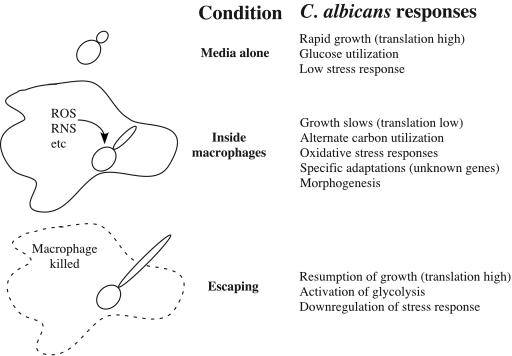
The late response in transcription is coincident with the morphogenetic switch from the yeast form to the filamentous form. The pattern involves a restoration in transcripts for the translational machinery, activation of glycolysis, and downregulation of the stress response (Fig. 7). These changes did not occur in control cultures undergoing the yeast-to-hypha switch. Moreover, a mutant that remains in the yeast form (Δcph1Δefg1) fails to reprogram and does not manifest the late response. These data provide strong evidence for the separation of the metabolic and morphogenetic programs during the course of infection and suggests that initiation of the late program of gene expression is dependent on the proper occurrence of postphagocytosis events.
FIG. 7.
Summary of C. albicans phagocytic responses. Phagocytosis induces C. albicans to adopt a slower growth program and to induce stress responses and alternate carbon metabolism compared to cells in medium alone. The morphogenetic response then allows escape, and these adaptations are reversed.
The primary environmental niche for C. albicans is generally considered to be the intestinal tract of warm-blooded animals (29). One manifestation of the adaptation to survival in this environment may be in “Candida-specific” genes, i.e., genes that lack homology to genes in other organisms. If the Candida-specific genes are really adaptations to the host environment, we might expect to see them overrepresented in relevant conditions. Indeed, in our annotation such unique genes represent 18% of the genome as a whole, but twice that (36%) of the macrophage-signature cluster. This set of genes (which, importantly, lack human homologs), are ideal candidates for development as antifungal targets.
The transcriptional plasticity of C. albicans is likely to be critical to its ability to survive in diverse environments. C. albicans survives as a commensal in the gastrointestinal tract and in the oral and vaginal cavities. It is probable that the phenotypes necessary for survival in the gut differ from those needed in the vagina, for example. Although global profiles are not available for cells grown in these sites, the transcriptional program for macrophage-ingested cells described here shares few similarities with those obtained from C. albicans cells in contact with neutrophils (32). This provides further support for the plasticity of these responses and indicates that C. albicans is not only able to undergo rapid large-scale changes but can also discern even subtle differences, such as whether it is being attacked by a macrophage or a neutrophil. In fact, it should be noted that the experiments described here use exclusively a cultured macrophage cell line, and we would anticipate an overlapping, but distinct, response in C. albicans cells phagocytosed by primary macrophages with greater antifungal activity. Such experiments are under way. The ability of C. albicans to make these changes during the switch to the early program is clearly critical to its survival in the macrophage because the Δcph1 Δefg1 mutant, which is frozen in the yeast form and the early program, survives, whereas S. cerevisiae does not. Moreover, since both organisms induce the glyoxylate pathway, this switch in metabolism is necessary but not sufficient for survival. Presumably, other components of the early pattern provide protection from the attack by the macrophage. Whole-genome arrays have permitted the identification of these systems, which can now be assessed for their role in pathogenesis.
Acknowledgments
We thank A. Johnson, D. Inglis, B. Braun, and A. Uhl for collaborating on microarray design and construction; they are also responsible for the majority of the genome annotation used in these analyses. S. Gordon, A. Andalis, and T. Galitski were instrumental in microarray printing. Other members of the Fink Lab, especially R. Wheeler, R. Prusty, and T. Reynolds, provided helpful advice. T. Patterson, J. Lopez-Ribot, and S. Redding collaborated on microarray construction.
We acknowledge the support of the Irvington Institute for Immunological Research (M.C.L.) and National Institutes of Health grant GM40266 (G.R.F.).
REFERENCES
- 1.Ausubel, F. M., B. Brent, R. E. Kingston, D. D. Moore, J. G. Seidman, J. A. Smith, and K. Struhl. 2000. Current protocols in molecular biology. John Wiley & Sons, Edison, N.J.
- 2.Bennett, R. J., M. A. Uhl, M. G. Miller, and A. D. Johnson. 2003. Identification and characterization of a Candida albicans mating pheromone. Mol. Cell. Biol. 23:8189-8201. [DOI] [PMC free article] [PubMed] [Google Scholar]
- 3.Birse, C. E., M. Y. Irwin, W. A. Fonzi, and P. S. Sypherd. 1993. Cloning and characterization of ECE1, a gene expressed in association with cell elongation of the dimorphic pathogen Candida albicans. Infect. Immun. 61:3648-3655. [DOI] [PMC free article] [PubMed] [Google Scholar]
- 4.Braun, B. R., and A. D. Johnson. 1997. Control of filament formation in Candida albicans by the transcriptional repressor TUP1. Science 277:105-109. [DOI] [PubMed] [Google Scholar]
- 5.Castro, M., N. V. Ralston, T. I. Morgenthaler, M. S. Rohrbach, and A. H. Limper. 1994. Candida albicans stimulates arachidonic acid liberation from alveolar macrophages through alpha-mannan and beta-glucan cell wall components. Infect. Immun. 62:3138-3145. [DOI] [PMC free article] [PubMed] [Google Scholar]
- 6.Densen, P., and G. L. Mandell. 1995. Granulocytic phagocytes, p. 78-101. In G. L. Mandell, R. G. Douglas, and J. E. Bennett (ed.), Principles and practices of infectious diseases. Churchill Livingstone, New York, N.Y.
- 7.DeRisi, J. L., V. R. Iyer, and P. O. Brown. 1997. Exploring the metabolic and genetic control of gene expression on a genomic scale. Science 278:680-686. [DOI] [PubMed] [Google Scholar]
- 8.Eisen, M. B., P. T. Spellman, P. O. Brown, and D. Botstein. 1998. Cluster analysis and display of genome-wide expression patterns. Proc. Natl. Acad. Sci. USA 95:14863-14868. [DOI] [PMC free article] [PubMed] [Google Scholar]
- 9.Enjalbert, B., A. Nantel, and M. Whiteway. 2003. Stress-induced gene expression in Candida albicans: absence of a general stress response. Mol. Biol. Cell 14:1460-1467. [DOI] [PMC free article] [PubMed] [Google Scholar]
- 10.Fradin, C., M. Kretschmar, T. Nichterlein, C. Gaillardin, C. d'Enfert, and B. Hube. 2003. Stage-specific gene expression of Candida albicans in human blood. Mol. Microbiol. 47:1523-1543. [DOI] [PubMed] [Google Scholar]
- 11.Gillum, A. M., E. Y. Tsay, and D. R. Kirsch. 1984. Isolation of the Candida albicans gene for orotidine-5′-phosphate decarboxylase by complementation of Saccharomyces cerevisiae ura3 and Escherichia coli pyrF mutations. Mol. Gen. Genet. 198:179-182. [DOI] [PubMed] [Google Scholar]
- 12.Gimeno, C. J., and G. R. Fink. 1994. Induction of pseudohyphal growth by overexpression of PHD1, a Saccharomyces cerevisiae gene related to transcriptional regulators of fungal development. Mol. Cell. Biol. 14:2100-2112. [DOI] [PMC free article] [PubMed] [Google Scholar]
- 13.Heymann, P., M. Gerads, M. Schaller, F. Dromer, G. Winkelmann, and J. F. Ernst. 2002. The siderophore iron transporter of Candida albicans (Sit1p/Arn1p) mediates uptake of ferrichrome-type siderophores and is required for epithelial invasion. Infect. Immun. 70:5246-5255. [DOI] [PMC free article] [PubMed] [Google Scholar]
- 14.Idnurm, A., and B. J. Howlett. 2002. Isocitrate lyase is essential for pathogenicity of the fungus Leptosphaeria maculans to canola (Brassica napus). Eukaryot. Cell 1:719-724. [DOI] [PMC free article] [PubMed] [Google Scholar]
- 15.Jouault, T., C. Fradin, P. A. Trinel, A. Bernigaud, and D. Poulain. 1998. Early signal transduction induced by Candida albicans in macrophages through shedding of a glycolipid. J. Infect. Dis. 178:792-802. [DOI] [PubMed] [Google Scholar]
- 16.Kèohler, J. R., and G. R. Fink. 1996. Candida albicans strains heterozygous and homozygous for mutations in mitogen-activated protein kinase signaling components have defects in hyphal development. Proc. Natl. Acad. Sci. USA 93:13223-13228. [DOI] [PMC free article] [PubMed] [Google Scholar]
- 17.Kibbler, C. C., S. Seaton, R. A. Barnes, W. R. Gransden, R. E. Holliman, E. M. Johnson, J. D. Perry, D. J. Sullivan, and J. A. Wilson. 2003. Management and outcome of bloodstream infections due to Candida species in England and Wales. J. Hosp. Infect. 54:18-24. [DOI] [PubMed] [Google Scholar]
- 18.Kontoyiannis, D. P., and R. E. Lewis. 2002. Antifungal drug resistance of pathogenic fungi. Lancet 359:1135-1144. [DOI] [PubMed] [Google Scholar]
- 19.Kuhn, K. M., J. L. DeRisi, P. O. Brown, and P. Sarnow. 2001. Global and specific translational regulation in the genomic response of Saccharomyces cerevisiae to a rapid transfer from a fermentable to a nonfermentable carbon source. Mol. Cell. Biol. 21:916-927. [DOI] [PMC free article] [PubMed] [Google Scholar]
- 20.Leberer, E., D. Harcus, I. D. Broadbent, K. L. Clark, D. Dignard, K. Ziegelbauer, A. Schmidt, N. A. Gow, A. J. Brown, and D. Y. Thomas. 1996. Signal transduction through homologs of the Ste20p and Ste7p protein kinases can trigger hyphal formation in the pathogenic fungus Candida albicans. Proc. Natl. Acad. Sci. USA 93:13217-13222. [DOI] [PMC free article] [PubMed] [Google Scholar]
- 21.Liu, H., J. Kohler, and G. R. Fink. 1994. Suppression of hyphal formation in Candida albicans by mutation of a STE12 homolog. Science 266:1723-1726. [DOI] [PubMed] [Google Scholar]
- 22.Lo, H. J., J. R. Kohler, B. DiDomenico, D. Loebenberg, A. Cacciapuoti, and G. R. Fink. 1997. Nonfilamentous Candida albicans mutants are avirulent. Cell 90:939-949. [DOI] [PubMed] [Google Scholar]
- 23.Lorenz, M. C., and G. R. Fink. 2001. The glyoxylate cycle is required for fungal virulence. Nature 412:83-86. [DOI] [PubMed] [Google Scholar]
- 24.Macphail, G. L., G. D. Taylor, M. Buchanan-Chell, C. Ross, S. Wilson, and A. Kureishi. 2002. Epidemiology, treatment and outcome of candidemia: a five-year review at three Canadian hospitals. Mycoses 45:141-145. [DOI] [PubMed] [Google Scholar]
- 25.McKinney, J. D., K. Honer zu Bentrup, E. J. Munoz-Elias, A. Miczak, B. Chen, W. T. Chan, D. Swenson, J. C. Sacchettini, W. R. Jacobs, Jr., and D. G. Russell. 2000. Persistence of Mycobacterium tuberculosis in macrophages and mice requires the glyoxylate shunt enzyme isocitrate lyase. Nature 406:735-738. [DOI] [PubMed] [Google Scholar]
- 26.Murad, A. M., C. d'Enfert, C. Gaillardin, H. Tournu, F. Tekaia, D. Talibi, D. Marechal, V. Marchais, J. Cottin, and A. J. Brown. 2001. Transcript profiling in Candida albicans reveals new cellular functions for the transcriptional repressors CaTup1, CaMig1, and CaNrg1. Mol. Microbiol. 42:981-993. [DOI] [PubMed] [Google Scholar]
- 27.Nantel, A., D. Dignard, C. Bachewich, D. Harcus, A. Marcil, A. P. Bouin, C. W. Sensen, H. Hogues, M. van het Hoog, P. Gordon, T. Rigby, F. Benoit, D. C. Tessier, D. Y. Thomas, and M. Whiteway. 2002. Transcription profiling of Candida albicans cells undergoing the yeast-to-hyphal transition. Mol. Biol. Cell 13:3452-3465. [DOI] [PMC free article] [PubMed] [Google Scholar]
- 28.Navas, M. A., S. CerdÂan, and J. M. Gancedo. 1993. Futile cycles in Saccharomyces cerevisiae strains expressing the gluconeogenic enzymes during growth on glucose. Proc. Natl. Acad. Sci. USA 90:1290-1294. [DOI] [PMC free article] [PubMed] [Google Scholar]
- 29.Odds, F. C. 1988. Candida and candidosis, 2nd ed. Bailliere Tindall, Philadelphia, Pa.
- 30.Planta, R. J., and W. H. Mager. 1998. The list of cytoplasmic ribosomal proteins of Saccharomyces cerevisiae. Yeast 14:471-477. [DOI] [PubMed] [Google Scholar]
- 31.Ramanan, N., and Y. Wang. 2000. A high-affinity iron permease essential for Candida albicans virulence. Science 288:1062-1064. [DOI] [PubMed] [Google Scholar]
- 32.Rubin-Bejerano, I., I. Fraser, P. Grisafi, and G. R. Fink. 2003. Phagocytosis by neutrophils induces an amino acid deprivation response in Saccharomyces cerevisiae and Candida albicans. Proc. Natl. Acad. Sci. USA 100:11007-11012. [DOI] [PMC free article] [PubMed] [Google Scholar]
- 33.Rude, T. H., D. L. Toffaletti, G. M. Cox, and J. R. Perfect. 2002. Relationship of the glyoxylate pathway to the pathogenesis of Cryptococcus neoformans. Infect. Immun. 70:5684-5694. [DOI] [PMC free article] [PubMed] [Google Scholar]
- 34.Schlosser-Silverman, E., M. Elgrably-Weiss, I. Rosenshine, R. Kohen, and S. Altuvia. 2000. Characterization of Escherichia coli DNA lesions generated within J774 macrophages. J. Bacteriol. 182:5225-5230. [DOI] [PMC free article] [PubMed] [Google Scholar]
- 35.Sherman, F. 1991. Getting started with yeast. Methods Enzymol. 194:3-21. [DOI] [PubMed] [Google Scholar]
- 36.Staab, J. F., and P. Sundstrom. 1998. Genetic organization and sequence analysis of the hypha-specific cell wall protein gene HWP1 of Candida albicans. Yeast 14:681-686. [DOI] [PubMed] [Google Scholar]
- 37.Stoldt, V. R., A. Sonneborn, C. E. Leuker, and J. F. Ernst. 1997. Efg1p, an essential regulator of morphogenesis of the human pathogen Candida albicans, is a member of a conserved class of bHLH proteins regulating morphogenetic processes in fungi. EMBO J. 16:1982-1991. [DOI] [PMC free article] [PubMed] [Google Scholar]
- 38.Tsong, A. E., M. G. Miller, R. M. Raisner, and A. D. Johnson. 2003. Evolution of a combinatorial transcriptional circuit: a case study in yeasts. Cell 115:389-399. [DOI] [PubMed] [Google Scholar]
- 39.Vereecke, D., K. Cornelis, W. Temmerman, M. Jaziri, M. Van Montagu, M. Holsters, and K. Goethals. 2002. Chromosomal locus that affects pathogenicity of Rhodococcus fascians. J. Bacteriol. 184:1112-1120. [DOI] [PMC free article] [PubMed] [Google Scholar]
- 40.Wang, Z. Y., C. R. Thornton, M. J. Kershaw, L. Debao, and N. J. Talbot. 2003. The glyoxylate cycle is required for temporal regulation of virulence by the plant pathogenic fungus Magnaporthe grisea. Mol. Microbiol. 47:1601-1612. [DOI] [PubMed] [Google Scholar]
- 41.Weinstein, M. P., M. L. Towns, S. M. Quartey, S. Mirrett, L. G. Reimer, G. Parmigiani, and L. B. Reller. 1997. The clinical significance of positive blood cultures in the 1990s: a prospective comprehensive evaluation of the microbiology, epidemiology, and outcome of bacteremia and fungemia in adults. Clin. Infect. Dis. 24:584-602. [DOI] [PubMed] [Google Scholar]


