Abstract
The development of biomaterials has significantly increased the potential for targeted drug delivery to a variety of cell and tissue types, including the pancreatic β-cells. In addition, biomaterial particles, hydrogels, and scaffolds also provide a unique opportunity to administer sustained, controllable drug delivery to β-cells in culture and in transplanted tissue models. These technologies allow the study of candidate β-cell proliferation factors using intact islets and a translationally relevant system. Moreover, determining the effectiveness and feasibility of candidate factors for stimulating β-cell proliferation in a culture system is critical before moving forward to in vivo models. Herein, we describe a method to co-culture intact mouse islets with biodegradable compound of interest (COI)-loaded poly(lactic-co-glycolic acid) (PLGA) microspheres for the purpose of assessing the effects of sustained in situ release of mitogenic factors on β-cell proliferation. This technique describes in detail how to generate PLGA microspheres containing a desired cargo using commercially available reagents. While the described technique uses recombinant human Connective tissue growth factor (rhCTGF) as an example, a wide variety of COI could readily be used. Additionally, this method utilizes 96-well plates to minimize the amount of reagents necessary to assess β-cell proliferation. This protocol can be readily adapted to use alternative biomaterials and other endocrine cell characteristics such as cell survival and differentiation status.
Keywords: Bioengineering, Issue 117, pancreatic islets, β-cell proliferation, biomaterials, cell culture, insulin, drug delivery
Introduction
Pancreatic β-cells are the only insulin-producing cells in the body and are critical to maintain blood glucose homeostasis. While healthy individuals have sufficient β-cell mass and function to properly regulate blood glucose, individuals with diabetes are characterized by insufficient β-cell mass and/or function1,2. It has been proposed that inducing β-cell proliferation can ultimately increase β-cell mass and restore glucose homeostasis in individuals with diabetes3. However, evaluation and validation of potential β-cell proliferative compounds in intact islets is necessary before effective therapies can be developed. Transplantation of cadaveric human islets into individuals with diabetes restores blood glucose homeostasis for some time, but the availability and success of this experimental procedure is hindered by a shortage of human islets available for transplant and by β-cell death in the islets after transplant4. Even with the discovery of factors that induce multiplication of insulin-producing cells, a major challenge still exists in delivering these factors to relevant sites in vivo. One strategy for sustained local delivery of β-cell proliferative compounds is poly(lactic-co-glycolic) acid (PLGA). PLGA has a history of use in FDA approved drug delivery products owing to its high safety, biodegradability, and extended release kinetics5. Specifically, PLGA is a copolymer of lactide and glycolide that degrades via hydrolysis with water either in vivo or in culture into lactic acid and glycolic acid, which are naturally occurring metabolites in the body. The encapsulated drug compound can be released in the surrounding environment by both diffusion and/or degradation-controlled release mechanisms. Encapsulation of COI provides protection against enzymatic degradation, improving the bioavailability of the reagent compared to unencapsulated COI5. We suggest that PLGA microspheres can be used to administer candidate compounds to intact islets in culture, and ultimately in vivo. Testing the efficacy of PLGA to administer β-cell mitogens to islets ex vivo is critical before transplantation protocols are explored.
Currently, there is no technique to measure β-cell proliferation in live animals. Experiments to assess effectiveness of potential proliferative compounds in vivo therefore require administration of these compounds to live animals, with subsequent dissection and processing of pancreata for immunolabeling. Such protocols are expensive and laborious, and require the compound to be administered systemically, without any guarantee that they will reach the islets. Conversely, several immortalized β-cell lines are available for the study of insulin-producing cells in culture, but these cell lines lack the islet architecture and environment found in living organisms6. Immortalized β-cell lines are also characterized as having a much higher degree of replication than endogenous β-cells in vivo, thus complicating analysis of compounds that induce proliferation. In this study, we describe a protocol that uses intact islets isolated from adult mice. Unlike β-cell lines, intact islets retain normal islet architecture. Likewise, in contrast to experiments conducted in vivo, administering proliferative compounds directly to cultured intact islets significantly reduces the quantity of reagents that is necessary to accurately measure β-cell proliferation.
The current study utilizes PLGA to administer a COI, in this example, recombinant human Connective Tissue Growth Factor (rhCTGF). The method described here confers a significant advantage over the administration of raw compound to cultured islets as it allows for a continual release of compound into the media. Notably, this assay can be modified to administer a wide variety of proteins and antibodies of interest to intact islets. Effects on other endocrine cell types, including α-cells, may also be analyzed.
Protocol
All procedures were approved and performed in accordance with the Vanderbilt Institutional Animal Care and Use Committee.
1. Labeling COI with Fluorophore (Optional)
Choose a fluorescent dye that will react with a free primary amine (e.g., on a protein), such as succinimidyl esters or fluorescein derivatives, to visualize microsphere cargo. Dissolve 8x molar excess (relative to moles of COI) of fluorophore into 200 µl of dimethyl sulfoxide (DMSO).
Resuspend 50 mg of COI up to a final volume of 800 µl in a vehicle solution (final concentration of 62.5 ng/ µl). The vehicle solution will vary depending on the source of the COI. NOTE: Typical vehicle solutions include phosphate buffered saline (PBS) or DMSO.
Add all of the fluorophore/DMSO solution from step 1.1 and resuspend the COI. Vortex overnight at 4 °C. Alternatively, vortex the reaction mixture at room temperature (RT) for 4 hr.
- Following the labeling reaction, remove excess fluorophore using a desalting column.
- Drain storage buffer from desalting column and rinse with 16 ml of deionized water. Discard all flow through.
- Add the fluorescently labeled COI to the desalting column. Elute with 1.2 ml of deionized water and collect the flow through.
- Repeat elution step an additional four times, each time adding 1.2 ml of deionized water and collecting the flow through as a separate sample.
- Freeze all collected flow through samples at -80 °C and lyophilize according to manufacturer's instructions for 24 hr.
2. COI-loaded Microsphere Preparation via the Water-in-oil-in-water Emulsion Solvent Evaporation Method
Add 1 mg of fluorescently labeled COI to 100 µl of deionized water to form the first water phase (W1).
Dissolve 65 mg of poly(lactic-co-glycolic acid) (50:50 Lactide:Glycolide, molecular weight 54,000 - 69,000) in 750 µl of dichloromethane in a microcentrifuge tube. Ultrasonicate for 10 - 30 sec (at 160 W) to completely dissolve the PLGA. This forms the oil phase (O).
Add all of the W1 phase generated in step 2.1 to the O phase in a drop-wise manner. Emulsify using a hand-held homogenizer at 20,000 rpm for 30 sec to form the W1/O phase.
Add all of the W1/O phase in a drop-wise manner to 15 ml of a 1% (weight/volume) aqueous poly(vinyl alcohol) (PVA) solution and emulsify using a hand-held homogenizer at 20,000 rpm for 30 sec.
Transfer all of the emulsion generated in step 2.4 to a 200 ml round-bottom flask and subject to a 635 mm Hg vacuum using a rotary evaporator for 1 hr to remove the solvent and generate the aqueous phase.
Aliquot 1 ml of the aqueous phase generated in step 2.5 to fourteen microcentrifuge tubes. Centrifuging the aqueous solution in the microcentrifuge tubes at 7,500 x g for 8 min. NOTE: At this step, the microspheres are present in the remaining aqueous solution and are concentrated to the bottom of the microcentrifuge tube.
- Carefully remove 900 µl of aqueous solution from the microcentrifuge tubes with a micropipette, pipetting so as to not disturb the microspheres on the bottom of the microcentrifuge tube.
- Wash the microspheres of excess PVA by adding 1 ml deionized water to each microcentrifuge tube. Centrifuge at 7,500 x g for 8 min, and again carefully remove 900 µl of aqueous solution from the microcentrifuge tubes with a micropipette, pipetting so as to not disturb the microspheres on the bottom of the microcentrifuge tube. NOTE: The remaining 100 µl aqueous solution contains the generated microspheres.
Freeze the aqueous microsphere solution generated in step 2.7 at -80 °C.
Lyophilize microspheres using a lyophilizer according to manufacturer's instructions and store at -20 °C. NOTE: Measure loading efficiency of the COI by completely dissolving PLGA microspheres and determining the protein concentration relative to a standard curve of the free protein7.
As a negative control, generate a batch of "blank" microspheres (hereafter referred to as control microspheres). NOTE: The generation of control microspheres is identical to the generation of hydrophilic COI-loaded microspheres, with no COI addition to 800 µl of the vehicle solution in step 1.2. Match control particles in mass concentration of PLGA relative to COI-loaded PLGA microspheres for β-cell mitogen assay.
3. Preparation of Islet Culture Media and Pre-assay Media
Prepare 200 ml media for culture of intact mouse islets: RPMI 1640 media supplemented with 11 mM glucose, 10% horse serum, 100 U/ml penicillin G and 100 µg/ml streptomycin (hereafter referred to as islet culture media). NOTE 1: Unlike fetal bovine serum (FBS), horse serum lacks placental lactogen, an inducer of β-cell proliferation, which confounds analysis in the proliferation assay. NOTE 2: As the PLGA microspheres are not sterilized prior to use, the addition of antibiotics to the culture media is crucial to avoid microbiological contamination.
Remove 25 ml of islet culture media with an electronic pipettor and place in a 50 ml conical tube. Supplement the 25 ml of islet culture media in the 50 ml conical tube with a final concentration of 0.2 mM EGTA to generate the pre-assay media. NOTE: The addition of EGTA mildly loosens cell-cell contacts in the islets without altering islet architecture. This helps ensure the COI can reach the inner cells of the islet and helps prevent necrosis of the central islet.
4. Culturing of Intact Mouse Islets
Isolate intact islets from a pre-selected strain, sex, age, and genotype of mice following a routine collagenase digestion of the pancreas8,9. Pool together islets from like samples.
Aliquot 40 islets into one well of a 96-well tissue culture plate in 200 µl of islet culture media. Visually size-match islets per well (a precise assessment of islet equivalency (IEQ) between samples is not necessary). Fill empty wells immediately adjacent to wells with islets with 200 µl sterile water to provide an evaporation buffer. Culture islets in media overnight at 37 °C under an atmosphere of 95% air and 5% CO2.
5. Re-suspension of COI-loaded and Control PLGA Microspheres
After overnight islet recovery, resuspend microspheres by adding pre-assay media to one aliquot of lyophilized COI-loaded PLGA microspheres such that the final concentration of COI in the microcentrifuge tube is 10 ng/µl (the amount of COI present in a given amount of the generated microspheres was determined in step 2.9). Sonicate the resuspended COI-loaded microspheres in an ice water bath for 10 min at 160 W with 10 sec long pulses at 4 °C.
Resuspend control PLGA microspheres by adding the same volume of pre-assay media to an equal mass of lyophilized control PLGA microspheres. Sonicate the resuspended control PLGA microspheres in an ice water bath for 10 min at 160 W with 10 sec long pulses at 4 °C.
Visually confirm microspheres are dispersed by pipetting 2 µl of resuspended microspheres between two glass coverslips. Visualize microspheres using a 40X objective on an epifluorescence or brightfield microscope.
If significant aggregation of microspheres is still visible, sonicate in an ice water bath for an additional 10 min at 160 W with 10 sec long pulses at 4 °C and repeat visualization of resuspended microspheres.
6. Treatment of Islets with COI-loaded or Control PLGA Microspheres
For each well to be treated with COI-loaded microspheres, prepare assay media by diluting the 10 ng/µl of the resuspended COI microspheres with pre-assay media to a final volume of 100 µl and a pre-determined final concentration. NOTE: The optimal final concentration of the protein will vary for each COI used for this assay. Ideally, a range of final concentrations are tested.
For each well to be used as a control, prepare control assay media by diluting the resuspended control PLGA microspheres generated in step 5.2 with the same dilution volume used to dilute the COI-loaded microspheres in step 6.1. NOTE: Islets treated with control assay media will serve as a negative control to enable detection of any effect the blank microspheres may have on the experimental outcomes.
Carefully remove 100 µl of islet culture media from each well with a micropipette such that no islets are dislodged from the wells. Gently add 100 µl of assay media or 100 µl of control assay media to each well.
Incubate islets at 37 °C under an atmosphere of 95% air and 5% CO2 for three days.
7. Dispersing Intact Islets onto Microscope Slides
After three days, use a micropipette to carefully remove and discard assay media from all wells so as not to dislodge islets. Gently add 200 µl PBS to islets to wash them of the assay media. Carefully remove and discard PBS with a micropipette and gently wash islets again with an additional 200 µl PBS.
Remove and discard PBS with a micropipette and add 100 µl of 0.025% trypsin, 2 mM EDTA solution. Incubate at RT for 3 min. Pipet islets in the trypsin-EDTA solution up and down every 2 min until islets are visually confirmed to be dispersed into single cells (note that some small clumps of cells may still be present) using a light microscope. Transfer the entire volume of each well individually into labeled- microcentrifuge tubes.
Add 400 µl of islet culture media to each microcentrifuge tube to stop the trypsin-mediated cell dissociation.
Centrifuge samples for 5 min at 100 x g at 4 °C to pellet islet cells to the bottom of microcentrifuge tubes.
Gently remove supernatant using a micropipette, and resuspend pellet in 200 µl fresh islet culture media.
Using a cyto-centrifuge, centrifuge dissociated islets at 140 x g for 3 min onto charged microscope slides.
Air dry slides at RT for 10 min, then draw a box around the dissociated islets with a hydrophobic marking pen.
Fix cells with 75 µl 4% paraformaldehyde (PFA) at RT for 10 min. Remove 4% PFA and gently wash cells in 75 µl PBS two times. After washing, permeabilize cells with 75 µl 0.2% Triton X-100 in PBS for 10 min. Then, wash cells with 75 µl PBS. Caution: PFA is known to be allergenic, carcinogenic, and toxic.
8. Immunofluorescence Labeling of Dissociated Islets for Insulin and the Cell Proliferation Marker, Ki67
Prepare a humid chamber by placing two wet paper towels in the bottom of a 100 slide capacity microscope slide box.
Place slides flat down (cells facing up) in humid chamber. Prepare samples for labeling by aspirating the PBS from the previous wash step using a micropipette and adding 75 µl of blocking solution (5% Normal Donkey Serum (NDS) in PBS) to each slide to block non-specific binding of antibodies to the specimens. Incubate slides in blocking solution for 1 hr at RT.
Gently aspirate blocking solution using a micropipette and add 75 µl of primary antibody solution containing guinea pig anti-insulin and rabbit anti-Ki67 antibodies, diluted 1 µg/ml in 5% NDS in PBS. Incubate at RT for 1 hr. NOTE: Alternative markers of cell proliferation include proliferating cell nuclear antigen (PCNA) and phosphohistone H3 (PH3),
Gently aspirate primary antibody solution using a micropipette and rinse the cells with 75 µl PBS. Aspirate PBS using a micropipette and rinse cells two more times, each time with 75 µL PBS. Incubate each sample with 75 µl anti-guinea pig Cy5 secondary antibody and anti-rabbit Cy3 secondary antibody diluted to 2 µg/ml in blocking solution for 1 hr at RT. NOTE: Do not use secondary antibodies labeled with the same or spectrally overlapping fluorophore that was already used to label the microspheres.
Aspirate secondary antibody solution using a micropipette and incubate in 75 µl of 300 nM DAPI in PBS for 3 min at RT. Aspirate DAPI using a micropipette and rinse in deionized water for 5 min.
Gently aspirate water from samples using a micropipette. Spot a drop (roughly 100 µl) of fast-drying mounting solution containing antifade reagent on a glass coverslip. Invert the microscope slide sample side down, onto the mounting solution. Gently press the coverslip against the slide using a pipet tip to remove bubbles and wipe away excess liquid with a soft cleaning tissue. Allow coverslips to dry overnight at RT.
9. Image Acquisition and Analysis
Use an epifluorescence microscope with an attached camera for image acquisition. For each fluorophore, determine the optimal exposure time (typically 20 msec for DAPI, 40 - 80 msec for insulin, and 250 msec for Ki67). Ensure image acquisition parameters are equal for all samples.
For each sample, manually count 3,000 insulin-positive cells and of those count how many are also Ki67-positive. Only count insulin-positive cells that have a well-defined and clearly visible nucleus. Calculate the percentage of proliferating β-cells for each sample by dividing the number of Ki67-positive/insulin-positive cells by the total number of insulin-positive cells and multiplying by 100.
After determining the percentage of proliferating β-cells for each sample, determine if a statistically significant difference in β-cell proliferation exists between control and experimental samples by analyzing results with a one-way ANOVA using commercially available statistical software.
Representative Results
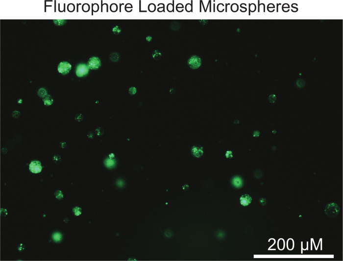
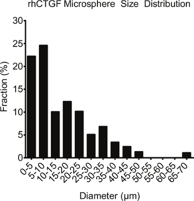
Figure 1 is a visual representation of the microspheres generated using the above protocol. The protocol described here yields rhCTGF-loaded microspheres of various sizes. The largest fraction of microspheres will be between 1 and 10 µm in diameter, though some microspheres may be larger (Figure 2). If desired, microsphere size can be tuned and optimized based on fabrication parameters such as homogenization speed and time, surfactant concentration used, and relative volumes of each water/oil/water phase10.
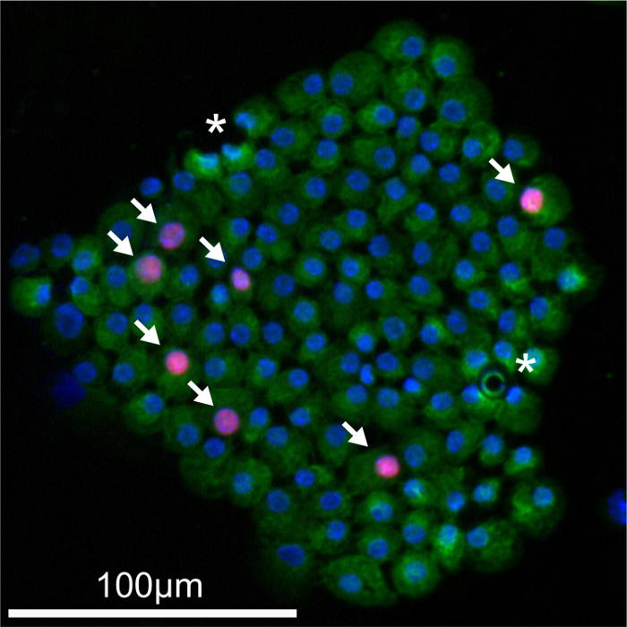
Following dispersal of intact islets treated with PLGA microspheres and subsequent immunolabeling, it is common to see regions of the sample devoid of any labeling in between cells (Figure 3). While most of the microspheres that are still intact after the culture treatment period are removed during the wash steps, some remain after the islets are spun onto the microscope slides. These microspheres cause the hole-like structures visualized during imaging. These residual microspheres typically do not interfere with subsequent quantification.
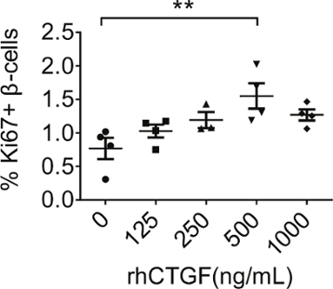
After imaging, the percentage of Ki67-positive/insulin-positive cells can be quantified by manually counting the total number of labeled cells, or using software image analysis. Previously, we have demonstrated that recombinant human Connective Tissue Growth Factor (rhCTGF) can stimulate mouse β-cell proliferation in intact islets ex vivo11. Using the aforementioned protocol, we generated PLGA microspheres containing rhCTGF (rhCTGF-PLGA). Treating intact islets with rhCTGF-PLGA microspheres for 3 days resulted in a similar increase in β-cell proliferation as previously reported with the raw protein, demonstrating that the protein did not lose any functionality during microsphere generation (Figure 4).
 Figure 1: Visual Representation of PLGA Microspheres. By incorporating a fluorescent dye into the manufacturing protocol, PLGA microspheres can be visualized using an epifluorescent microscope. Scale bar represents 200 μm. Please click here to view a larger version of this figure.
 Figure 2: Size Distribution of PLGA Microspheres. Although size distribution may vary, most microspheres will be less than 10 µm in diameter. Please click here to view a larger version of this figure.
 Figure 3:Immunofluorescent Visualization of Dispersed Proliferating β-cells. Dispersed islets are immunolabeled for the proliferation marker Ki67 (red) and insulin (green) to mark proliferating β-cells. Nuclei are labeled with DAPI (blue). Arrows indicate Ki67-positive/insulin-positive cells. Asterisks (*) indicate undegraded microspheres. Scale bar represents 100 μm. Please click here to view a larger version of this figure.
 Figure 4:Quantification and Analysis of β-cell Proliferation. Islets treated with different amounts of rhCTGF-PLGA microspheres. The number of Ki67-positive/insulin-positive cells was manually counted from all samples and expressed as a percentage of the total number of insulin-positive β-cells. X-axis corresponds to the final concentration of rhCTGF present in each treatment. Statistical significance was determined using a one-way ANOVA followed by Tukey's multiple comparison test. Statistical significance was set at p ≤ 0.05. Error bars represent standard error of the mean. Please click here to view a larger version of this figure.
Discussion
The study of β-cell proliferation in culture is typically hampered by several difficulties. First, immortalized β-cell lines are characterized by higher degrees of proliferation than what is found in endogenous β-cells in live islets. Additionally, these immortalized cell lines lack the normal architecture critical for normal β-cell function. These two facts make it difficult to determine if results obtained using immortalized β-cell lines will hold true when tested in vivo or in whole islets. Our described protocol, which uses freshly isolated intact mouse islets, circumvents these issues as the islet architecture is maintained and β-cell proliferation is comparable to that found in vivo.
One significant concern when culturing intact islets is the possibility that cells within the islet core will undergo hypoxia-induced necrosis or will not be exposed to rhCTGF. For this reason, a final concentration of 0.1 mM EGTA is added to the media to loosen cell-cell contacts without disrupting islet architecture. We have previously published that the addition of EGTA alone can increase β-cell proliferation, presumably due to increased access of nutrients and mitogens in the media to the islet core12. The increase in β-cell proliferation in response to EGTA could also be due to the decrease in cell-cell contact itself13,14. The media described in this protocol is also supplemented with horse serum instead of the more traditional fetal bovine serum. This substitution is made due to the presence of placental lactogen in fetal bovine serum, which can stimulate β-cell proliferation on its own, potentially complicating analysis in the proliferation assay.
Determination of the relative toxicity of the PLGA microspheres (with or without the COI) to the islet cells can be assessed through immunofluorescence analysis of markers of cell death or DNA damage, including TUNEL or γ-H2AX15,16. Although the addition of PLGA microspheres has not shown any obvious detrimental effects to islets ex vivo, users should be aware of the effects the microspheres can have on image analysis. As demonstrated in Figure 3 and mentioned in the results section, undegraded microspheres can be apparent as dark spots within immunofluorescent images, with more microspheres appearing when increasing concentrations are utilized.
It should be noted that the described protocol has been tailored to work with isolated mouse islets. As such, it is unclear if identical conditions will work with islets harvested from alternative organisms, such as rat and humans. Notably, islets harvested from different organisms often have different optimal culturing conditions and differing degrees of β-cell proliferation17,18.
Numerous biomaterials are potentially available to administer rhCTGF to isolated islets, including poly(thioketal-urethane) and poly(propylene sulfide)19. We chose to focus on PLGA due to several notable qualities that it possesses. First, PLGA is a relatively affordable reagent and the microspheres can be generated with standard equipment (centrifuge, homogenizer, lyophilizer) available at most research institutions. PLGA is an artificial polymer that has precedent for successful use in FDA approved devices owing to its biocompatibility, biodegradability, and its ability to modulate compound release rates20. PLGA degrades by hydrolysis in the presence of water, releasing its cargo in the process. The chemical composition of PLGA particles can be tailored such that a compound is released in vivo over the period of several weeks. PLGA has also been used in preclinical animal trials and human clinical therapies to locally administer compounds to various organs and tissues21,22. Other hydrophobic polymers can also be used in the generation of biodegradable microspheres. These polymers can respond to specific environmental stimuli, such as pH, temperature, and the presence of reactive oxygen species23,24. Thus, researchers should carefully consider which biomaterial is most appropriate for their studies.
Any researcher utilizing PLGA, or other biomaterials, to study β-cell proliferation ex vivo should be aware of potential differences between the effects of encapsulated COI compared to unencapsulated COI. For example, the delivery of rhCTGF via PLGA could change the degree of and timing of β-cell proliferation induction compared to treatment with unencapsulated protein. Ongoing studies in our lab are currently examining these potential differences.
The analysis of β-cell proliferation in cultured islets is a powerful model for the identification and mechanistic analysis of β-cell mitogens. Using the assay described here we show a relatively quick and cost-effective method for administering a COI to isolated cultured islets with an extended release delivery vehicle. This assay should be applicable to nearly any COI of interest, and our choice to focus on CTGF is purely based on its previously published ability to stimulate β-cell proliferation in vivo and ex vivo11. Additionally, this assay can validate the effectiveness and safety of using PLGA as a delivery method before advancing to models where islets are transplanted in living organisms. Overall, the described protocol provides a novel way to administer compounds to cultured islets with a broader impact on measuring the safety of PLGA to transplantable tissue.
Disclosures
The authors have nothing to disclose.
Acknowledgments
The authors would like to thank Bethany Carboneau (Vanderbilt University) for critical reading of this manuscript. We also thank Anastasia Coldren (Vanderbilt University Medical Center Islet Procurement and Analysis Core) for islet isolations, and Dr. Alvin C. Powers (Vanderbilt University Medical Center) and Dr. David Jacobson (Vanderbilt University) for use of their centrifuge and tissue culture facility. This research involved use of the Islet Procurement and Analysis Core of the Vanderbilt Diabetes Research and Training Center supported by NIH grant DK20593. This work was supported by an American Heart Association Postdoctoral Fellowship (14POST20380262) to R.C.P., and grants from the Juvenile Diabetes Research Foundation (1-2011-592), and Department of Veterans Affairs (1BX00090-01A1) to M.A.G.
References
- Butler AE, et al. Beta-cell deficit and increased beta-cell apoptosis in humans with type 2 diabetes. Diabetes. 2003;52(1):102–110. doi: 10.2337/diabetes.52.1.102. [DOI] [PubMed] [Google Scholar]
- Levy J, Atkinson AB, Bell PM, McCance DR, Hadden DR. Beta-cell deterioration determines the onset and rate of progression of secondary dietary failure in type 2 diabetes mellitus: the 10-year follow-up of the Belfast Diet Study. Diabet Med. 1998;15(4):290–296. doi: 10.1002/(SICI)1096-9136(199804)15:4<290::AID-DIA570>3.0.CO;2-M. [DOI] [PubMed] [Google Scholar]
- Bouwens L, Rooman I. Regulation of pancreatic beta-cell mass. Physiol Rev. 2005;85(4):1255–1270. doi: 10.1152/physrev.00025.2004. [DOI] [PubMed] [Google Scholar]
- McCall M, Shapiro AM. Update on islet transplantation. Cold Spring Harb Perspect Med. 2012;2(7):007823. doi: 10.1101/cshperspect.a007823. [DOI] [PMC free article] [PubMed] [Google Scholar]
- Danhier F, et al. PLGA-based nanoparticles: an overview of biomedical applications. J Control Release. 2012;161(2):505–522. doi: 10.1016/j.jconrel.2012.01.043. [DOI] [PubMed] [Google Scholar]
- Skelin M, Rupnik M, Cencic A. Pancreatic beta cell lines and their applications in diabetes mellitus research. ALTEX. 2010;27(2):105–113. doi: 10.14573/altex.2010.2.105. [DOI] [PubMed] [Google Scholar]
- Rui J, et al. Controlled release of vascular endothelial growth factor using poly-lactic-co-glycolic acid microspheres: in vitro characterization and application in polycaprolactone fumarate nerve conduits. Acta Biomater. 2012;8(2):511–518. doi: 10.1016/j.actbio.2011.10.001. [DOI] [PMC free article] [PubMed] [Google Scholar]
- Lacy PE, Kostianovsky M. Method for the isolation of intact islets of Langerhans from the rat pancreas. Diabetes. 1967;16(1):35–39. doi: 10.2337/diab.16.1.35. [DOI] [PubMed] [Google Scholar]
- Szot GL, Koudria P, Bluestone JA. Murine pancreatic islet isolation. J Vis Exp. 2007. p. e255. [DOI] [PMC free article] [PubMed]
- Mukherjee B, Santra K, Pattnaik G, Ghosh S. Preparation, characterization and in-vitro evaluation of sustained release protein-loaded nanoparticles based on biodegradable polymers. Int J Nanomedicine. 2008;3(4):487–496. doi: 10.2147/ijn.s3938. [DOI] [PMC free article] [PubMed] [Google Scholar]
- Riley KG, et al. Connective tissue growth factor modulates adult beta-cell maturity and proliferation to promote beta-cell regeneration in mice. Diabetes. 2015;64(4):1284–1298. doi: 10.2337/db14-1195. [DOI] [PMC free article] [PubMed] [Google Scholar]
- Mosser RE, Gannon M. An assay for small scale screening of candidate beta cell proliferative factors using intact islets. Biotechniques. 2013;55(6):310–312. doi: 10.2144/000114115. [DOI] [PMC free article] [PubMed] [Google Scholar]
- Carvell MJ, Marsh PJ, Persaud SJ, Jones PM. E-cadherin interactions regulate beta-cell proliferation in islet-like structures. Cell Physiol Biochem. 2007;20(5):617–626. doi: 10.1159/000107545. [DOI] [PubMed] [Google Scholar]
- Wakae-Takada N, Xuan S, Watanabe K, Meda P, Leibel RL. Molecular basis for the regulation of islet beta cell mass in mice: the role of E-cadherin. Diabetologia. 2013;56(4):856–866. doi: 10.1007/s00125-012-2824-6. [DOI] [PMC free article] [PubMed] [Google Scholar]
- Gavrieli Y, Sherman Y, Ben-Sasson SA. Identification of programmed cell death in situ via specific labeling of nuclear DNA fragmentation. J Cell Biol. 1992;119(3):493–501. doi: 10.1083/jcb.119.3.493. [DOI] [PMC free article] [PubMed] [Google Scholar]
- Kuo LJ, Yang LX. Gamma-H2AX - a novel biomarker for DNA double-strand breaks. In Vivo. 2008;22(3):305–309. [PubMed] [Google Scholar]
- Daoud J, Rosenberg L, Tabrizian M. Pancreatic islet culture and preservation strategies: advances, challenges, and future outlook. Cell Transplant. 2010;19(12):1523–1535. doi: 10.3727/096368910X515872. [DOI] [PubMed] [Google Scholar]
- Carter JD, Dula SB, Corbin KL, Wu R, Nunemaker CS. A practical guide to rodent islet isolation and assessment. Biol Proced Online. 2009;11:3–31. doi: 10.1007/s12575-009-9021-0. [DOI] [PMC free article] [PubMed] [Google Scholar]
- Xu Q, He C, Xiao C, Chen X. Reactive Oxygen Species (ROS) Responsive Polymers for Biomedical Applications. Macromol Biosci. 2016;16(5):635–646. doi: 10.1002/mabi.201500440. [DOI] [PubMed] [Google Scholar]
- Makadia HK, Siegel SJ. Poly Lactic-co-Glycolic Acid (PLGA) as Biodegradable Controlled Drug Delivery Carrier. Polymers (Basel) 2011;3(3):1377–1397. doi: 10.3390/polym3031377. [DOI] [PMC free article] [PubMed] [Google Scholar]
- Cui F, Shi K, Zhang L, Tao A, Kawashima Y. Biodegradable nanoparticles loaded with insulin-phospholipid complex for oral delivery: preparation, in vitro characterization and in vivo evaluation. J Control Release. 2006;114(2):242–250. doi: 10.1016/j.jconrel.2006.05.013. [DOI] [PubMed] [Google Scholar]
- Kavanaugh TE, Werfel TA, Cho H, Hasty KA, Duvall CL. Particle-based technologies for osteoarthritis detection and therapy. Drug Deliv Transl Res. 2015. [DOI] [PMC free article] [PubMed]
- Joshi RV, Nelson CE, Poole KM, Skala MC, Duvall CL. Dual pH- and temperature-responsive microparticles for protein delivery to ischemic tissues. Acta Biomater. 2013;9(5):6526–6534. doi: 10.1016/j.actbio.2013.01.041. [DOI] [PMC free article] [PubMed] [Google Scholar]
- Poole KM, et al. ROS-responsive microspheres for on demand antioxidant therapy in a model of diabetic peripheral arterial disease. Biomaterials. 2015;41:166–175. doi: 10.1016/j.biomaterials.2014.11.016. [DOI] [PMC free article] [PubMed] [Google Scholar]


