Abstract
Numerous qualitative and quantitative techniques can be used to test sensory nerves and pain in both research and clinical settings. The current study demonstrates a quantitative sensory testing protocol using techniques to measure tactile sensation and pain threshold for pressure and heat using portable and easily accessed equipment. These techniques and equipment are ideal for new laboratories and clinics where cost is a concern or a limiting factor. We demonstrate measurement techniques for the following: cutaneous mechanical sensitivity on the arms and legs (von-Frey filaments), radiant and contact heat sensitivity (with both threshold and qualitative assessments using the Visual Analog Scale (VAS)), and mechanical pressure sensitivity (algometer, with both threshold and the VAS). The techniques and equipment described and demonstrated here can be easily purchased, stored, and transported by most clinics and research laboratories around the world. A limitation of this approach is a lack of automation or computer control. Thus, these processes can be more labor intensive in terms of personnel training and data recording than the more sophisticated equipment. We provide a set of reliability data for the demonstrated techniques. From our description, a new laboratory should be able to set up and run these tests and to develop their own internal reliability data.
Keywords: Behavior, Issue 118, human, nerve, pain, sensation, cost-efficient, sensory, quantitative sensory testing
Introduction
Chronic pain conditions are a worldwide clinical problem. More than 1.5 billion people worldwide suffer from chronic pain, and approximately 5% of the global population suffers from neuropathic pain, with incidence rates increasing with age1. In America, it is estimated that pain affects more people than diabetes, heart disease, and cancer, combined2. While awareness of this problem is increasing, treatments are not always successful, can be expensive, and may have serious side effects, including addiction. Research on treatments is ongoing, but as pain varies greatly between individuals, pain measurement for research or diagnosis can be problematic. In particular, the reliance on qualitative approaches, such as the Visual Analog Scale (VAS), for determining treatment efficacy has been problematic because of the subjective and personal nature of pain3. As more research laboratories and smaller clinics around the world answer questions about and treat pain, measures that are accurate, consistent, portable, quantitative, and affordable are in great demand.
A key distinction in pain measurement is acute versus chronic pain. Acute pain is a normal response to injury, infection, or another noxious stimulus. Acute pain normally resolves with treatment and time, and the pain location is usually site-specific. Chronic pain, however, can be related to an initial bout of acute pain, or it can be idiopathic. Chronic pain may relate to the site of injury, but it is often widespread throughout the body4. Chronic pain can last for weeks, months, and even years, causing substantial physical, psychological, and monetary burdens on patients and their families, employers, and societies. The ability to identify and quantify pain is critical for correct diagnosis, evaluation of ongoing treatment, and development of new analgesic treatments. Quantitative and qualitative sensory testing are thus critical for diagnosis and treatment.
Several methods can be used to examine peripheral sensation and pain: nerve conduction velocity (NCV), somatosensory evoked potentials (SEP), skin biopsies, and quantitative sensory testing (QST). Clinicians also routinely use bedside neurologic sensory testing, but this testing is not calibrated and does not use a standardized set of instructions5. Exams of NCV and SEP can be informative, but compared to QST, they require highly specialized equipment, typically only examine large nerve fibers, only measure loss of function, and do not test the entire somatosensory system6,7. Skin biopsies are used to assess nerve fiber density, but compared to QST, they are invasive and require tissue processing and microscopy time, which could take several days to accomplish8. Furthermore, the biopsy only examines a small, specific area of the somatosensory system and does not test nerve function. QST measurements overcome most of the limitations of other testing methods. Recently, standardized normative data for QSTs have been made available, which further add to their utility to assess pain and neural sensation9-11. We therefore focus the current protocol on QST measures for chronic pain.
New technologies have made the assessment of pain and physical sensation (e.g., pressure and heat) precise and reliable within well-equipped laboratories that have established internal protocols12. Many of these technologies, however, are not easily portable and are cost-prohibitive for new or small research laboratories and medical clinics. Additionally, protocols for technology use are not standardized across laboratories13, which can affect reliability. Therefore, the goal of this manuscript is to demonstrate effective and reliable pain and sensory measures that can be conducted with equipment that is available in most clinics or research laboratories. The rationale for the development of the current protocol is that while many people suffer from chronic pain conditions, and accurate assessment of pain is needed for diagnosis and treatment, there are no published protocols with visual demonstrations of assays.
An example of a nearly fully automated device for testing acute pain is the Neuro Sensory Analyzer, which can reliably assess thermal pain sensation, as demonstrated by Angst et al. following a cutaneous burn in human subjects14. The unit is modular, and additional sensory testing devices can be added. In their study, Angst et al. also demonstrate pressure sensory testing with the use of Punctuated Pressure Probes, which were custom built. While these probes should offer more consistent results, few laboratories or clinics have them.
The current protocol demonstrates QST measures for chronic pain: von Frey filaments for cutaneous sensory testing, a radiant ("Hargreaves" method) and contact heat technique, and pressure algometry for deep tissue pain. These QST measurements are not unique. Rather, they are the most common and generally accepted measurements for human sensory testing in medical clinics, hospitals, and research laboratories13,15,16. Mechanical and thermal stimulation are used to examine cutaneous and deep sensation. These measures, furthermore, include the evaluation of both small and large fiber sensitivity for normal sensation and pain. To assess deep tissue pain (muscle), pressure algometry is used, which is the most frequently applied technique for the quantification of pain in soft tissue such as muscles17,18. Both A-delta and C fibers mediate pain induced by pressure stimulation19. Stimulation of both fibers is an advantage and a disadvantage, in that it examines multiple pathways, making it a good overall measure, but it is also less specific. To examine touch sensitivity, mechanical stimulation of skin with von Frey filaments is used because they are one of the most commonly used sensory devices in pain and medical neural clinics. Von Frey filaments stimulate A-beta fibers,20 but are not specific as both low threshold mechanoreceptors and nociceptors can be activated21. The use of these filaments has been criticized, mainly because of potential variability of the application procedure (degree of filament indention or accidental movement of the hand) and concerns that the mechanical filament characteristics may change over time22,23. This protocol addresses these issues by providing detailed instructions with a script and calibration of filaments.
For thermal pain, radiant heat using the "Hargreaves" method (visible light and ramping temperature) and a heat block to examine contact heat are used. Contact and radiant heat activate thermal receptors differently and can even confound one another. It has been shown that dynamic contact can inhibit thermal nociception24. This is similar to the concept of thermal referral, in which touch contributes to normal temperature perception25-27. Therefore, one measure of thermal sensation and two measures of thermal pain are included. First, radiant heat is used to determine the threshold for temperature change detection (starting from room temperature). Second, the radiant heat source is used to determine the threshold for heat pain. The detection of warm thermal change (non-nociceptive) is mediated in part by transient receptor potential (TRP) channels on C fibers, while heat pain is mediated by TRPV1/V2 and other higher-threshold channels on C and A-delta fibers28-30. At threshold determination, rapid skin heating activates first A-delta fibers, corresponding to the "first pain," followed by a C fiber-mediated "second pain," described as "throbbing, burning, or swelling"31. Heating gives a preferential activation of C fibers and is the best evaluation of second pain32. In the contact heat assay, a constant nociceptive temperature is applied to determine the qualitative intensity and affective aspects of pain.
Another variable considered in developing the QST protocol is anatomical location. For acute or location-specific pain, the anatomical site of the pain is typically used for testing. Because the protocol was designed with chronic pain conditions in mind, we take a more global approach. The protocol assesses sensation on the forearm and leg instead of the hand, as it has been shown that heat pain thresholds are significantly higher on the hand than on the forearm33 and that thermal nociception can be perceived on the hand, although less frequently and less intensely than on the forearm24. While the protocol was designed for the majority of chronic pain conditions, we caution users that some chronic pain conditions affect specific anatomical regions, and this should be taken into account when modifying the protocol for a specific patient population.
While these QST measures are the most commonly used and are accepted as some of the most reliable, they are inexpensive and common enough that most clinics and research laboratories might already have access to them, can afford them, and can transport them. This QST protocol is useful to any laboratory or clinic where measures are needed for humans with chronic pain. To date, there are currently no published visual reports demonstrating a protocol for the use and reliability of these measures. Based upon this protocol demonstration and tips on improving reliability, a laboratory or clinic could easily examine their own test-retest reliability. Because many clinics will need to utilize several technicians to measure all patients, inter-rater reliability data would be useful in selecting a protocol. We include a small set of data that suggests that the protocol has good reliability, but each clinic and laboratory is strongly advised to use this as an example, as each clinic and each patient population with chronic pain is unique.
Notes on injury risk for sensory and pain testing:
Risk of injury related to cutaneous mechanical testing is extremely rare and unlikely. Mechanical testing is safe and widely used. Risks to the individual are minimal because 1) this is not a painful or noxious stimulus; 2) subjects are instructed that they may stop any procedure at any time, with no adverse consequences; and 3) the level of sensation experienced by subjects is well below their tolerance level and threshold for pain.
Risk of injury related to thermal pain testing is minimal. Thermal testing is safe and widely used. While thermal testing does produce pain, risks to the individual are minimal because 1) the pain is transient in nature and generally subsides immediately after the procedure; 2) subjects are instructed that they may stop any procedure at any time, with no adverse consequences; and 3) the level of pain experienced by subjects is below their tolerance level. With Hargreaves thermal stimulation, there is a very slight risk of receiving a burn, but this is minimized by the following: 1) the positive lockout of stimulus parameters above 50 °C; 2) the built-in shutdown system in the stimulator that prevents the delivery of prolonged or high-intensity stimuli (20 sec); and 3) the electronic thermometer that measures the temperature at the glass surface before and during each use (see below in the instrument section). Pain threshold trials will proceed only if the temperature detected at the 20 sec cutoff is ≤50 °C.
Risk of injury related to pressure pain testing is minimal. Pressure testing is safe and widely used. While pressure testing does produce pain, risks to the individual are minimal because 1) the pain is transient in nature and generally subsides immediately after the procedure; 2) subjects are instructed that they may stop any procedure at any time, with no adverse consequences; 3) the level of pain experienced by subjects is below their tolerance level; and 4) the pain applied is never more than that subject's pain threshold, which is well below any pressure that could cause damage. A rare side effect of pressure testing is bruising at the stimulus site. In this situation, a subject should not be retested at the bruised site. The chance for bruising can be minimized by study exclusion of individuals that bruise easily or are taking blood thinners.
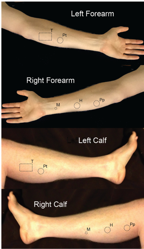
During the enrollment period, participants are given a full description of all sensory and pain measures that will be used. With initial consent, all participants are allowed to experience all sensory and pain measures before full enrollment. All sensory and pain assays are based on well-established assays used in both healthy human participants and in chronic pain patients34. All assays involve either innocuous (non-painful stimuli) or acute noxious stimuli (painful stimuli) that do not damage tissue. The time between different tests is >5 min, to allow the subject to rest and to reduce the potential for sensory fatigue or sensitization. The sequential order of tests is the same during each testing session. Specific sites of testing are limited to the T1 dermatome on the left and right forearms and L3/S2 dermatome on the left and right calves. All sites for testing are marked with a marker, and individual sites are spread out to avoid overlapping receptive field activation (Figure 1). See the Materials and Equipment Table for the full materials list. For retest reliability studies, individual subjects were tested by two experimenters in a single day.
Protocol
All tests with human subjects should be approved by the Institutional Review Board at the individual institution. All testing described for the current study was approved by the Duquesne University Institutional Review Board for human subject research. Training for and descriptions of each measure are as follows:
1. Cutaneous Mechanical Sensitivity Assay13
NOTE: Enrolled participants are asked to sit in a chair, with support provided for the extremity to be tested. The assay involves determining the sensitivity threshold for innocuous cutaneous stimulation. Stimulation is provided with standard sensory evaluator von Frey filaments (see the equipment section). These small nylon filaments each apply a single force (ranging from 0.078 mN (0.008 g) to 4.08 mN (1.0 g)).
Before the start of the first experimental trial, allow the participant to feel and manipulate the filaments. Give the filament to the participant and let them gently bend it against the skin of their hand.
During each trial, ask the subject to look away from their forearm or calf. Apply the filament to the subject's forearm or calf until it bows, and ask if they feel the filament.
- Starting with the smallest filament (0.078 mN; below the sensory threshold for human detection), conduct five trials on the subject's forearm or calf for each filament.
- With each filament (e.g., the 0.078 mN filament), apply the filament four times in the "positive" trials.
- For the other trial, do not apply the filament, but still ask the subject if they feel the filament. NOTE: This "negative" trial will be randomly inserted with the four "positive" trials and is designed to test for false responses (i.e., the subject thinks they feel something even though no stimulus is applied). This is necessary for sensory threshold testing, because random noise in the sensory system and/or other stimuli (e.g., a light breeze) can cause a false response.
If a subject detects ≥3 of the positive trials and 0 negative trials for a filament, then record that filament as the subject's "mechanical sensory threshold" on the data form.
For a single filament, if the subject detects <2 of the real trials and/or >0 of the false trials, then start another round of 5 trials with the next-biggest filament until the sensory threshold is reached. NOTE: Sensory thresholds vary for human participants, but in experience, they typically range from 1.57-9.81 mN (data not shown). This force is enough to feel light innocuous pressure. Typical testing time for each body part (forearm and calf) is about 5 min. It is also possible to measure needle-like pain with these filaments, but this usually entails using larger-diameter filaments.
2. Radiant Heat Sensitivity Assay35
NOTE: Enrolled participants are asked to sit in a chair, with support provided for the extremity to be tested. The assay involves determining the sensitivity threshold for non-painful heat change and for painful thermal stimulation. Stimulation is provided with a radiant heat device35. This device uses a focused light beam to slowly heat a subject's skin through a piece of 0.64-mm-thick safety glass (see below in the instrument section).
Before the start of the training and experimental trials, show the device to the subject; allow them to feel the stimulus with their hand.
Ask the subject to rest their forearm or calf on the room temperature glass plate, which should be covered with a rubber-insulating sheet except for the small window for stimulus presentation. NOTE: The insulating sheet allows the subject to focus on the stimulus presentation without the cooling sensation associated with placing one's body against a room-temperature object.
Using a mirror, position the light source under a marked area on the subject's forearm or calf (Figure 1). NOTE: When the leg or arm is raised from the surface of the glass, the thermal stimulus automatically stops and the time since the beginning of the trial is then recorded as the "latency to respond."
Complete two trials for each test on each limb (innocuous temperature detection and pain threshold) in two distinct marked areas to avoid retesting at a single site.
- For the innocuous temperature detection trial, ask the subject to raise their leg or arm or depress the "stop" button when they feel the temperature change.
- Set the device so that the typical withdrawal threshold occurs at approximately 10 sec into the trial and so that the device shuts off after 20 sec. To accomplish this withdrawal threshold, set the device to ramp the temperature such that the stimulus reaches 47 °C at 10 sec. NOTE: For innocuous temperature detection trials, the typical temperature on the glass at threshold is 37 °C (99 °F). In the pain threshold trial, subjects are told to raise their leg or arm or depress the "stop" button when they feel the stimulus transition from "innocuous warmth or heat" to "painful heat." The typical temperature on the glass at threshold is ~47 °C (121 °F). The maximum temperature of the trial at the 20 sec cutoff time point is 50 °C, which is well below the cumulative temperature that causes tissue damage in humans36.
- Use the constant temperature assay28 (heat block) to evaluate both the quality and unpleasantness of thermal pain. Using the heat block, set the temperature to 45 °C for the stimulus. NOTE: 45 °C is a standard temperature that is the typical minimal stimulus necessary to feel thermal pain and is known to activate TRPV1 nociceptive receptors29.
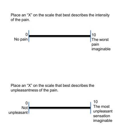
- Before step 2.7.2, explain the standard 0-10 VAS and show a 10 cm line to the subject. Inform the subject that on the "quality scale," "0" represents "no pain" and "10" represents "the worst pain imaginable," and that on the "unpleasantness scale," "0" represents "not unpleasant" and "10" represents "the most unpleasant sensation imaginable" (Figure 2).
- Apply the stimulus (3 cm x 5 cm heating block) for 3 sec to the marked location on the left forearm or calf (as shown in Figure 1 at the site marked "T").
- Immediately following the stimulus, ask the subject to evaluate the quality and unpleasantness of the pain using a standard 0-10 VAS.
3. Pressure Sensitivity Assay13,37
NOTE: Enrolled participants are asked to sit in a chair, with support provided for the extremity to be tested. The assay involves determining the sensitivity threshold for painful pressure stimulation and then determining the quality and unpleasantness of that same pressure in a separate trial. Stimulation is provided with a standard clinical pressure algometer (see below in the instrument section in the Materials and Equipment Table). This device consists of a 2 cm probe connected to a pressure meter.
Before the first training and experimental trial, allow the subject to apply the stimulus to themselves under careful supervision.
- For the pain threshold trial, place the probe on the subject's forearm or calf and apply pressure gradually.
- Complete two trials each to the forearm and calf at two distinct sites (on each limb) to avoid damage to a single area.
- During a trial, apply pressure gradually, until the stimulus transitions from "innocuous pressure" to "painful pressure". Ask the subject to say "stop" at this point and remove the stimulus from the subject's forearm or calf.
- Remove the algometer from the subject. The device automatically records the greatest pressure applied. Record this as the "pressure pain threshold" for the trial.
- Inform the subject that the constant pressure trials are next.
- After determining the pressure threshold for the subject, apply an additional trial on the opposite limb to determine the subject's pain associated with a painful pressure stimulus (in a third testing site). Match the exact stimulus to the subject's pain threshold determined during the baseline trials (e.g., if baseline trials for the forearm indicated a pressure threshold of 50 N, then the subject will be asked to evaluate the pain of that stimulus).
- In this trial, ask the subject to evaluate the quality and unpleasantness of a pressure stimulus given for 3 sec.
- Use a standard 0-10 VAS. Inform the subject that on the "quality scale," "0" represents "no pain" and "10" represents "the worst pain imaginable." On the "unpleasantness scale," "0" represents "not unpleasant" and "10" represents "the most unpleasant sensation imaginable."
- During this trial, apply a painful stimulus, and then ask the subject to evaluate that pain (on the two VASs described above).
4. Reliability Study
NOTE: To examine the reliability of the protocol, we conducted a small study to compare the ratings of subjects between one male and one female examiner.
Recruit participants by posting flyers. Have the interested volunteers who meet the inclusion criteria (Supplemental 1) participate in an orientation session where the study is described and the testing techniques are demonstrated.
Ask potential volunteers questions and have them read and sign the informed consent documents approved by the University IRB. NOTE: The two examiners for this study were laboratory technicians (one male and one female) who were trained by study investigators who have experience from the clinic and laboratory in pain measurement and management and in neural sensation.
Test all subjects with two different examiners, with the two examinations 30 min apart.
To assess inter-rater reliability for this study, calculate intraclass correlation coefficients (model 3,2) [ICC(3,2)] using a two-way mixed analysis of variance (ANOVA) with absolute agreement for each dependent variable (eight total)38. A statistical software can be used for all statistical analyses.
Representative Results
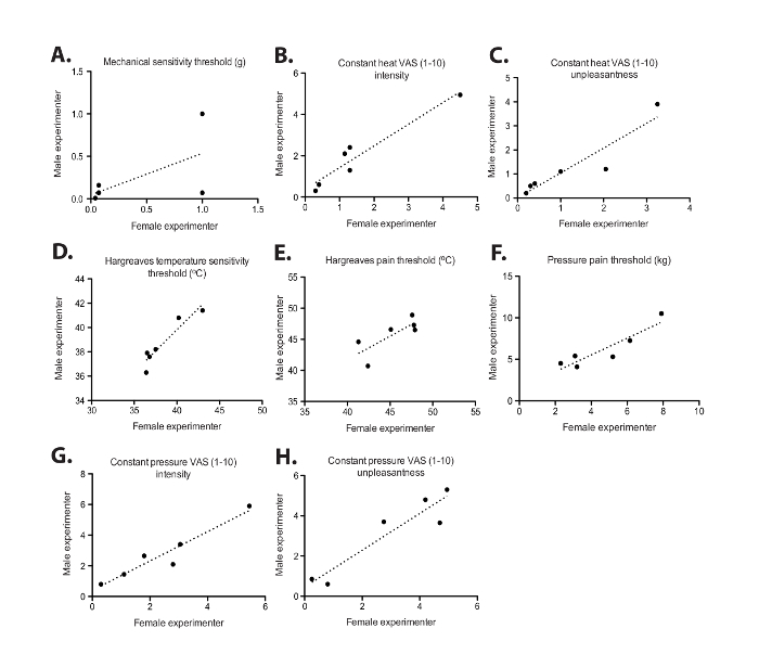
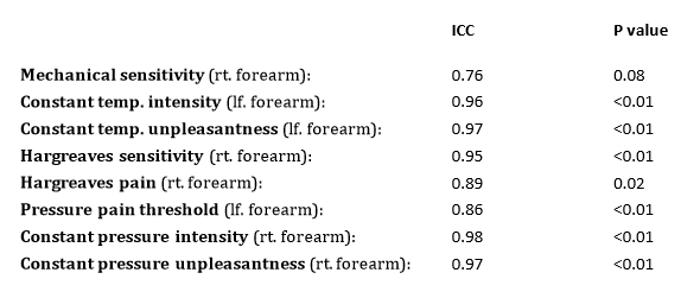
Here, we describe the implementation of cost-effective qualitative and quantitative assays to measure innocuous sensation and pain in human participants using the VAS (Figure 2). The visual representation is important, because accurate and precise results of these examinations are dependent upon correct and consistent protocol execution by the technician. Additionally, it is valuable to know if multiple technicians performing the technique as described can collect reproducible data. While it was not the intention of this study to complete a comprehensive reliability analysis (i.e., we did not perform a statistical correction for multiple testing), the results demonstrate measurement consistency and provide an example analysis of what each laboratory to newly adopt this technique might perform to quantify reliability. To test the inter-experimenter reliability of the assays, two individual experimenters (one male and one female), tested six subjects. All subjects completed the study with no adverse events. Subjects were tested by the two experimenters on a single day, with 30 min between the tests. The order of experimenter testing (male first versus female first) was randomized across the six subjects. The subjects' average age was 21.8 years (SD = 2.0) and the average BMI was 23.5 (SD = 3.3); three of the six subjects were female. As seen in Table 1 and Figure 3, inter-experimenter reliability was strong for mechanical, thermal, and pressure testing. Intraclass correlation [ICC(3,2)] average measures for all inter-experimenter reliability data were above 0.7. In addition, inter-experimenter reliability [ICC(3,2)] average measures were all statistically significant, except for the mechanical sensitivity test (p = 0.075).
 Figure 1: Illustration of sensory testing sites on the left and right forearm and calf. Sites for individual testing are marked with a standard surgical marker. M = mechanical; H = Hargreaves radiant heat; Pp = Pressure pain; Pt = Pressure pain threshold; T = Constant temperature pain. Please click here to view a larger version of this figure.
 Figure 2: Illustration of the Visual Analog Scale (VAS). This figure represents a standard 0-10 visual analog scale (VAS) with a 10-cm line. This scale is used to represent the quality of pain, where "0" represents "no pain" and "10" represents "the worst pain imaginable," and the unpleasantness of pain, where "0" represents "not unpleasant" and "10" represents "the most unpleasant sensation imaginable." Please click here to view a larger version of this figure.
 Figure 3: Evaluation of inter-experimenter reliability of sensory testing in human participants. Individual subjects (n = 6) were assayed for (A) mechanical sensation (p = 0.075), (B) constant heat visual analog scale (VAS) intensity (p = 0.001), (C) constant heat VAS unpleasantness (p = 0.001), (D) radiant heat temperature sensitivity (p = 0.003), (E) radiant heat pain threshold (p = 0.021), (F) pressure threshold (p = 0.002), (G) constant pressure VAS intensity (p = 0.001), and (H) constant pressure VAS unpleasantness (p = 0.001) on a single day (>30 min between tests) by two separate experimenters. P-values represent intraclass correlation coefficient significance. The dotted lines are lines of best fit. Please click here to view a larger version of this figure.
 Table 1: Intraclass correlation coefficients [ICC(3,2)] for the seven pain and sensitivity measures. Individual subjects (n = 6) were assayed by two investigators (one male and one female). All tests were performed on the same day (30 min apart). ICC(3,2) and corresponding P-values are given for each measure. Please click here to view a larger version of this table.
Discussion
We have demonstrated cost-effective and simple qualitative and quantitative sensory tests that can be used to assess mechanical sensation, thermal sensation and pain, and pressure pain in human subjects. The value of these assays is their ease of implementation and low amount of necessary training time. Each experimenter received a minimal amount of training (one trial observation and one trial implementation). Thus, multiple technicians could be trained in one day. The results suggest strong inter-experimenter and within-subject reliability. Depending on the number of tests that each lab uses, statistical correction for multiple ICC testing is advisable.
One measure of inter-experimenter reliability, mechanical sensitivity, did not reach statistical significance (ICC = 0.76, p = 0.08). We reexamined the data collection procedure laboratory notes for two of the subjects and found no abnormalities in data collection. While it is likely that a larger sample size would have reached statistical significance, we think that this is noteworthy for three reasons. First, pain, a subjective experience, is difficult to measure, and efforts to standardize testing cannot be overemphasized. Second, the possibility of a gender bias in pain testing should be considered when conducting these measures. Finally, there is a possibility of a proportional bias, in that at the end of the spectrum considered "high sensitivity," the tests may become less reliable. A more extensive study would need to be conducted to correctly ascertain if this bias exists.
Critical steps in ensuring consistency are reading from a script when explaining tests to a participant; checking the force exerted by monofilaments; and making efforts to ensure that that the intensity, frequency, duration, and localization of the experimental stimuli involved are precisely controlled. Additionally, room temperature could be a factor while measuring sensation, so room temperature should be controlled and recorded. For these results, although it is impossible with this design to truly disambiguate within-subject and intra-experimenter reliability, the fact that a subject demonstrates consistent thresholds suggests that these assays are stable enough for use in clinical and research trials. Furthermore, this is an important finding because it demonstrates a lack of retesting sensitization or sensitivity fatigue.
Most importantly for large clinics, these data show that multiple trained experimenters can reliably implement these tests, and that gender differences between experimenters and subjects or patients are unlikely to affect the results. The protocol is thus broadly applicable to clinics or research laboratories where employee turn-over and the training of new technicians occurs, as this is unlikely to affect the results of the QST assays.
An important limitation of the current study is its sampling of healthy volunteers. There are numerous chronic pain syndromes, and each patient population is unique. Rather than limit our study to one type or classification of chronic pain, we decided to test healthy volunteers as a general model. Each clinic or laboratory is advised to conduct their own internal analysis for a specific patient population.
The overall significance of the protocol is that these assays are reasonably priced and easy to include in typical sensory testing protocols (research or clinical); they are also reliable, even across examiners. The only real limitation is the need for some training and for manual recording of all data. We did not find troubleshooting or modifications to be necessary, so long as the proper equipment is available.
Disclosures
The authors have nothing to disclose.
Acknowledgments
The authors acknowledge the following funding sources: the Duquesne University Faculty Development Fund grants awarded to Kimberly Szucs, PhD and Alex Kranjec, PhD and to Benedict Kolber, PhD and Matthew Kostek, PhD. We also acknowledge Rachel Sweetnich for experimental assistance and funding from the Duquesne University Pain Undergraduate Research Experience program awarded to Sweetnich (Mentors: Szucs and Kostek).
References
- Analysts GI. Pain Management - A Global Strategic Business Report. Global Industry Analysts. 2012. p. 727.
- Medicine A.A.o.P., editor. AAPM Facts and Figures on Pain. 2015.
- Loeser JD, Treede RD. The Kyoto protocol of IASP Basic Pain Terminology. Pain. 2008;137(3):473–477. doi: 10.1016/j.pain.2008.04.025. [DOI] [PubMed] [Google Scholar]
- Clauw DJ. Fibromyalgia: a clinical review. JAMA. 2014;311(15):1547–1555. doi: 10.1001/jama.2014.3266. [DOI] [PubMed] [Google Scholar]
- Haanpaa M, et al. NeuPSIG guidelines on neuropathic pain assessment. Pain. 2011;152(1):14–27. doi: 10.1016/j.pain.2010.07.031. [DOI] [PubMed] [Google Scholar]
- Cruccu G, et al. Recommendations for the clinical use of somatosensory-evoked potentials. Clin Neurophysiol. 2008;119(8):1705–1719. doi: 10.1016/j.clinph.2008.03.016. [DOI] [PubMed] [Google Scholar]
- Backonja MM, et al. Value of quantitative sensory testing in neurological and pain disorders: NeuPSIG consensus. Pain. 2013;154(9):1807–1819. doi: 10.1016/j.pain.2013.05.047. [DOI] [PubMed] [Google Scholar]
- Mainka T, Maier C, Enax-Krumova EK. Neuropathic pain assessment: update on laboratory diagnostic tools. Curr Opin Anaesthesiol. 2015;28(5):537–545. doi: 10.1097/ACO.0000000000000223. [DOI] [PubMed] [Google Scholar]
- Maier C, et al. Quantitative sensory testing in the German Research Network on Neuropathic Pain (DFNS): somatosensory abnormalities in 1236 patients with different neuropathic pain syndromes. Pain. 2010;150(3):439–450. doi: 10.1016/j.pain.2010.05.002. [DOI] [PubMed] [Google Scholar]
- Magerl W, et al. Reference data for quantitative sensory testing (QST): refined stratification for age and a novel method for statistical comparison of group data. Pain. 2010;151(3):598–605. doi: 10.1016/j.pain.2010.07.026. [DOI] [PubMed] [Google Scholar]
- Pfau DB, et al. Quantitative sensory testing in the German Research Network on Neuropathic Pain (DFNS): reference data for the trunk and application in patients with chronic postherpetic neuralgia. Pain. 2014;155(5):1002–1015. doi: 10.1016/j.pain.2014.02.004. [DOI] [PubMed] [Google Scholar]
- Olesen AE, Andresen T, Staahl C, Drewes AM. Human experimental pain models for assessing the therapeutic efficacy of analgesic drugs. Pharmacol Rev. 2012;64(3):722–779. doi: 10.1124/pr.111.005447. [DOI] [PubMed] [Google Scholar]
- Rolke R, et al. Quantitative sensory testing: a comprehensive protocol for clinical trials. Eur J Pain. 2006;10(1):77–88. doi: 10.1016/j.ejpain.2005.02.003. [DOI] [PubMed] [Google Scholar]
- Angst MS, Tingle M, Phillips NG, Carvalho B. Determining heat and mechanical pain threshold in inflamed skin of human subjects. J Vis Exp. 2009. p. e1092. [DOI] [PMC free article] [PubMed]
- Dyck PJ, et al. Cool, warm, and heat-pain detection thresholds: testing methods and inferences about anatomic distribution of receptors. Neurology. 1993;43(8):1500–1508. doi: 10.1212/wnl.43.8.1500. [DOI] [PubMed] [Google Scholar]
- Tena B, et al. Reproducibility of Electronic Von Frey and Von Frey monofilaments testing. Clin J Pain. 2012;28(4):318–323. doi: 10.1097/AJP.0b013e31822f0092. [DOI] [PubMed] [Google Scholar]
- Staahl C, Christrup LL, Andersen SD, Arendt-Nielsen L, Drewes AM. A comparative study of oxycodone and morphine in a multi-modal, tissue-differentiated experimental pain model. Pain. 2006;123(1-2):28–36. doi: 10.1016/j.pain.2006.02.006. [DOI] [PubMed] [Google Scholar]
- Reddy KS, Naidu MU, Rani PU, Rao TR. Human experimental pain models: A review of standardized methods in drug development. J Res Med Sci. 2012;17(6):587–595. [PMC free article] [PubMed] [Google Scholar]
- Adriaensen H, Gybels J, Handwerker HO, Van Hees J. Nociceptor discharges and sensations due to prolonged noxious mechanical stimulation--a paradox. Hum Neurobiol. 1984;3(1):53–58. [PubMed] [Google Scholar]
- Burke D, Mackenzie RA, Skuse NF, Lethlean AK. Cutaneous afferent activity in median and radial nerve fascicles: a microelectrode study. J Neurol Neurosurg Psychiatry. 1975;38(9):855–864. doi: 10.1136/jnnp.38.9.855. [DOI] [PMC free article] [PubMed] [Google Scholar]
- Woolf CJ, Max MB. Mechanism-based pain diagnosis: issues for analgesic drug development. Anesthesiology. 2001;95(1):241–249. doi: 10.1097/00000542-200107000-00034. [DOI] [PubMed] [Google Scholar]
- Wylde V, Palmer S, Learmonth ID, Dieppe P. Test-retest reliability of Quantitative Sensory Testing in knee osteoarthritis and healthy participants. Osteoarthritis Cartilage. 2011;19(6):655–658. doi: 10.1016/j.joca.2011.02.009. [DOI] [PubMed] [Google Scholar]
- Geber C, et al. Test-retest and interobserver reliability of quantitative sensory testing according to the protocol of the German Research Network on Neuropathic Pain (DFNS): a multi-centre study. Pain. 2011;152(3):548–556. doi: 10.1016/j.pain.2010.11.013. [DOI] [PubMed] [Google Scholar]
- Green BG. Temperature perception on the hand during static versus dynamic contact with a surface. Atten Percept Psychophys. 2009;71(5):1185–1196. doi: 10.3758/APP.71.5.1185. [DOI] [PMC free article] [PubMed] [Google Scholar]
- Green BG. Referred thermal sensations: warmth versus cold. Sens Processes. 1978;2(3):220–230. [PubMed] [Google Scholar]
- Green BG, Lederman SJ, Stevens JC. The effect of skin temperature on the perception of roughness. Sens Processes. 1979;3(4):327–333. [PubMed] [Google Scholar]
- Stevens JC, Green BG, Krimsley AS. Punctate pressure sensitivity: effects of skin temperature. Sens Processes. 1977;1(3):238–243. [PubMed] [Google Scholar]
- Fowler CJ, Sitzoglou K, Ali Z, Halonen P. The conduction velocities of peripheral nerve fibres conveying sensations of warming and cooling. J Neurol Neurosurg Psychiatry. 1988;51(9):1164–1170. doi: 10.1136/jnnp.51.9.1164. [DOI] [PMC free article] [PubMed] [Google Scholar]
- Tominaga M. TRP Ion Channel Function in Sensory Transduction and Cellular Signaling Cascades. In: Liedtke WB, Heller S, editors. Frontiers in Neuroscience. Boca Raton: CRC Press/Taylor & Francis; 2007. [PubMed] [Google Scholar]
- Yarnitsky D, Ochoa JL. Warm and cold specific somatosensory systems. Psychophysical thresholds, reaction times and peripheral conduction velocities. Brain. 1991;114(Pt 4):1819–1826. doi: 10.1093/brain/114.4.1819. [DOI] [PubMed] [Google Scholar]
- Hughes AM, Rhodes J, Fisher G, Sellers M, Growcott JW. Assessment of the effect of dextromethorphan and ketamine on the acute nociceptive threshold and wind-up of the second pain response in healthy male volunteers) Br J Clin Pharmacol. 2002;53(6):604–612. doi: 10.1046/j.1365-2125.2002.01602.x. [DOI] [PMC free article] [PubMed] [Google Scholar]
- Handwerker HO, Kobal G. Psychophysiology of experimentally induced pain. Physiol Rev. 1993;73(3):639–671. doi: 10.1152/physrev.1993.73.3.639. [DOI] [PubMed] [Google Scholar]
- Taylor DJ, McGillis SL, Greenspan JD. Body site variation of heat pain sensitivity. Somatosens Mot Res. 1993;10(4):455–465. doi: 10.3109/08990229309028850. [DOI] [PubMed] [Google Scholar]
- Drury DG, Stuempfle KJ, Shannon R, Miller J. An investigation of exercise-induced hypoalgesia after isometric and cardiovascular exercise. Journal of Exerc Physiol. 2004;7(4) [Google Scholar]
- Sternberg WF, Bokat C, Kass L, Alboyadjian A, Gracely RH. Sex-dependent components of the analgesia produced by athletic competition. J Pain. 2001;2(1):65–74. doi: 10.1054/jpai.2001.18236. [DOI] [PubMed] [Google Scholar]
- Yarmolenko PS, et al. Thresholds for thermal damage to normal tissues: an update. Int J Hyperthermia. 2011;27(4):320–343. doi: 10.3109/02656736.2010.534527. [DOI] [PMC free article] [PubMed] [Google Scholar]
- Kinser AM, Sands WA, Stone MH. Reliability and validity of a pressure algometer. J Strength Cond Res. 2009;23(1):312–314. doi: 10.1519/jsc.0b013e31818f051c. [DOI] [PubMed] [Google Scholar]
- Portney LG, Watkins MP. Foundations of Clinical Research: Applications to Practice. Prentice Hall; 2009. [Google Scholar]


