Abstract
Cholecystokinin (CCK) is a neuropeptide that affects growth rate in chickens by regulating appetite. CCK peptides exert their function by binding to two identified receptors, CCKAR and CCKBR in the GI tract and the brain, respectively, as well as in other organs. In mammals, CCK/CCKAR interactions affect a number of immunological parameters, including regulation of lymphocytes and functioning of monocytes. Thus, food intake and growth can potentially be altered by infection and the resulting inflammatory immune response. It is uncertain, however, whether chicken express CCKAR in immune organs and cells, and, if so, whether CCKAR expression is regulated by pathogen derived inflammatory stimuli. Herein, we identify expression of CCKAR protein in chicken peripheral blood mononuclear cells (PBMC) including monocytes, and expression of the CCKAR gene in PBMC, thymus, bursa, and spleen, in selected commercial and pure chicken breeds. Further, stimulation with various types of E. coli heat-labile enterotoxins or lipopolysaccharide significantly regulated expression of CCKAR on monocytes in the different breeds. Ligation of CCKAR with antibodies in PBMC induced mobilization of Ca2+, indicating that CCKAR is signal competent. Injection with polyinosinic: polycytidylic acid (poly I:C), a synthetic analogue of double stranded viral RNA that binds Toll-Like Receptor-3 (TLR3), also regulated gene expressions of CCKAR and proinflammatory cytokines, in the different breeds. Interestingly, variations in the expression levels of proinflammatory cytokines in the different breeds were highly correlated with CCKAR expression levels. Taken together, these findings indicate that the physiological function of CCKAR in the chicken is tightly regulated in immune organs and cells by external inflammatory stimuli, which in turn regulate growth. This is the first report CCKAR expression in immune organs and cells, in any species, and the initial observation that CCKAR is regulated by inflammatory stimuli associated with bacterial and viral infection.
Keywords: Appetite, CCKAR, Gangliosides, E. coli heat-labile enterotoxins, Immune regulation
Introduction
Cholecystokinin (CCK) is a key regulatory neuropeptide produced mainly in the upper part of the intestine by a small subset of cells during feeding. While neurons expressing agouti-related protein (AGRP) and proopiomelanocortin (POMC) in the hypothalamus are important in the control of appetite, CCK and CCK receptor (CCKR) interactions in the periphery act as a satiety feedback mechanism transmitting signals from the intestine in respect to the level of food intake [1-3]. In rodents, there are two types of CCKRs, CCKAR (also CCKR1) and CCKRB (also CCKR2) have been identified in pancreatic acini and the brain, respectively [4,5]. Whereas CCKAR is highly selective for binding a biologically active sulfated form of CCK (CCK-8), CCKBR binds sulfated, non-sulfated CCK, and gastrin [6]. Similar to the expression state in mammals, CCKRs are also expressed in various tissues in the chicken [7,8] and may perform a similar biological function. Specifically, CCKAR is widely expressed in tissues of chicken including the cerebellum, hypothalamus, small intestine, cecum, pancreas and gallbladder whereas CCKBR expression is highest in the brain and proventriculus. Recently, a region downstream the CCKAR gene and levels of CCKAR expression in nonimmune organs were implicated in variation in growth among various breeds, resulting in altered sensitivity to the CCK satiety signal [7].
While CCKAR expression was demonstrated in tissues and organs involved in food intake, control of appetite, and growth, no evidence exists in the chicken in relation to its possible distribution in immune organs and immune cells. This potential distribution is important to address since previous experiments in mammals have shown that food intake and growth were affected by infection and the resulting inflammatory immune response, which in turn control both the biological function and the expression level of CCK [9-11]. Thus, intestinal inflammation resulted in reduced food intake that was associated with increased expression of CCK [10]. On the other hand, in the chicken, pathogens or agents that mimic pathogens components were shown to regulate expression of proinflammatory cytokines and growth, in experimentally challenged animals [11-15]. Since the expression levels of CCKAR in nonimmune organs are likely responsible for variation in growth and body weight among chicken breeds [7], CCKAR expression in immune organs would presumably provide a target for regulation of the expression by infection and/or the resulting proinflammatory cytokines.
In this study, we confirmed expression of CCKAR protein in chicken PBMC including monocytes, and revealed that CCKAR is regulated by various types of E. coli heat-labile enterotoxins [specifically the B subunit of LT-I (LT-IB5), a type I heat-labile enterotoxin, and the type II heat-labile enterotoxin LT-IIa holotoxin and its B subunit (LT-IIaB5)] [16-21], and lipopolysaccharide (LPS). CCKAR gene expression was demonstrated in various types of immune organs, and its level was regulated by poly I:C, a compound mimicking viral double stranded RNA. Poly I:C also regulated gene expression levels of proinflammatory cytokines which were highly correlated with levels of expression of CCKAR.
Materials and Methods
Chicken breeds
This study was conducted on commercial chicken strains, including Pearl White Leghorn, Red Ranger Broilers and Egyptian Fayoumi (pure breed). Birds were purchased one day old from Murray McMurray Hatchery, Iowa, USA. The chicks were a mix of males and females. Chicks were vaccinated by Marek's and Coccidia vaccines at the source. Chicks were kept in cages in the poultry house at Tuskegee University, College of Veterinary Medicine, AL, USA. Temperature in the house was maintained based on age of the bird. Starter diet and water were available ad libitum. The animal protocol was approved by Tuskegee University Animal Care and Use, and Biosafety Committees.
Reagents
Alexa Fluor 488 rabbit anti CCKAR polyclonal antibody (Bioss Inc., # bs 11514R, A488, USA) and biotin conjugated rabbit anti CCKAR polyclonal antibody (Bioss Inc., # bs 11514R, USA). The bs11514R antibodies are raised against a synthetic peptide, and are cross reactive against mouse, rat, human and chicken CCKAR. Mouse IgG FITC antibody (Southern Biotech., # 0102 02, USA), and PE conjugated mouse anti chicken monocytes/macrophages antibody (Southern Biotech., # 8420 09, USA). For calcium experiments, Fura Red, AM, cell permeant (Life Technologies, # F3021, USA), ionomycin (life technology # 124222, USA) and streptavidin from Streptomyces avidinii (Sigma Aldrich, # S4762, USA) were used. For cell culture, RPMI 1640 medium modified with 20 mM HEPES (Sigma # H0887, USA) and 200 mM L glutamine (Sigma # G7513, USA), penicillin/streptomycin (Sigma # P4333, USA), β-mercaptoethanol (Sigma # M6250, USA), HyClone™ fetal bovine serum (FBS) (Life Science, # SH30396.03, Canada), and Hanks’ Balanced Salt Solution (Sigma # H8264, USA) were employed. For harvesting cultured PBMC, Trypsin/EDTA solution (Sigma, # T4049, USA) was used. For RNA extraction from tissue and PBMC, TRI Reagent (TRI Reagent, Sigma, # 93289, USA), and Histopaque® 1077 (Sigma, # 10771, USA) were used. For conventional PCR, Taq PCR Master Mix (2X) (QIAGEN, # 201443, USA), Gelpilot Mid-Range Ladder (100) (QIAGEN, # 239135, USA), Gelpilot® DNA loading Dye, 5x (QIAGEN, # 239901, USA) and Agarose (Biomatik, # A2127, USA). For qPCR analysis, RT2 HT First Strand Kit (QIAGEN, # 330411, USA) and RT2 SYBR Green ROX qPCR Mastermix (QIAGEN, # 330520, USA), and molecular biology water (non Diethylpyrocarbonate (DEPC) treated, DNAse, RNAse and protease free) (Sigma# W4502) were used. For injection, poly (I:C) (InvivoGen, # tlrl pic, USA) was used. For stimulation of PBMC, lipopolysaccharide (LPS) from E. coli 0111:B4 was purchased from Sigma Aldrich (USA). Recombinant E. coli heat-labile enterotoxin B subunits (LT-IB5) expressed in the yeast Pichia pastoris (Sigma-Aldrich, >90% pure), and highly purified recombinants LT-IIa, and LT-IIaB5 were used. Recombinant preparation of the B-subunits of LT-II were obtained from E. coli and purified to >90% by affinity chromatography using a His Bind resin column and gel filtration chromatography [22-24].
Primers
Conventional PCR primers and the β-actin primer (Table 1) were designed using Geneious R8 and Primer Blast NCBI, respectively. The amplification of the correct amplicon was confirmed by the sequence and alignment using Sequences Nucleotide BLAST NCBI. For qPCR primer assays (Table 1) were purchased from QIAGEN.
Table 1.
CCKAR and cytokines primers used in conventional and real time PCR.
| A. Primers used in conventional PCR | ||||
|---|---|---|---|---|
| Forward primer (3 ------ 5) | Reverse primer (5 ------ 3) | TM | Size | Exon/boundary |
| GTGTGCATATGTCTGAATGTGTG | TTGGGAATGAGGGTGAATGG | 64 | 559 | Exon 1 and 2 |
| TGCATGCCATTCACCCTCAT | AATGAGGCCATACGCAACCA | 60 | 417 | Exon 2, 3 and 4 |
| GGATAATAATGATGGTTGCGTATGG | GTAGGACAGCAGGTGGATAAAG | 64 | 413 | Exon 5 |
| B. Primers used in Real time PCR | ||||
| Gene ID | Catalog No. | Ref Seq Accession no. | Amplicon Size (bp) | Company |
| CCKAR | PPG07425A | NM_001081501.1 | 74 | QIAGEN |
| IL-1β | PPG00540A | NM_204524.1 | 138 | QIAGEN |
| IL-6 | PPG00665C | NM_204628.1 | 123 | QIAGEN |
| IFN-γ | PPG01291A | NM_205149.1 | 72 | QIAGEN |
| GAPDH | PPG00285B | NM_204305.1 | 115 | QIAGEN |
| Actin Beta | F:CGGACTGTTACCAACACCC R:ATCCTGAGTCAAGCGCCAAA |
NM_205518.1 | 116 | IDT |
Isolation of PBMC
Blood was collected into tubes containing EDTA from the jugular vein under sterile conditions. Blood was diluted with equal volume of phosphate-buffered saline (PBS, pH 7.4) and layered onto equal volume of Histopaque®1077. The suspension was spun down by centrifugation at 400 xg for 30 min at Room Temperature (RT). After centrifugation, the upper layer was carefully aspirated and discarded. The opaque interphase, containing PBMC, was carefully transferred with Pasteur pipette into clean sterile conical centrifuge tubes. After addition of PBS, cells were spun down for 10 min at 400 xg. The supernatant was aspirated and discarded. Washing was repeated twice before suspending the cells in PBS.
Assay for effects of toxins on PBMC
PBMC were harvested from pooled blood samples from 3 birds from each breed. PBMC (1 × 10⁶) were incubated with 5 μg/ml and 2.5 μg/ml LT-IB5 and LT-IIaB5, respectively. LT- IIa was used at pre-optimized dose of 100 ng; LPS was used at a concentration of 1 μg/ml. Untreated cells (medium) were included as a control. After 24 h incubation at 37°C, the medium was removed and retained for subsequent analysis. The remaining cells were harvested from the plate by Trypsin/EDTA, and washed with PBS. To minimize unspecific binding of antibodies to cells, 1% FBS was added to cells in 200 μl PBS containing 0.5% FBS and 0.01 % NaN3. After adding anti CCKAR antibody (1/50) and anti-monocytes KUL01 (1/400), the mixture was incubated on ice for 50 min, and analyzed by flow cytometry (Becton Dickenson, FlowJo software. Tree Star, OR). Controls included unlabeled cells and cells stained with Mouse IgG FITC antibody as a negative control for the FIITC anti-CCKAR antibody.
Assay for mobilization of calcium in PBMC
PBMC from 3 broilers and 3 layers were each pooled and stimulated in vitro overnight with LPS to increase CCKAR expression, as shown above (Figure 1), or left unstimulated. LPS-untreated cells (medium) were included as control. To increase CCKAR expression, cells were stimulated with 1μg/ml LPS before analysis of the calcium response was performed. Cells at 5 × 10⁶/ml were loaded with pre optimized concentration of Fura Red/AM (4 μg/ml) in Hanks Balanced Salt Solution, which contains Ca+2, in the presence of 5% FBS. Cells and dye were incubated for 45 min at 37°C. After washing, the cells were aliquoted to 1 × 10⁶/0.5 ml buffer and rested for 30 min at RT. Cells were then pre-incubated with biotin conjugated anti-CCKAR (1/50) for 5 min at 37°C and immediately transferred to flow cytometer for analysis. A baseline measurement was obtained for the first 25 s, after which streptavidin was added at 10 μg/ml, and the acquisition continued for the total duration of 3 min. Positive and negative controls included ionomycin added at 2 μl from 3 mM reconstituted solution, and cells incubated with the antibody or streptavidin alone, respectively.
Figure 1.
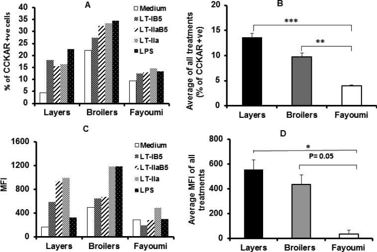
Stimulation of chicken monocytes significantly increases CCKAR protein expression. PBMC were cultured with the E. coli enterotoxin LT-IIa or the B-subunit of LT-I and LT-II (LT-IB5 and LT-IIaB5, respectively), or LPS. Cells were harvested, and labelled with FITC anti-CCKAR and PE anti-KUL01 (for monocytes) and analyzed by flow cytometry. The percentage of cells expressing CCKAR was determined by gating out low FSC/high SSC cells (dead cells) [17] and platelets, and by gating on CCKAR+ and KUL01+cells. The total number of cells expression CCKAR (Figure 1A) and Mean Fluorescence Intensity (MFI) (Figure 1C) are shown. The results represent average of two experiments. In B and D, the average effects of all stimulants (− medium) on number of cells expressing CCKAR (Figure 1B) and level of CCKAR (Figure 1D) in the different breeds is compared. *, ** and *** denotes statistical significance (one-way Anova) with a p<0.05, p <0.01 and p<0.001, respectively.
Sample collection
For tissue collection, birds of 20 day old were euthanized by terminal exposure to CO2. For collection of PBMC, birds were bled by the heart puncture after death for RNA isolation. Tissue samples were collected from Bursa of Fabricius, thymus, and spleen. The body weight of each bird was recorded before euthanasia and collection of samples. Tissue samples were placed in 1.5 ml Eppendorf tubes, immediately frozen in liquid nitrogen, and stored at −80°C until processed in the laboratory.
RNA isolation and reverse transcription
Total RNA was extracted from 30-50 mg of tissue by the TRI reagent, pelleted and eluted in RNase free water. The integrity of the RNA was examined by gel electrophoresis and the concentration was measured by Nano drop ND1000 (NanoDrop Technologies Inc., Wilmington, USA). Two μg of RNA sample was reverse transcribed using the RT2 HT First Strand Kit. The cDNA product was verified by conventional PCR using CCKAR primers, and analyzed by agarose gel electrophoresis.
Conventional PCR
Three primers were designed to flank exon 1, 2, 3, 4, and 5 of CCKAR gene. Four μl of cDNA, 12.5 μl Taq Mastermix, 0.5 μl of foreword and reverse primers (10 μM), and molecular biology water were added to a total volume of 25 μl. Thermal cycling conditions were: an initial denaturation at 95°C for 3 min, followed by 45 cycles of 95°C for 30 s, annealing for 30 s at temperature according to the primer (see above), extension at 72°C for 50 s, and final extension at 72°C for 5 min. The amplified products were examined by gel electrophoresis, sequenced, and aligned in the data base.
Real time PCR
CCKAR, β-actin, GAPDH, IL1-β, IL-6, IFN-γ specific primers were used to amplify the gene products. For this purpose, 1 μl of cDNA, and Real time PCR kits [RT2 SYBR Green ROX qPCR Mastermix (2) (QIAGEN) were used. Amplification was carried out by using CCKAR primer assay (QIAGEN), and duplicate samples for each tissue from four birds and from each breed. Samples from two replicates were analyzed using MxPro QPCR software. The thermal cycling conditions were: initial denaturation at 95°C for 10 min, followed by 45 cycles at 95°C for 15 s, annealing/extension for 1 min at 60°C. DNA produced by Real time PCR reactions was resolved on a Stratagene 300 P Real time PCR system (Agilent Technologies, USA). Assays were analyzed using MxPro QPCR software (Agilent Technologies, USA). CT values for each sample were determined and used in the calculation of “fold change” based on the Livac method [25], mRNA expressions for each tissue were normalized using β actin and GAPDH.
Poly I:C injection and its effects on CCKAR and cytokines gene expression levels
20 days old birds were injected with poly I:C (100 μg/kg bwt). 10 birds from each breed, were injected IP with Poly I:C (100 μg/kg bw). Four additional birds from each breed were injected with PBS (controls). Tissue samples were collected after 24 h from all organs. Total RNA was extracted and Real time PCR analysis was performed as described above.
Statistical analysis
Statistical analysis of the data was performed using GraphPad Prism 6 software (GraphPad software Inc.). One-way ANOVA was used to examine the breed effect on CCKAR expression. Two-way ANOVA was used to examine the breed and organ effects on CCKAR expression. For statistical significance between breeds, one-way ANOVA was followed by the unpaired t-test, and two-way ANOVA was followed by the Holm Sidak method. Linear regression analysis was performed to determine the association between expressions of CCKAR and cytokines.
Results
Stimulation of chicken PBMC by toxins significantly increases CCKAR protein expression
CCK/CCKAR interactions in mammals or their derived cells have been shown to affect a number of immunological parameters, including regulation of lymphocytes and monocytes function [26-28]. However, there is no evidence in chicken how CCKAR expression is regulated by external stimuli including infectious agents and their products. Since the immune response, particularly the inflammatory response to infectious agents may have dramatic consequences on appetite and growth we examined the effect of a set of bacterial toxins on protein expression of the CCKA receptor in PBMC and on the surface of monocytes in different breeds of chicken.
Figure 1 shows CCKAR expression in unstimulated (medium) and stimulated PBMCs. Following stimulation with LPS, LT-IB5, LT-IIaB5, and LT-IIa, for 24 h at 37°C, cells were labelled with FITC anti CCKAR and the chicken monocyte/macrophage KUL01 Abs, and analyzed by flow cytometry. The data in Figure 1A is showing the percentage of treated PBMC including monocytes expressing CCKAR. CCKAR expression increased in all breeds compared to cells incubated with the medium alone. However, the noticeable increase in CCKAR expression is shown in the commercial layers and broilers compared to the pure breed Fayoumi, when treated and untreated cells were compared. For statistical analysis and to minimize bias effects of one treatment, the added effects of all inflammatory mediators (minus medium) were calculated (Figure 1B). This was statistically highly significant in the formers (One-way ANOVA, p=0.01 for broilers and p=0.001 for layers) compared to the Fayoumi breed. A representative contour double staining analysis of the cells from one experiment is shown (Figure 1 supplement). As shown, compared to medium, the toxins and especially LPS increased the number of cells expressing CCKAR in PBMC. Further, LPS had notable stimulation effect on monocytes in layers and broilers but no effect in the Fayoumi.
Because the effects of the holotoxin, B subunits and LPS could also result in an increase in the level (rather than number) of CCKAR on cells that already express detectable amount of the receptor, we performed histogram analysis of the data in which traces of stimulated and unstimulated cells were superimposed. An increase in the mean fluorescence intensity was then recorded. The overall effect in Figures 1C and 1D show similar patterns to those in Figures 1A and 1B, noticeably an increase in the level of CCKAR in the commercial breeds compared to Fayoumi. The added effects of PBMC stimulation by all bacterial toxins (minus medium) was also statistically significant or nearly significant in the formers (One-way ANOVA, p=0.02 for layers and p=0.05 for broilers).
Chicken CCKAR is a signaling competent receptor on PBMC
To ensure that CCKAR on PBMC is functional, the ability of anti CCKAR antibody to induce mobilization of calcium was investigated (Figure 2). Calcium ions (Ca2+) are universal intracellular messengers that control a variety of different physiological processes in the cells such as neurotransmitters release, muscle contraction, cell proliferation, gene transcription, etc. [29-31]. In immune cells, elevation of calcium is generally an indication of cell activation.
Figure 2.
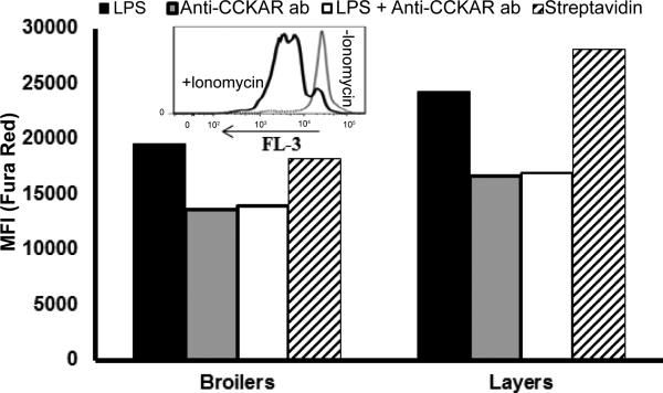
Mobilization of Ca2+ following ligation of CCKAR. PBMC were isolated from pooled blood samples from 3 birds. To stimulate expression of CCKAR, cells were pre cultured for 24 h with LPS. Cells were then loaded with Fura Red Ca+2 dye. To induce mobilization of calcium, biotin conjugated anti CCKAR antibody and streptavidin were employed. Shown is a representative of one of two experiments with similar results. LPS indicates cells stimulated with LPS alone (no anti CCKAR); anti CCKAR indicates non LPS stimulated cells ligated with anti CCKAR + streptavidin; LPS + anti CCKAR ab indicates LPS stimulated cells ligated with anti CCKAR antibody + streptavidin, and streptavidin indicates cells to which streptavidin alone was added.
The capacity of the cells to flux Ca2+ was initially evaluated by adding ionomycin, an effective ionophore that raises intracellular Ca2+ levels (insert in Figure 1). Cells stimulated with LPS in the absence of anti CCKAR ab were taken as a base level of fluorescence (Figure 2). Ligation of LPS-stimulated cells with anti CCKAR antibody followed by streptavidin induced mobilization of Ca2+ (decrease in fluorescence is a property of Fura Red in loaded cells with the dye). However, even in the absence of prior LPS stimulation, ligation of CCKAR by the antibody and streptavidin induced a similar decrease in the level of fluorescence. Addition of streptavidin alone or anti CCKAR antibody without ligation with streptavidin did not induce mobilization of calcium. These results demonstrate that anti CCKAR antibody induces mobilization of Ca2+ in chicken PBMC suggesting that chicken CCKAR is signaling competent.
Chicken CCKAR is expressed in immune organs and PBMC in different breeds
Having demonstrated CCKAR protein expression on monocytes and PBMC, mRNA gene expression was then examined in PBMC and in several immune organs including the bursa, thymus, and spleen. Since maturation of the bursa and its maximum size occurs between 2 to 4 weeks after hatching, an event that is also mimicked by the development of the thymus, conventional PCR followed by Real time PCR analysis of CCKAR gene expression were performed in 20 days old chicks from each breed. Conventional PCR (Figure 3 supplementary material) was performed as an initial screening for CCKAR gene expression in chicken immune organs before qPCR analysis. RNA was extracted from different tissues and from PBMC and reverse transcribed into cDNA. This was further used in PCR and real time PCR. For each procedure, specific primers were used for amplification (Table 1).
Amplification of CCKAR gene in PBMC, spleen, bursa and thymus in tissues and cells isolated from the layers are shown (Figure 3, supplementary material). A similar DNA profile was obtained from a transcript analysis of the other two breeds (data not shown). The product size from Exon 1 was 559 bp; Exon 2, 3 and 4 sizes were 417 bp, and Exon 5 size was 413 bp. The amplicons were subsequently sequenced and aligned on the database to the CCKAR gene sequence. An alignment match of 99% was present. This is the first report that demonstrates the presence of the CCKAR gene message in chicken PBMC, bursa, spleen and thymus.
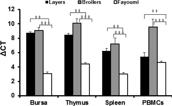
To address expression level of the CCKAR gene, Real time PCR was performed, and levels were compared in different breeds (Figure 3). While differences in CCKAR expression in PBMC and the immune organs were not statistically significant between broilers and layers, expression was significantly higher in Fayoumi compared to the other breeds (Two-way ANOVA, p<0.01 and p<0.001 compared to layers and broilers, respectively). This is shown by the significantly lower value of ΔCT (Figure 3). There was no effect of the organs on CCKAR expression in the different breeds (Two-way ANOVA, p>0.05). These results demonstrated that CCKAR was expressed in multiple immune organs and in PBMC, and that the levels of expression varied among breeds.
Figure 3.
Expression of CCKAR message in immune organs and PBMC. RNA was extracted from different tissues and from PBMC from four 20 day old birds, and reverse transcribed to cDNA. Amplification was performed using Real time PCR system. Shown are averages +/− SEM of ΔCT values from 4 birds for each breed, calculated by determining the CT value of CCKAR compared to that of the house keeping gene, β actin. Two-way ANOVA followed by two tailed unpaired t-test were used to determine the statistical analysis. ** and *** denotes significance with p<0.01 and p<0.001, respectively.
Poly I:C injection regulates CCKAR and cytokines gene expressions in immune organs and PBMC
Inflammatory mediators such as bacterial toxins can influence expression of CCKAR (Figure 1). We further examined how poly I:C, a synthetic analogue of double stranded RNA, that binds TLR3 and induce production of proinflammatory cytokines, regulates CCKAR and cytokines gene expressions in immune organs and in PBMC (Figure 4). Twenty day old chicks were injected with 100 μg/kg bwt of poly I:C. Control birds were injected with PBS only. RNA was extracted from the different tissues and from PBMC and reverse transcribed. Real time PCR was performed in samples containing cDNA from each tissue.
Figure 4.
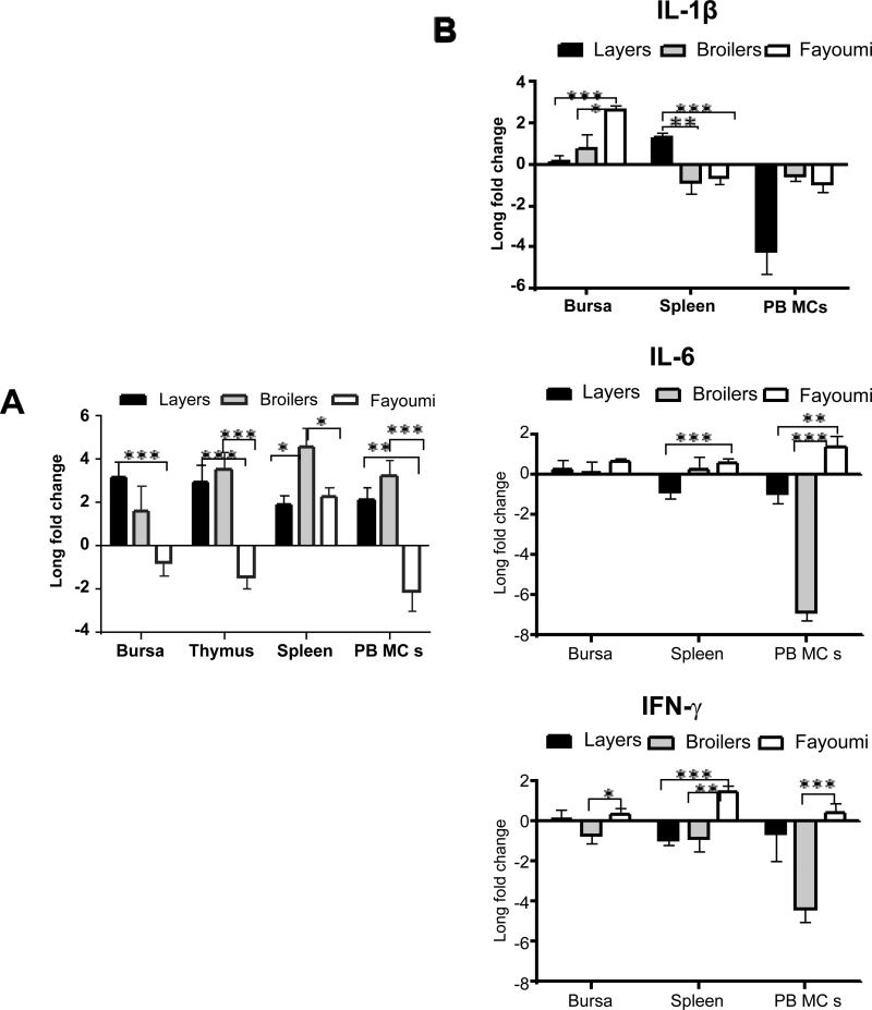
Poly I:C injection regulates CCKAR and cytokines gene expressions in Immune Organs and PBMC. RNAs from different tissues were extracted and reverse transcribed to cDNA. The cDNA was used to analyze gene expression by Real time PCR. In A: Log 2 fold change of CCKAR gene expression after poly I:C injection is shown. In B: Log 2 fold change of cytokines gene expression after poly I:C injection is shown. Two-way ANOVA was used to test the effect of breeds, organs, and both on CCKAR and cytokines. For comparative analysis of significance between breeds two-way ANOVA was followed by the Holm Sidak method. *, ** and *** denotes significance with p <0.05, p<0.01 and p<0.001, respectively. Shown are the averages of log 2 fold change in birds +/− SEM.
The injection of poly:IC increased the level of expression of CCKAR in all immune organs in the broilers and layers chicks, whereas a decrease in expression was noticeable in all organs in the Fayoumi with the exception of the spleen in an increase occurred. The breeds and the organs effects on the level of CCKAR expression were statistically significant (Two-way ANOVA, for breeds p<0.0001; for organs p<0.05). There was also significant effect of the interactions between organs and breeds on CCKAR expression (p<0.05). The difference in CCKAR expression levels between layers and Fayoumi were statistically significant in the bursa, thymus and PBMC, but not in the spleen (Two-way ANOVA, p<0.001 for the bursa and thymus, and p<0.01 for PBMC) (Figure 4A). Further, the difference in the expression level between broilers and Fayoumi were statistically significant in the spleen, thymus and PBMC, but not in the bursa (Two-way ANOVA, p<0.001 for thymus and PBMC, and p<0.05 for spleen). Finally, CCKAR expression level was significantly different in the broilers compared to the layers, but only in the spleen (p<0.05). Thus poly I:C injection differentially regulated CCKAR expression in the different breeds; it downregulated expression in Fayoumi, in the bursa, thymus and PBMC, while it increased expression level in the layers and broilers in these organs. In the spleen, however, poly I:C increased the expression of CCKAR in all breeds.
To determine if expression of CCKAR was correlated with expression of inflammatory cytokines, we also measured the expression of IL-1β, IL-6 and IFN-γ in various tissues in the different breeds. Figure 4B shows effects of poly I:C injection on the expression level of inflammatory cytokines in the different immune organs, and for each breed. Because there is no reported known function of inflammatory cytokines in the thymus, this organ was not analyzed.
For IL-1β, there was significant effect of the breed, the organs, and both on this cytokine (two-way ANOVA, p<0.05 for the breed; p<0.0001 for the organs; p<0.0001 for both). Analysis of IL-1β expression in the bursa revealed significantly higher expressions in Fayoumi compared to layers and broilers (Two-way ANOVA, p<0.001 compared to layers and p<0.05 compared to broilers). On the other hand, in the spleen, IL-1β was downregulated in the broilers and Fayoumi while it was significantly increased in the layers (p<0.001 and p<0.01 compared to Fayoumi and broilers, respectively). In PBMC, IL-1β was downregulated in all breeds. Thus, in Fayoumi poly I:C injection significantly increased expression of IL-1β in the bursa, and downregulated the expression in the spleen and PBMC. Further, with the exception of a significant increase of IL-1β expression in the spleen, in the layers, expression levels in the bursa and PBMC in the layers and broilers were either not significant or are downregulated.
For IL-6, there was significant effect of the breed, the organs, and both on this cytokine (two-way ANOVA, p<0.0001 for each). Poly I:C injection increased its expression in the bursa in all breads. However, this was not significantly different among the breeds. Further, poly I:C downregulated IL-6 expression in the spleen in the layers whereas its expression was significantly higher in Fayoumi compared to layers (p<0.001). In PBMC, poly I:C injection downregulated expression of IL-6 in the broilers and layers whereas in Fayoumi this was significantly higher (p<0.001 and p<0.01, respectively). Thus, in Fayoumi poly I:C increased expression of IL-6 in the bursa, spleen and PBMC. On the other hand, in the layers and broilers, the expressions were downregulated or were no significant, except in the bursa where IL-6 expression is increased.
For IFN-γ, there was significant effect of the breed, the organs, and both on this cytokine (two-way ANOVA, p<0.0001 for the breed; p<0.01 for the organs; p<0.01 for both). The general trend was little or downregulation of the expression in the layers and the broilers while IFN-γ expression was significantly higher in Fayoumi in the bursa, spleen and PBMC compared to broilers (p<0.05 in the bursa, p<0.05 and p<0.01 in the spleen, and p<0.001 in the PBMC). IFN-γ expression in the Fayoumi was also significantly higher compared to the layers, but only in the spleen (p<0.001).
In conclusion, poly I:C injection regulated, though variably in different breeds, expression levels of CCKAR and inflammatory cytokines in immune organs. Nevertheless, most of the increases in cytokines expression occurred in Fayoumi while CCKAR expression level in this breed decreased. A similar inverse relationship trend may be applied to the layers and broilers wherein no significant effect, or a decrease in cytokines expression levels, was associated with an increase of those of CCKAR.
Variations in the expression levels of inflammatory cytokines are predicted from those of CCKAR in immune organs
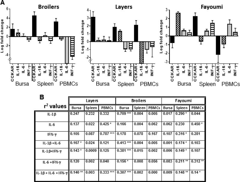
To better visualize the directions of CCKAR and cytokines expressions in the bursa, spleen and PBMC, in each breed, a summary graph depicted from Figure 4 is shown (Figure 5A).
Figure 5.
Variations In the expression levels of inflammatory cytokines are predicted from those of CCKAR in immune organs. A: summary of the effect of poly I:C injection on CCKAR and cytokines gene expression in the different breeds. B: regression analysis of the association between CCKAR and cytokines gene expressions. Analysis was performed at 90% confidence level. Shown are the r2 values. *, **, and *** denote statistical significance at p<0.1, p<0.05 and p<0.01, respectively.
For linear regression analysis of the expressions (Figure 5B), the question posed in the test was “what would best predict variations in the level of each or combination of the cytokines from CCKAR expression levels in each organ following poly I:C injection”?. The r2 value, a measurement of the linear relationship between these 2 parameters, and the p value set at 90% confidence level, are reported.
In the layers, a significant association was found between the level of CCKAR expression with each of those of the IL-6 and IFN-γ in PBMC (p<0.1, and p<0.001, respectively). Additionally, a significant association occurred when the added effects of multiple cytokines was analyzed. Thus, CCKAR expression level was associated with IL-1β+IL-6, IL-1β+IFN-γ, and with all 3 cytokines, in the bursa (p<0.1, p<0.1 and p=0.05, respectively). In PBMC, CCKAR expression level was also associated with the combined levels of the 3 cytokines (p<0.01).
In the broilers, a significant association was only found in the bursa. In this case, the trend of CCKAR gene expression was associated with IL-1β, IL-1β+IL-6, IL-1β+IFN-γ, IL-6+IFN-γ and with all 3 cytokines (p<0.01, p<0.01, p<0.01, p<0.1, and p<0.01, respectively).
In the Fayoumi, an association between CCKAR and cytokines expression levels was found only in the spleen and PBMC. In the spleen, this was significant for each of IL-1β and IFN-γ (p=0.1 and p<0.1, respectively); IL-1β+I-6 (p<0.1); IL-1β+IFN-γ (p<0.1); IL-6+IFN-γ (p<0.05); and with all 3 cytokines (p<0.01). On the other hand, in PBMC, a significant association was found with IL-6 (p=0.1); IL-6+IFN-γ (p<0.05); and all 3 cytokines (p<0.1).
In summary, expression levels of inflammatory cytokines could be predicted from the levels of CCKAR expressed in immune organs and PBMC. Noteworthy, the strongest association between these 2 parameters in all breeds was evident in the combined expressions of IL-1β+IL-6+IFN-γ. Nevertheless, good associations could also be found with single cytokines or a mix of 2 cytokines. It should be noted that a similar regression analysis in which cytokines were chosen as a fixed parameter with variable CCKAR expression levels resulted in a similar linear relationship (not shown). These results support statistically significant association between expression of CCKAR and the proinflammatory cytokines, following poly I:C injection.
Discussion
In this paper, we postulated that CCKAR expression in the chicken extends beyond organs directly involved in the control of appetite and growth, and includes immune organs and cells. We moreover hypothesized that external pathogen derived stimuli and endogenous cytokines regulate CCKAR expression level. These notions were confirmed by our data. We demonstrate that CCKAR protein is expressed in PBMC and is a competent signaling receptor. Further, inflammatory stimuli such as bacterial toxins and LPS, and poly I:C, regulate CCKAR protein level, and its message is detected in PBMC, spleen, bursa, and thymus, respectively. Moreover, poly I:C regulates expression of proinflammatory cytokines in those organs in the different breeds examined. Finally, an association between expression levels of CCKAR and those of cytokines is demonstrated.
A large number of studies in mammalian species addressed the role of CCK in mediating satiety signal feedback in unchallenged animals. Moreover, the effects of pathogens and inflammatory agents on CCK level, food intake, and growth have also been examined. Thus, CCK had pleiotropic effects on mammalian immune cells, including suppression of B cell costimulatory molecules, maturation of dendritic cells, murine macrophages, human neutrophils, and proinflammatory cytokines [10,28,32-35]. These studies highlighted the crosstalk between pathogens, CCK, and the immune system.
In the chicken, on the other hand, there is a lack of understanding of the mechanisms involved in the crosstalk between CCK, pathogens and the immune response [14,36,37]. Because the expression level of CCKAR on various cells could affect the physiological function of CCK and its outcome, studies that investigate CCKAR expression in challenged and unchallenged animals would be of great importance. Recently [7], addressed the role of a region downstream the CCKAR gene, and levels of CCKAR expression in nonimmune organs. These were reported responsible for variations in growth among chicken strains, resulting in altered sensitivity to the CCK satiety signal. Thus, by using 16 generations of cross between fast and slow growing strains of chicken, a region downstream the CCKAR gene was responsible for up to 19% difference in body weight. Moreover, when levels of CCKAR mRNA were measured after injection of CCK, these were lower in the high growth compared to the low growth strains whereas it was intermediate in the heterozygotes. These results have been interpreted in light of the modern changes in appetite introduced in the domestication of chicken.
To address the possible crosstalk between pathogen derived stimuli, CCKAR, and cytokines, we selected 3 breeds that represent high growth broilers (Red Ranger Broilers), relatively low growth layers (Pearl White Leghorn) and a pure breed (Egyptian Fayoumi). The pure breed Egyptian Fayoumi is generally well known for its resistance to disease [38,39], as such would be ideal to compare the effects of those stimuli on CCKAR expression levels in this breed with the other two breeds. Figure 1 shows comparative CCKAR protein expression data of the 3 breeds as a result of stimulation of PBMC with various forms of E. coli - labile enterotoxins and LPS. In this regard, LT-IIa, LT-IIaB5, and LT-IB5 bind to ganglioside receptors expressed on surface of immune cells, and induce signals resulting in activation of antigen presenting cells (including monocytes), T cells, and secretion of cytokines [16-21]. The results in Figure 1 clearly show a striking difference in CCKAR protein expression among the different breeds after stimulation. The most notable is the significantly lower expression of the receptor in Fayoumi compared to the other 2 breeds. Considering that LT-IB5, LT-IIa, LT-IIab5, and LPS each binds to a different receptor; GM1, GD1b, a complex of GD1b and TLR2, and TLR4, respectively [16,20,22], this result is a strong indication of CCKAR low expression and unresponsiveness to these bacterial stimuli in the Fayoumi birds. Conversely, in the broilers and layers, signaling pathways involved in the response to LPS and the enterotoxins appear to intersect with those involved in the upregulation of CCKAR. CCKAR is a G protein-coupled 7-transmembrane spanning receptor. Further experiments addressed CCKAR signaling competence in PBMC (Figure 2). Ligation of CCKAR with specific antibody induced mobilization of calcium. This result is consistent with those in the mammalian counterpart where CCKAR signals involved a number of pathways that ultimately generate calcium, NF-kB and PKC [6].
Having demonstrated CCKAR protein expression in PBMC, we hypothesized that CCKAR is expressed in immune organs such as the spleen, bursa and thymus, as well as in PBMC. The data from Figure 3, using both PCR and Real time PCR, demonstrated the presence of the gene message in those organs and in PBMC. CCKAR normal expression in the Fayoumi birds was significantly lower compared to the broilers and layers, consistent with its protein expression, at least in monocytes in PBMC (Figure 1). In this regard, the expression of receptors other than CCKAR on immune cells which are involved in the control of food intake at the hypothalamic level is documented in mammals. For example, a previous report indicated that immune cells expressed leptin and ghrelin receptors on human T lymphocytes and monocytes by which they exert mutually antagonistic effects on the secretion of proinflammatory cytokines [40]. Similarly, in a mouse model of induced inflammation, CCKAR antagonist restored food intake that was dependent on CD4+ T cells and IL-4Rα [10].
Further indication of the influence of agents that produce proinflammatory cytokines on CCKAR expression is shown in Figure 4 and Figure 5. Poly I:C is a synthetic analogue of double stranded viral RNA that binds TLR3, and induce chicken proinflammatory cytokines including IL-1β, IL-6, and type 1 IFN-s [41,42]. Poly I:C was also reported to act synergistically with CpG-ODN (binds to TLR21 the avian equivalent to mammalian TLR9) to increase the production of NO and expression of inducible nitric oxide synthase [41] and the magnitude of IFN-y and IL-10 mRNA [43]. These results indicated that that CpG-ODN and poly I:C synergize to promote a Th1-biased inflammatory. Further, the synergistic effects of CPG-ODN and poly I:C was evident in stimulation of IL-4 and IL-10 suggesting a feedback mechanism to the inflammatory Th1 response to these agonists [43]. Nevertheless, stimulation by each of these agonists alone resulted in differential outcomes of cytokines expression [43].
In the current study; however, it was unclear whether the injection of poly I:C regulated other factors which may be implicated in the increase of CCKAR expression. One possibility is that poly I:C induced expression of proinflammatory cytokines that acted on cells expressing CCKAR. Analysis of the expressions of IL-1β, IL-6 and IFN-γ revealed that most of the increases in cytokine expressions appeared to occur in the Fayoumi birds, while at the same time CCKAR expressions decreased in those shown organs (Figure 4B). Nevertheless, in the Fayoumi in the spleen CCKAR expression increased and IL-1β decreased. Similarly, in PBMCs, CCKAR expression decreased while IL-1B expression decreased. The increase in proinflammatory cytokines concomitant with a decrease in CCKAR expression in this breed may be responsible for its reported resistance to disease [38,39,44], and is consistent with the CCK inhibitory function in mammalian immune cells [10,11,34]. A similar inverse trend between CCKAR expression and those of some proinflammatory cytokines is shown for the layers and broilers (Figure 5A). However, in this case, except for IL-1 in the spleen, the increases in the level of cytokines were either minimal or their levels were downregulated suggesting the possibility that these two breeds are less able to fight some pathogen insults, represented here by poly I:C, compared to the pure Fayoumi breed. Moreover, the presence of an association between CCKAR expression levels and those of proinflammatory cytokines following poly I:C injection (Figure 5B) suggests their mutual regulation.
Analysis of the effects of organs on normal CCKAR expression showed no statistical significance (Figure 3B) whereas after injection of poly I:C both the breed and organs had significant effects on the levels of expression of CCKAR and cytokines (Figure 4). Although there was a variation in how each of the immune organs and breeds influenced the level of expression, nevertheless this study highlights the importance of the breed and immune organ in considering future challenge experiments that address chicken growth. For example, areas of future investigation will be the type and role of cells that express CCKAR in determining outcome of cytokine expression in these organs, and whether CCKAR expression affects expression of other cytokines that may play an important role in stimulating chicken growth or their immunity against microbial challenge. Moreover, similar investigation of CCKAR expression in immune organs in other species may point out to evolutionary conservative traits.
In conclusion, the results in this study demonstrate expression of CCKAR in immune organs and cells in a variety of selected breeds of chicken. CCKAR in PBMC, and presumably in the spleen, bursa and thymus, is signaling-competent. CCKAR is regulated in these organs by the selected external bacterial and viral stimuli. Further, the association between CCKAR and proinflammatory cytokines suggests that CCKAR expression is tightly regulated by these pathogen stimuli. The sensitivity of CCKAR expression appears to vary among the different chicken breeds. The results implicate the involvement of immune organs in the regulation of CCKAR expression by agents that induce proinflammatory cytokines.
Supplementary Material
Acknowledgements
Research contributed by T.O. Nashar to this study was supported by grants from the United States Department of Education (P031B120901) and United States National Institute of Health RCMI core laboratory facilities grant (G12MD00758-23), and a PhD fellowship to S. Elkassas from the Egyptian Ministry of Higher Education. Research contributed by T.D. Connell and G. Hajishengallis is supported by United States National Institute of Health grant R01 DE017138. We are indebted to Dr. John Heath for help in the statistical analysis and Mr Jason White for managing the core facilities.
Footnotes
Authors Agreement
All authors agree about the content of the article and have no conflict of interest.
References
- 1.Stengel A, Taché Y. Interaction between gastric and upper small intestinal hormones in the regulation of hunger and satiety: ghrelin and cholecystokinin take the central stage. Curr Protein Pept Sci. 2011;12:293–304. doi: 10.2174/138920311795906673. [DOI] [PMC free article] [PubMed] [Google Scholar]
- 2.Dockray GJ. Cholecystokinin. Curr Opin Endocrinol Diabetes Obes. 2012;19:8–12. doi: 10.1097/MED.0b013e32834eb77d. [DOI] [PubMed] [Google Scholar]
- 3.Wynne K, Stanley S, McGowan B, Bloom S. Appetite control. J Endocrinol. 2005;184:291–318. doi: 10.1677/joe.1.05866. [DOI] [PubMed] [Google Scholar]
- 4.Saito A, Sankaran H, Goldfine ID, Williams JA. Cholecystokinin receptors in the brain: characterization and distribution. Science. 1980;208:1155–1156. doi: 10.1126/science.6246582. [DOI] [PubMed] [Google Scholar]
- 5.Sankaran H, Goldfine ID, Deveney CW, Wong KY, Williams JA. Binding of cholecystokinin to high affinity receptors on isolated rat pancreatic acini. J Biol Chem. 1980;255:1849–1853. [PubMed] [Google Scholar]
- 6.Singh P. Role of Annexin-II in GI cancers: interaction with gastrins/ progastrins. Cancer Lett. 2007;252:19–35. doi: 10.1016/j.canlet.2006.11.012. [DOI] [PMC free article] [PubMed] [Google Scholar]
- 7.Dunn IC, Meddle SL, Wilson PW, Wardle CA, Law AS, et al. Decreased expression of the satiety signal receptor CCKAR is responsible for increased growth and body weight during the domestication of chickens. Am J Physiol Endocrinol Metab. 2013;304:E909–E921. doi: 10.1152/ajpendo.00580.2012. [DOI] [PMC free article] [PubMed] [Google Scholar]
- 8.Ohkubo T, Shamoto K, Ogino T. Structure and tissue distribution of Cholecystokinin-1 receptor in chicken. J Poult Sci. 2007;44:98–104. [Google Scholar]
- 9.Leslie FC, Thompson DG, McLaughlin JT, Varro A, Dockray GJ, et al. Plasma cholecystokinin concentrations are elevated in acute upper gastrointestinal infections. QJM. 2003;96:870–871. doi: 10.1093/qjmed/hcg140. [DOI] [PubMed] [Google Scholar]
- 10.McDermott JR, Leslie FC, D'Amato M, Thompson DG, Grencis RK, et al. Immune control of food intake: enteroendocrine cells are regulated by CD4+ T lymphocytes during small intestinal inflammation. Gut. 2006;55:492–497. doi: 10.1136/gut.2005.081752. [DOI] [PMC free article] [PubMed] [Google Scholar]
- 11.Zhang JG, Cong B, Li QX, Chen HY, Qin J, et al. Cholecystokinin octapeptide regulates lipopolysaccharide-activated B cells co-stimulatory molecule expression and cytokines production in vitro. Immunopharmacol Immunotoxicol. 2011;33:157–163. doi: 10.3109/08923973.2010.491079. [DOI] [PubMed] [Google Scholar]
- 12.He H, Genovese KJ, Swaggerty CL, MacKinnon KM, Kogut MH. Co-stimulation with TLR3 and TLR21 ligands synergistically up-regulates Th1-cytokine IFN-gamma and regulatory cytokine IL-10 expression in chicken monocytes. Dev Comp Immunol. 2012;36:756–760. doi: 10.1016/j.dci.2011.11.006. [DOI] [PubMed] [Google Scholar]
- 13.Kaiser P, Rothwell L, Galyov EE, Barrow PA, Burnside J, et al. Differential cytokine expression in avian cells in response to invasion by Salmonella typhimurium, Salmonella enteritidis and Salmonella gallinarum. Microbiol. 2000;12:3217–3226. doi: 10.1099/00221287-146-12-3217. [DOI] [PubMed] [Google Scholar]
- 14.Klasing KC, Laurin DE, Peng RK, Fry DM. Immunologically mediated growth depression in chicks: influence of feed intake, corticosterone and interleukin-1. J Nutri. 1987;117:1629–1637. doi: 10.1093/jn/117.9.1629. [DOI] [PubMed] [Google Scholar]
- 15.Xing Z, Schat KA. Expression of cytokine genes in Marek's disease virus-infected chickens and chicken embryo fibroblast cultures. Immunology. 2000;100:70–76. doi: 10.1046/j.1365-2567.2000.00008.x. [DOI] [PMC free article] [PubMed] [Google Scholar]
- 16.Connell TD. Cholera toxin, LT-I, LT-IIa and LT-IIb: the critical role of ganglioside binding in immunomodulation by type I and type II heat-labile enterotoxins. Expert Rev Vaccines. 2007;6:821–834. doi: 10.1586/14760584.6.5.821. [DOI] [PMC free article] [PubMed] [Google Scholar]
- 17.El-Kassas S, Faraj R, Martin K, Hajishengallis G, Connell TD, et al. Cell clustering and delay/arrest in T-cell division implicate a novel mechanism of immune modulation by E. coli heat-labile enterotoxin B-subunits. Cell Immunol. 2015;295:150–162. doi: 10.1016/j.cellimm.2015.02.014. [DOI] [PMC free article] [PubMed] [Google Scholar]
- 18.Hajishengallis G, Tapping R, Martin MH, Nawar H, Lyle EA, et al. Toll-like receptor 2 mediates cellular activation by the B subunits of type II heat-labile enterotoxins. Infect Immun. 2005;73:1343–1349. doi: 10.1128/IAI.73.3.1343-1349.2005. [DOI] [PMC free article] [PubMed] [Google Scholar]
- 19.Hajishengallis G, Connell TD. Type II heat-labile enterotoxins: structure, function, and immunomodulatory properties. Vet Immunol Immunopathol. 2013;152:68–77. doi: 10.1016/j.vetimm.2012.09.034. [DOI] [PMC free article] [PubMed] [Google Scholar]
- 20.Nashar TO, Williams NA, Hirst TR. Importance of receptor binding in the immunogenicity, adjuvanticity and therapeutic properties of cholera toxin and Escherichia coli heat-labile enterotoxin. Med Microbiol Immunol. 1998;187:3–10. doi: 10.1007/s004300050068. [DOI] [PubMed] [Google Scholar]
- 21.Williams NA, Hirst TR, Nashar TO. Immune modulation by the cholera-like enterotoxins: from adjuvant to therapeutic. Immunol Today. 1999;20:95–101. doi: 10.1016/s0167-5699(98)01397-8. [DOI] [PubMed] [Google Scholar]
- 22.Cody V, Pace J, Nawar HF, King-Lyons N, Liang S, et al. Structure-activity correlations of variant forms of the B pentamer of Escherichia coli type II heat-labile enterotoxin LT-IIb with Toll-like receptor 2 binding. Acta Crystallogr D Biol Crystallogr. 2012;68:1604–1612. doi: 10.1107/S0907444912038917. [DOI] [PMC free article] [PubMed] [Google Scholar]
- 23.Hajishengallis G, Nawar H, Tapping RI, Russell MW, Connell TD. The Type II heat-labile enterotoxins LT-IIa and LT-IIb and their respective B pentamers differentially induce and regulate cytokine production in human monocytic cells. Infect Immun. 2004;72:6351–6358. doi: 10.1128/IAI.72.11.6351-6358.2004. [DOI] [PMC free article] [PubMed] [Google Scholar]
- 24.Lee CH, Nawar HF, Mandell L, Liang S, Hajishengallis G, et al. Enhanced antigen uptake by dendritic cells induced by the B pentamer of the type II heat-labile enterotoxin LT-IIa requires engagement of TLR2. Vaccine. 2010;28:3696–3705. doi: 10.1016/j.vaccine.2010.03.016. [DOI] [PMC free article] [PubMed] [Google Scholar]
- 25.Livak KJ, Schmittgen TD. Analysis of relative gene expression data using real-time quantitative PCR and the 2(-Delta Delta C(T)) Method. Methods. 2001;25:402–408. doi: 10.1006/meth.2001.1262. [DOI] [PubMed] [Google Scholar]
- 26.Carrasco M, Hernanz A, La Fuente MD. Effect of cholecystokinin and gastrin on human peripheral blood lymphocyte functions, implication of cyclic AMP and interleukin 2. Regul Pept. 1997;70:135–142. doi: 10.1016/s0167-0115(97)00025-6. [DOI] [PubMed] [Google Scholar]
- 27.Cong B, Li SJ, Yao YX, Zhu GJ, Ling YL. Effect of cholecystokinin octapeptide on tumor necrosis factor a transcription and nuclear factor-kB activity induced by lipopolysaccharide in rat pulmonary interstitial macrophages. World J Gastroenterol. 2002;8:718–723. doi: 10.3748/wjg.v8.i4.718. [DOI] [PMC free article] [PubMed] [Google Scholar]
- 28.Zhang JG, Cong B, Jia XX, Li H, Li QX, et al. Cholecystokinin octapeptide inhibits immunoglobulin G1 production of lipopolysaccharide-activated B cells. Int Immunopharmacol. 2011;11:1685–1690. doi: 10.1016/j.intimp.2011.05.027. [DOI] [PubMed] [Google Scholar]
- 29.Berridge MJ. Inositol trisphosphate and calcium signalling. Nature. 1993;361:315–325. doi: 10.1038/361315a0. [DOI] [PubMed] [Google Scholar]
- 30.Berridge MJ, Bootman MD, Lipp P. Calcium--a life and death signal. Nature. 1998;395:645–648. doi: 10.1038/27094. [DOI] [PubMed] [Google Scholar]
- 31.Petersen OH, Petersen CC, Kasai H. Calcium and hormone action. Annu Rev Physiol. 1994;56:297–319. doi: 10.1146/annurev.ph.56.030194.001501. [DOI] [PubMed] [Google Scholar]
- 32.Carrasco M, Rio MD, Hernanz A, la Fuente MD. Inhibition of human neutrophil functions by sulfated and nonsulfated cholecystokinin octapeptides. Peptides. 1997;18:415–422. doi: 10.1016/s0196-9781(96)00338-5. [DOI] [PubMed] [Google Scholar]
- 33.De la Fuente M, Campos M, Del Rio M, Hernanz A. Inhibition of murine peritoneal macrophage functions by sulfated cholecystokinin octapeptide. Regul Pept. 1995;55:47–56. doi: 10.1016/0167-0115(94)00091-b. [DOI] [PubMed] [Google Scholar]
- 34.Han DY, Cong B, Li SJ, Liu XL, Ma CL, et al. Effect of CCK-8 on IL-12 secreted in murine bone marrow-derived dendritic cell induced by LPS. Chinese Pharmacol Bullet. 2008;3:357–361. [Google Scholar]
- 35.Ni ZY, Cong B, Li SJ, Ma CL, Yan YX, et al. Effects of cholecystokinin octapeptide on expression of IL-1ß, IL-6, IL-4 and IL-10 in lipopolysaccharide-attacked mice. Chin J Pathophysiol. 2007;23:331–335. [Google Scholar]
- 36.Rodriguez-Sinovas A, Fernandez E, Manteca X, Fernandez AG, Gonalons E. CCK is involved in both peripheral and central mechanisms controlling food intake in chickens. Am J Physiol. 1997;272:R334–340. doi: 10.1152/ajpregu.1997.272.1.R334. [DOI] [PubMed] [Google Scholar]
- 37.Savory CJ, Gentle MJ. Effects of food deprivation, strain, diet and age on feeding responses of fowls to intravenous injections of cholecystokinin. Appetite. 1983;4:165–176. doi: 10.1016/s0195-6663(83)80029-4. [DOI] [PubMed] [Google Scholar]
- 38.Liu W, Kaiser MG, Lamont SJ. Natural resistance-associated macrophage protein 1 gene polymorphisms and response to vaccine against or challenge with Salmonella enteritidis in young chicks. Poult Sci. 2003;82:259–266. doi: 10.1093/ps/82.2.259. [DOI] [PubMed] [Google Scholar]
- 39.Redmond SB, Chuammitri P, Andreasen CB, Palic D, Lamont SJ. Chicken heterophils from commercially selected and non-selected genetic lines express cytokines differently after in vitro exposure to Salmonella enteritidis. Vet Immunol Immunopathol. 2009;132:129–134. doi: 10.1016/j.vetimm.2009.05.010. [DOI] [PubMed] [Google Scholar]
- 40.Dixit VD, Schaffer EM, Pyle RS, Collins GD, Sakthivel SK, et al. Ghrelin inhibits leptin- and activation-induced proinflammatory cytokine expression by human monocytes and T cells. J Clin Invest. 2004;114:57–66. doi: 10.1172/JCI21134. [DOI] [PMC free article] [PubMed] [Google Scholar]
- 41.He H, MacKinnon KM, Genovese KJ, Kogut MH. CpG oligodeoxynucleotide and double-stranded RNA synergize to enhance nitric oxide production and mRNA expression of inducible nitric oxide synthase, pro-inflammatory cytokines and chemokines in chicken monocytes. Innate Immun. 2011;17:137–144. doi: 10.1177/1753425909356937. [DOI] [PubMed] [Google Scholar]
- 42.Parvizi P, Mallick AI, Haq K, Schlegel B, Sharif S. A Toll-like receptor 3 agonist (polyI:C) elicits innate host responses in the spleen and lungs of chickens. Can J Vet Res. 2012;76:230–234. [PMC free article] [PubMed] [Google Scholar]
- 43.Swaggerty CL, Kaiser P, Rothwell L, Pevzner IY, Kogut MH. Heterophil cytokine mRNA profiles from genetically distinct lines of chickens with differential heterophil-mediated innate immune responses. Avian Pathol. 2006;35:102–108. doi: 10.1080/03079450600597535. [DOI] [PubMed] [Google Scholar]
- 44.Seliger C, Schaerer B, Kohn M, Pendl H, Weigend S, et al. A rapid high-precision flow cytometry based technique for total white blood cell counting in chickens. Vet Immunol Immunopathol. 2012;145:86–99. doi: 10.1016/j.vetimm.2011.10.010. [DOI] [PubMed] [Google Scholar]
Associated Data
This section collects any data citations, data availability statements, or supplementary materials included in this article.


