Persoonia : Molecular Phylogeny and Evolution of Fungi logoLink to Persoonia : Molecular Phylogeny and Evolution of Fungi
. 2016 Dec 21;37:218–403. doi: 10.3767/003158516X694499

Fungal Planet description sheets: 469–557

PW Crous 1,2,, MJ Wingfield 3, TI Burgess 4, GEStJ Hardy 4, C Crane 5, S Barrett 6, JF Cano-Lira 7, JJ Le Roux 8, R Thangavel 9, J Guarro 7, AM Stchigel 7, MP Martín 10, DS Alfredo 11, PA Barber 12, RW Barreto 13, IG Baseia 14, J Cano-Canals 15, R Cheewangkoon 16, RJ Ferreira 17, J Gené 7, C Lechat 18, G Moreno 19, F Roets 20, RG Shivas 21, JO Sousa 14, YP Tan 21, NP Wiederhold 22, SE Abell 23, T Accioly 14, JL Albizu 24, JL Alves 13, ZI Antoniolli 26, N Aplin 25, J Araújo 27, M Arzanlou 28, JDP Bezerra 29, J-P Bouchara 30, JR Carlavilla 19, A Castillo 19, VL Castroagudín 31, PC Ceresini 31, GF Claridge 32, G Coelho 33, VRM Coimbra 34, LA Costa 35, KC da Cunha 36, SS da Silva 35, R Daniel 37, ZW de Beer 2, M Dueñas 10, J Edwards 38, P Enwistle 39, PO Fiuza 35, J Fournier 40, D García 7, TB Gibertoni 34, S Giraud 30, M Guevara-Suarez 7, LFP Gusmão 35, S Haituk 16, M Heykoop 19, Y Hirooka 41, TA Hofmann 42, J Houbraken 1, DP Hughes 27, I Kautmanová 43, O Koppel 44, O Koukol 45, E Larsson 46, KPD Latha 47, DH Lee 48, DO Lisboa 13, WS Lisboa 13, Á López-Villalba 19, JLN Maciel 49, P Manimohan 47, JL Manjón 19, S Marincowitz 2, TS Marney 21, M Meijer 1, AN Miller 50, I Olariaga 51, LM Paiva 29, M Piepenbring 52, JC Poveda-Molero 53, KNA Raj 47, HA Raja 54, A Rougeron 30, I Salcedo 51, R Samadi 28, TAB Santos 35, K Scarlett 55, KA Seifert 44, LA Shuttleworth 37, GA Silva 29, M Silva 13, JPZ Siqueira 7, CM Souza-Motta 29, SL Stephenson 56, DA Sutton 22, N Tamakeaw 16, MT Telleria 10, N Valenzuela-Lopez 7, A Viljoen 57, CM Visagie 44, A Vizzini 58, F Wartchow 59, BD Wingfield 48, E Yurchenko 60, JC Zamora 61, JZ Groenewald 1
PMCID: PMC5315290  PMID: 28232766

Abstract

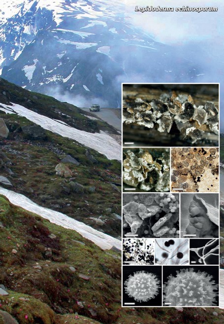
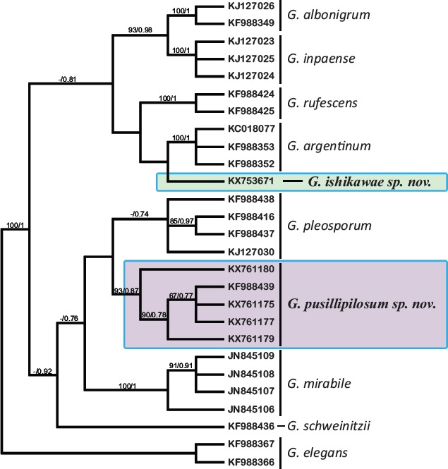
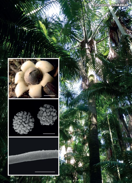
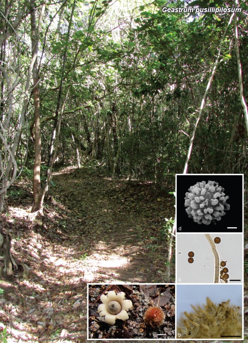
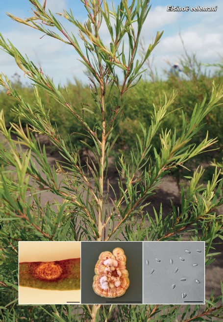
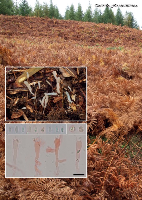
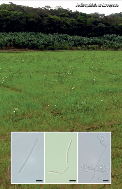
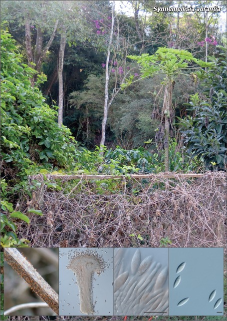
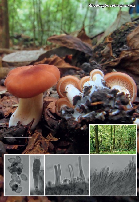
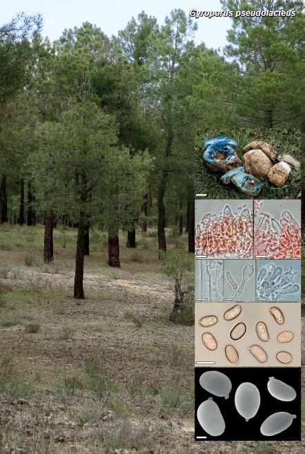
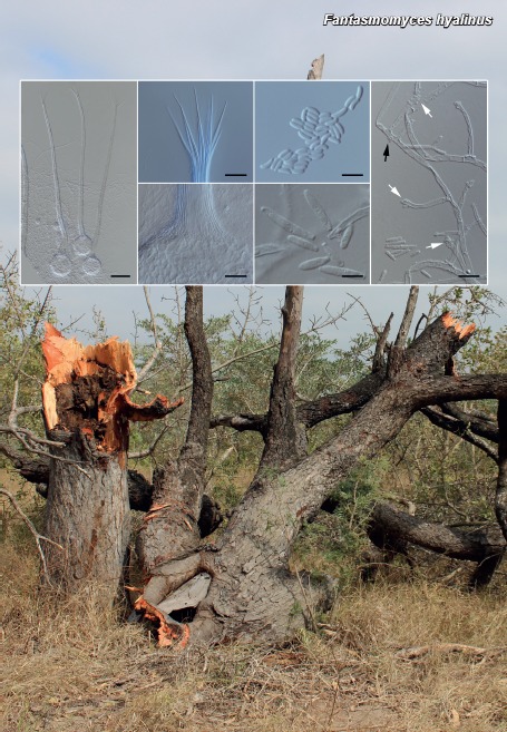
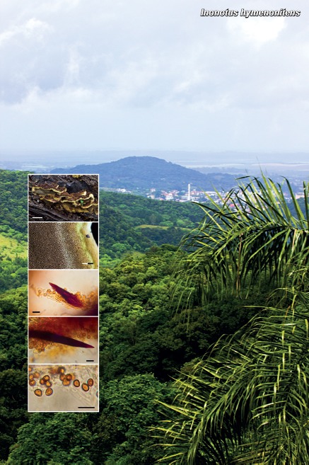
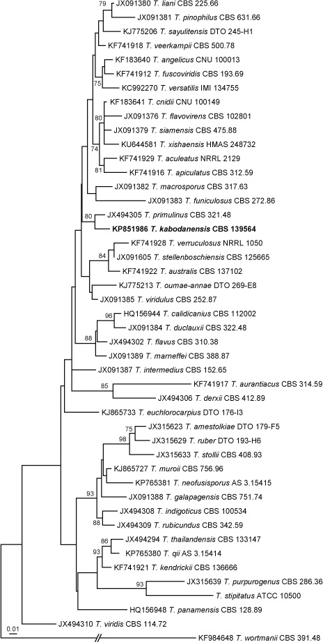
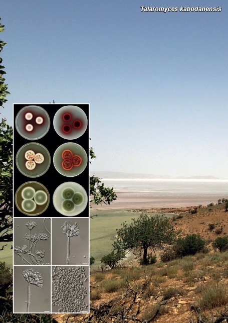
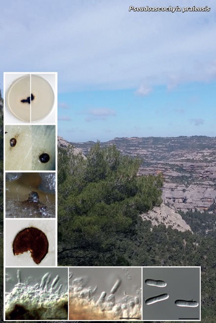
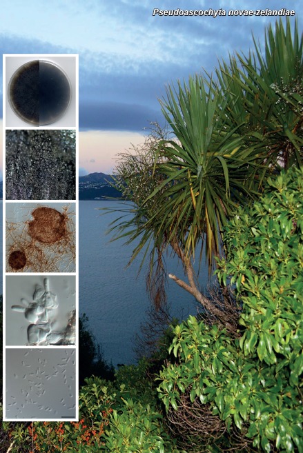
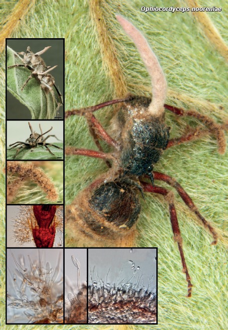
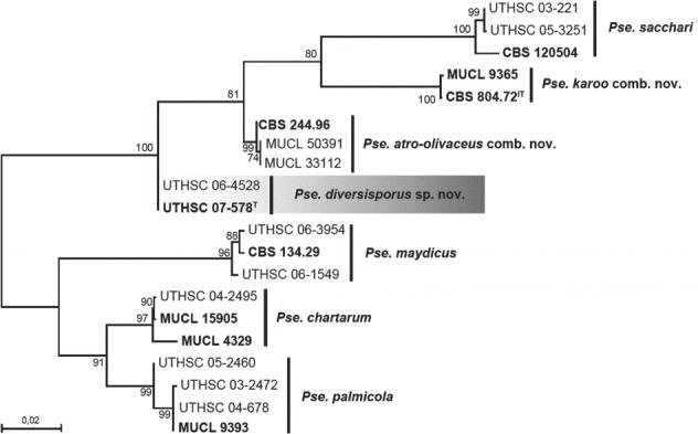
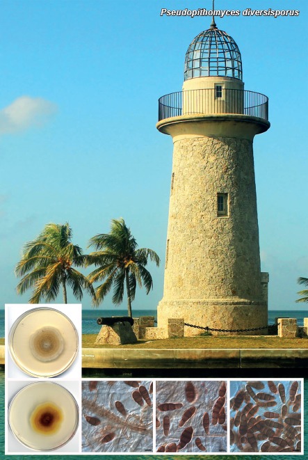
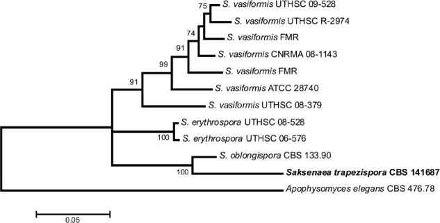
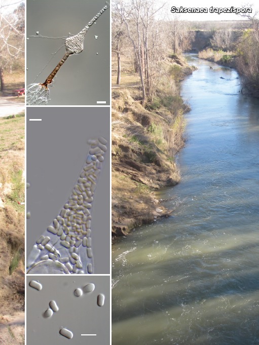
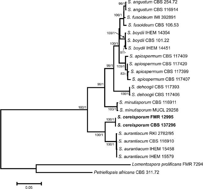
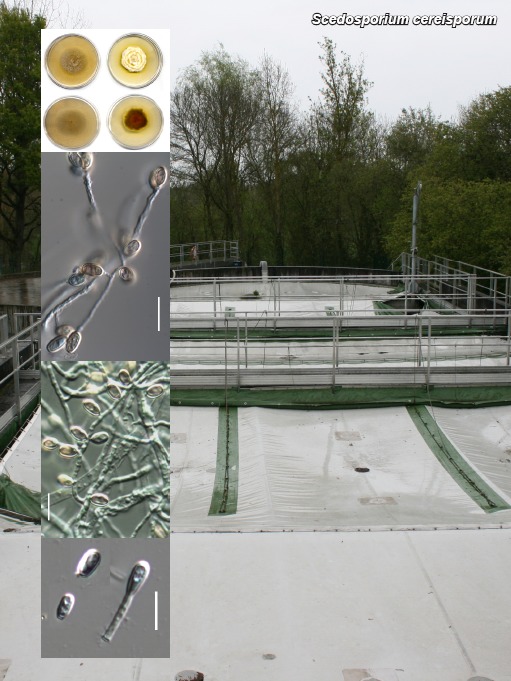
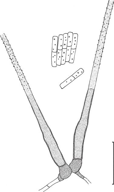
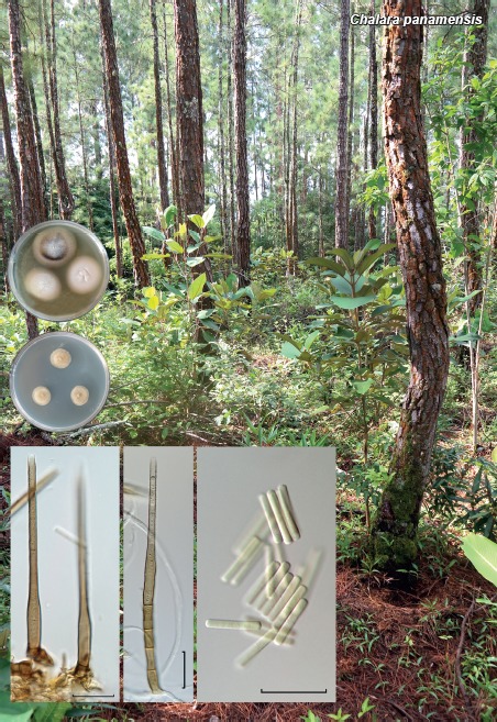
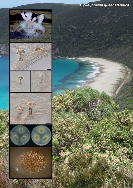
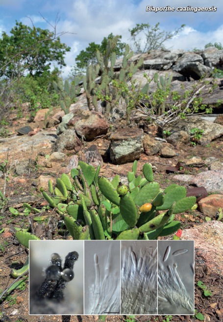
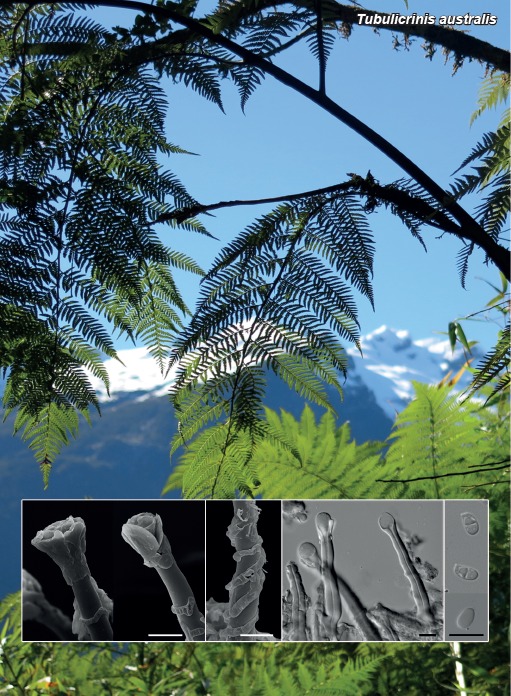
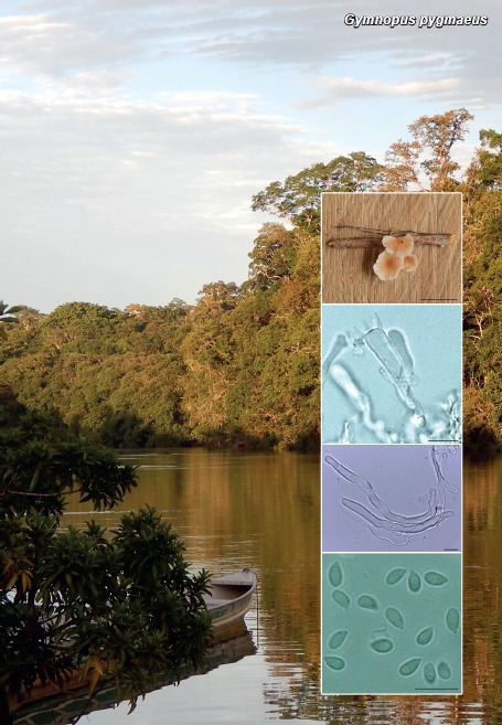
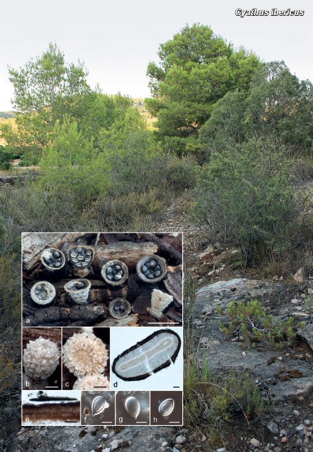
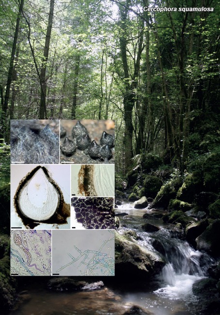
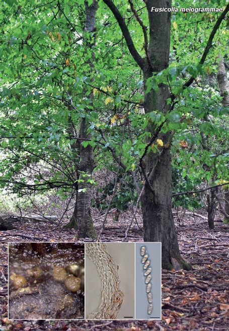
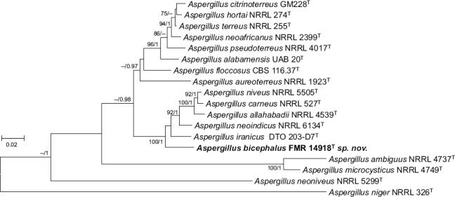
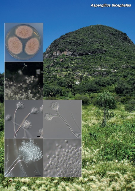
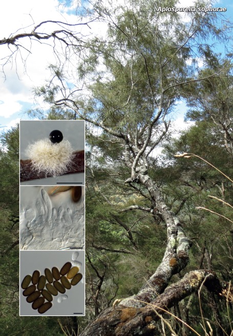
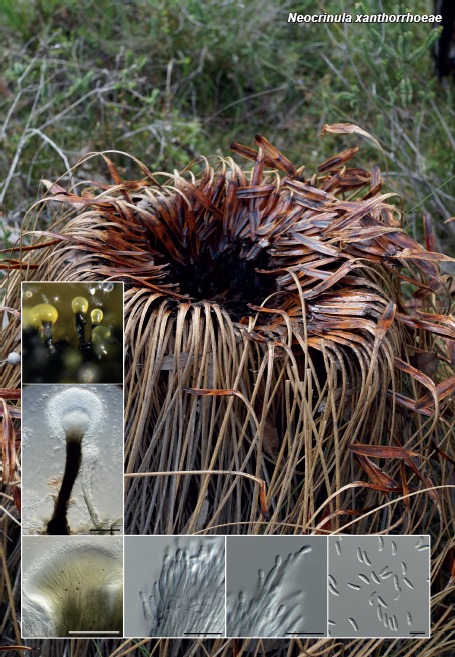
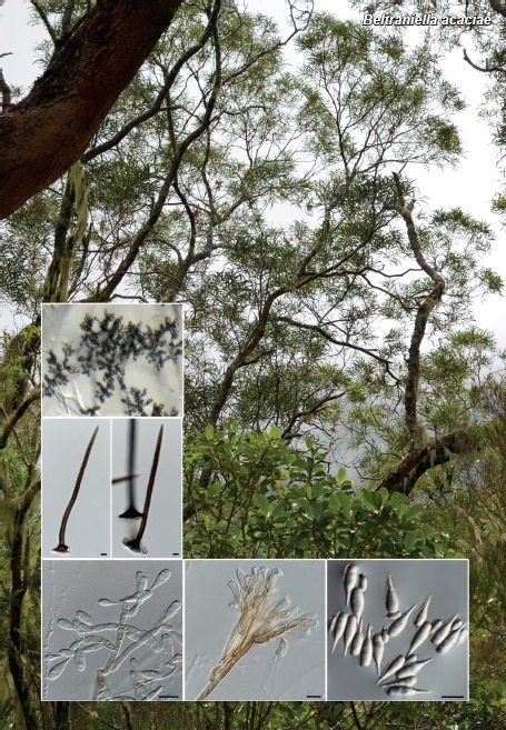
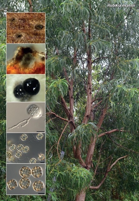
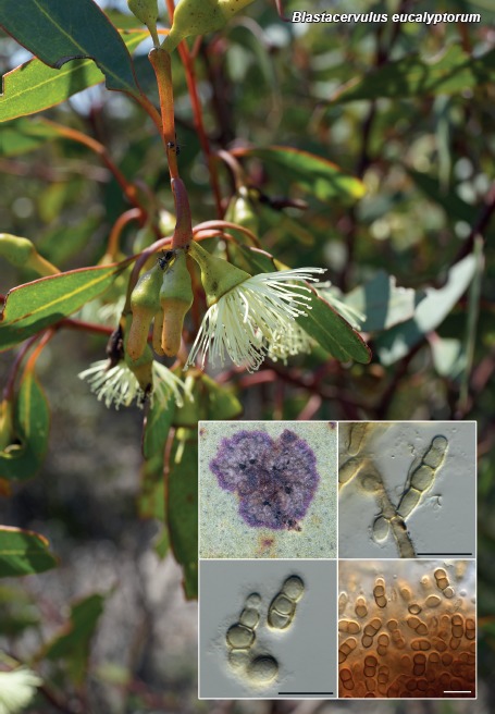
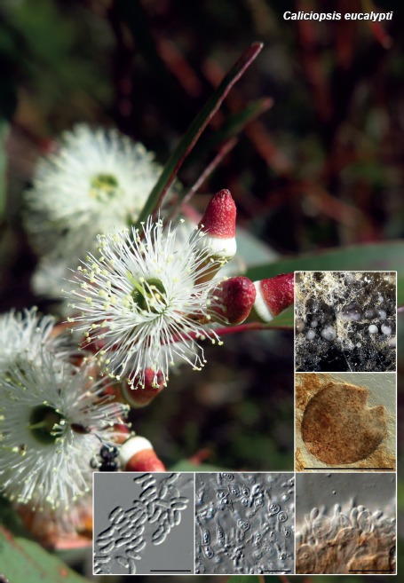
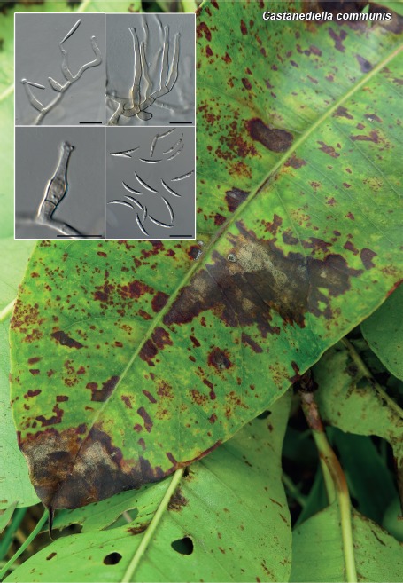
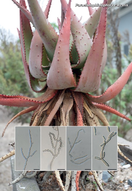
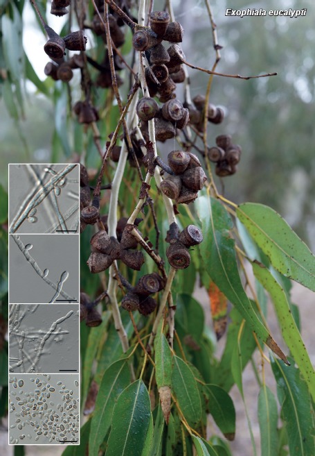
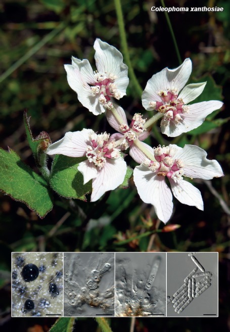
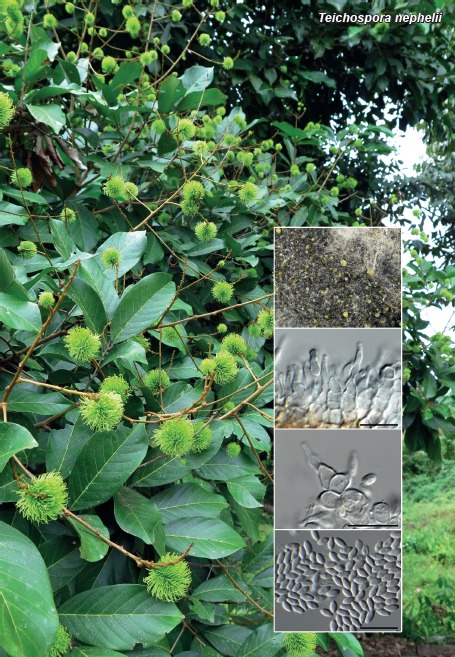
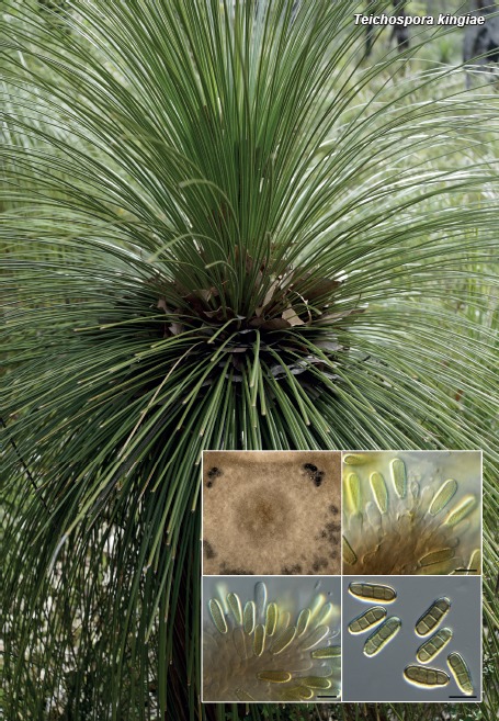
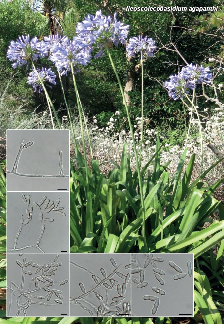
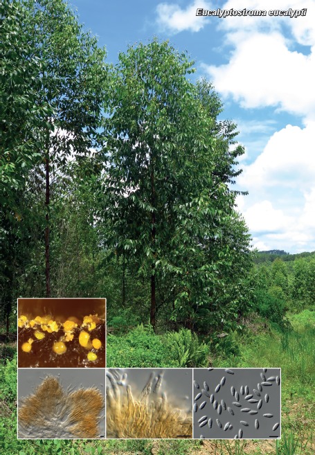
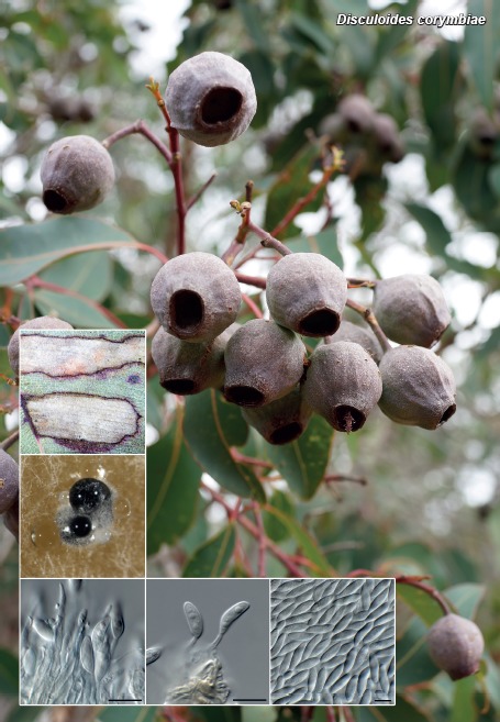
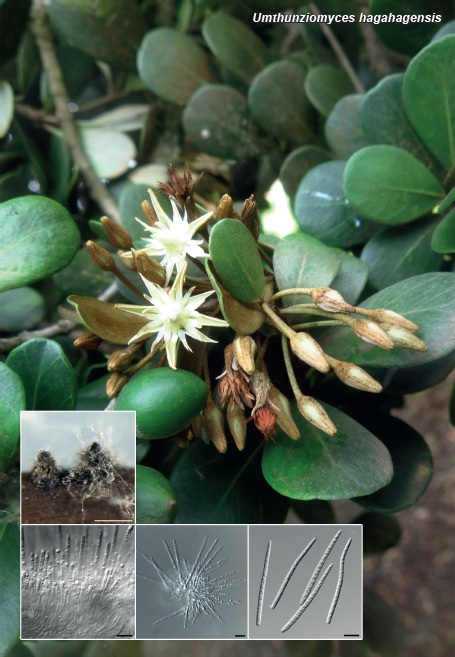
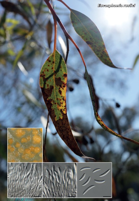
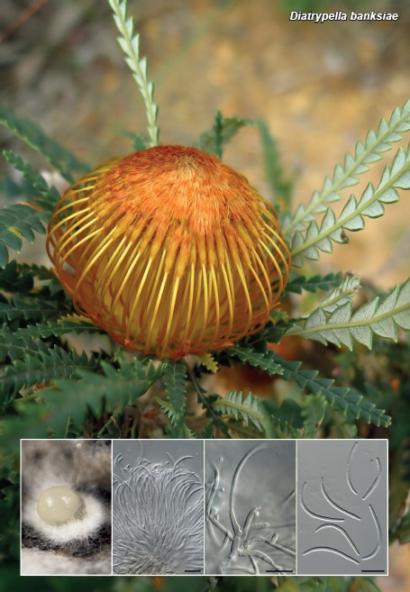
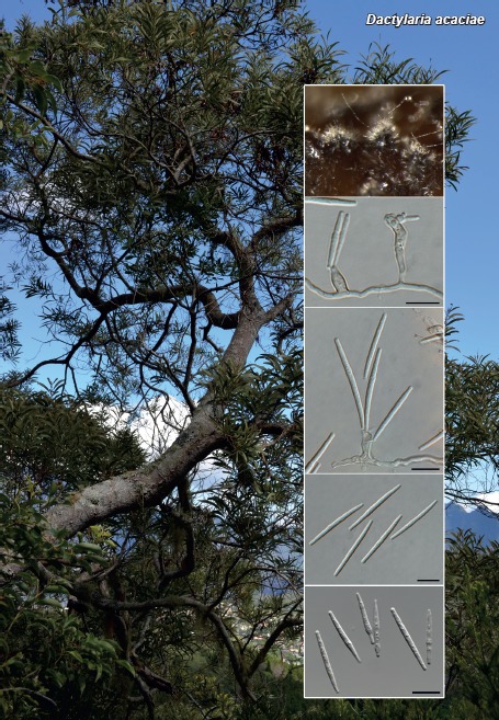
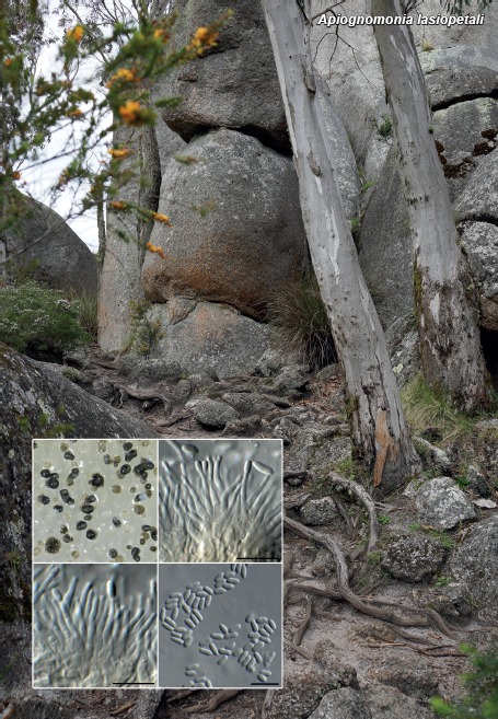
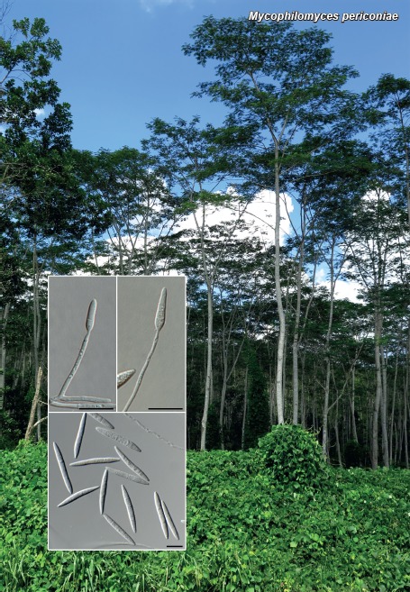
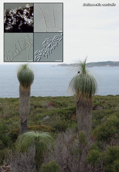
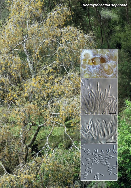
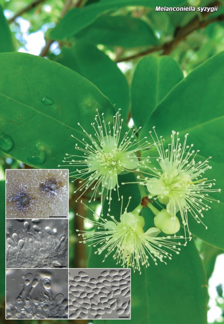
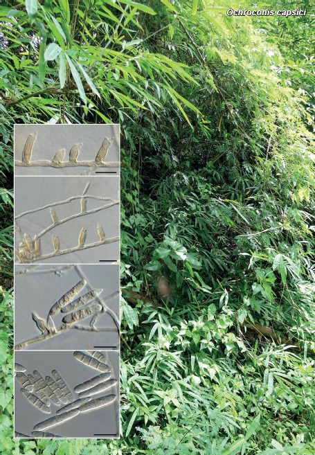
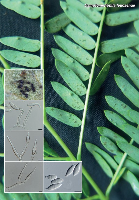
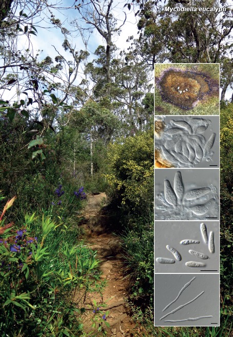
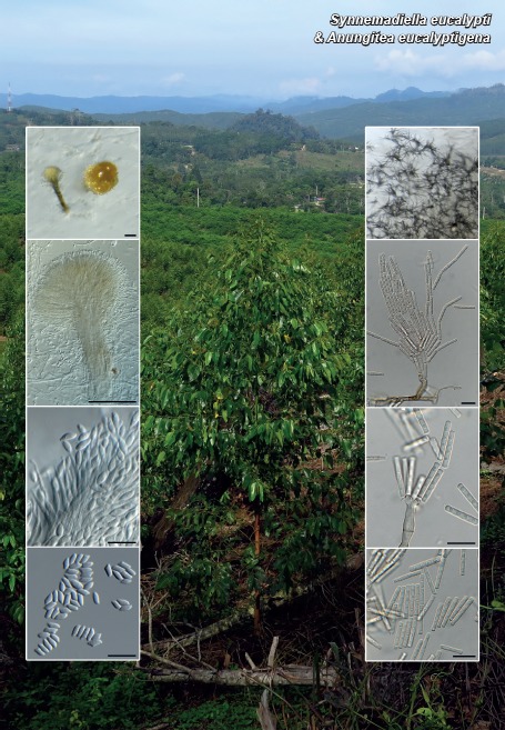
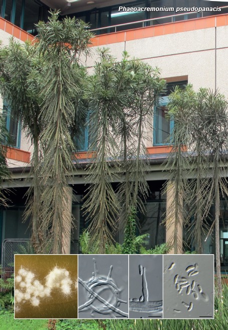
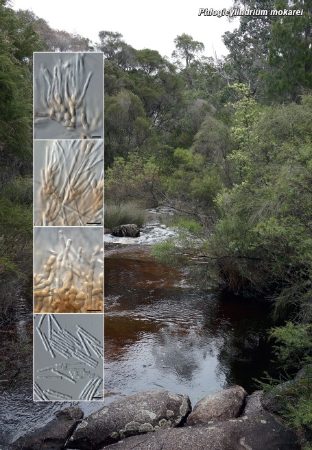
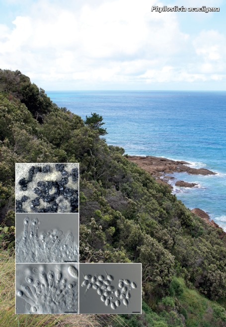
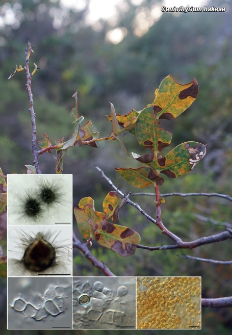
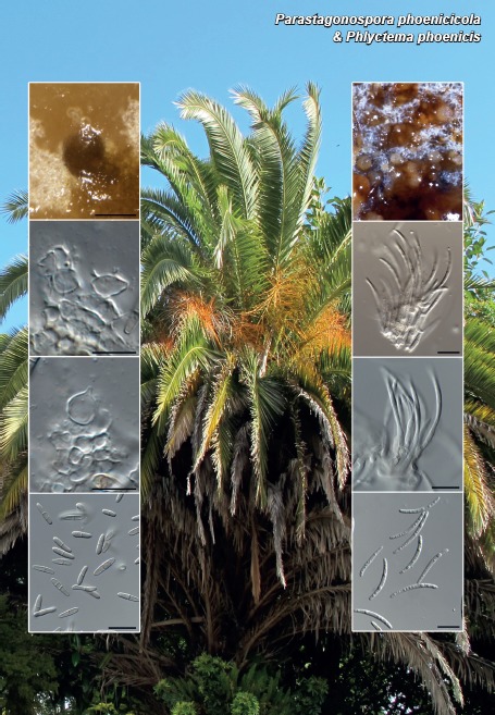
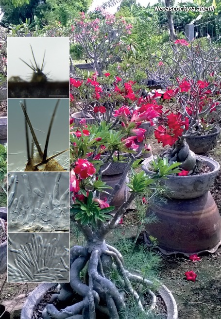
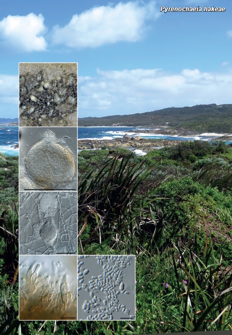
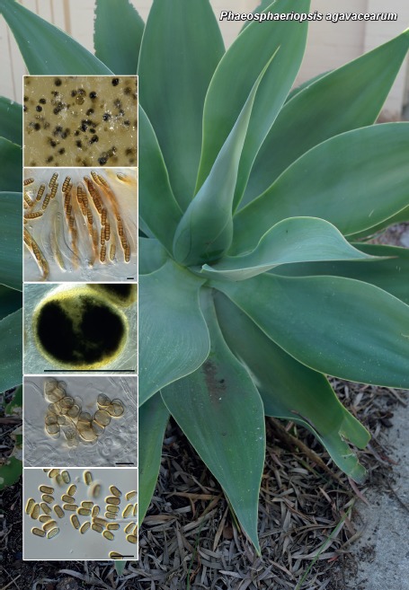
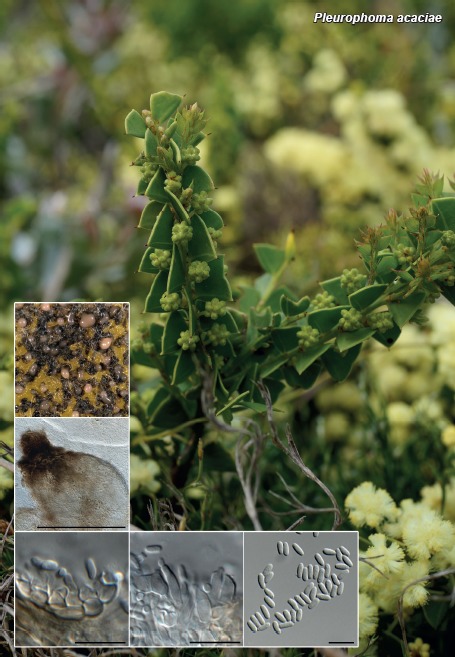
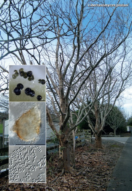
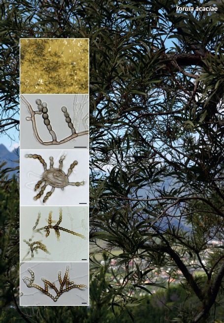
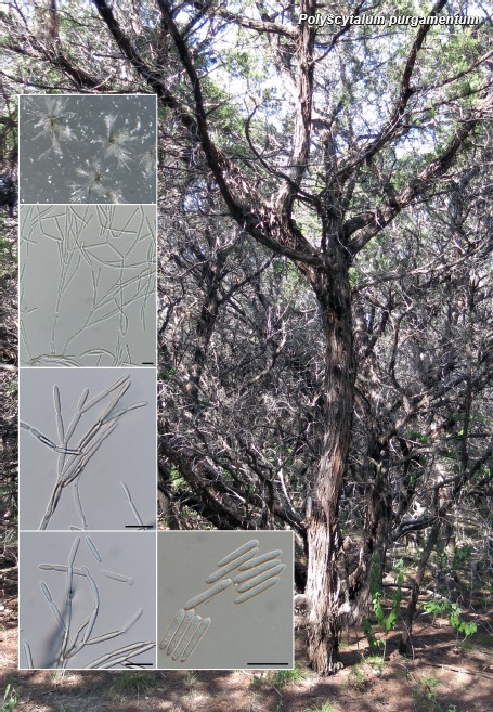
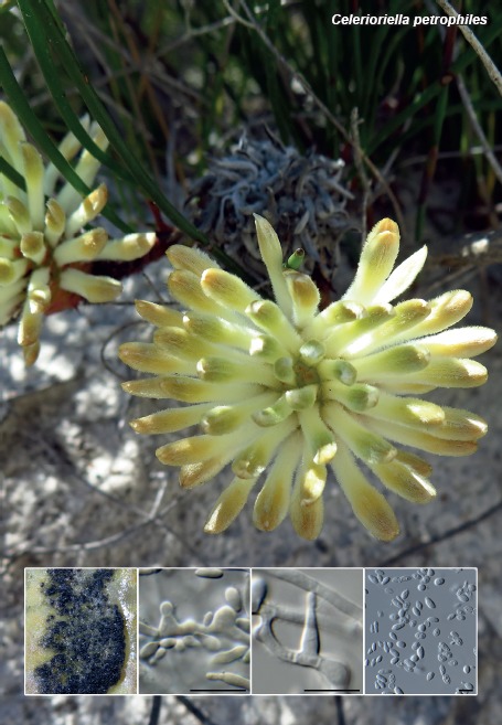
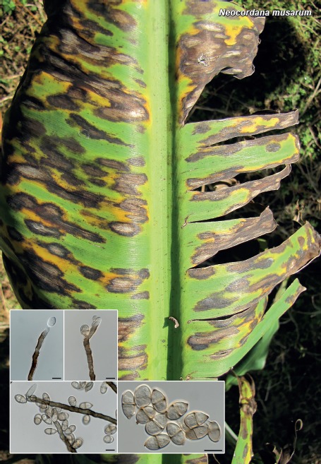
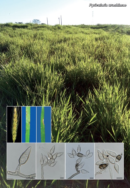
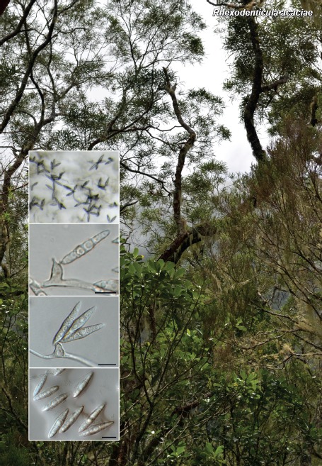
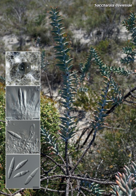
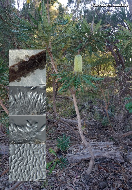
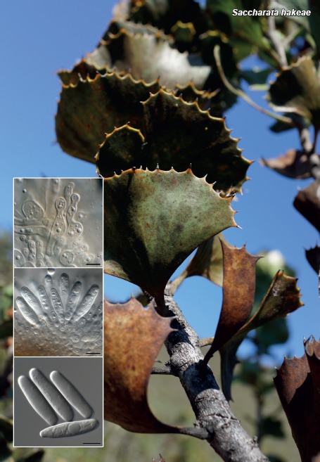
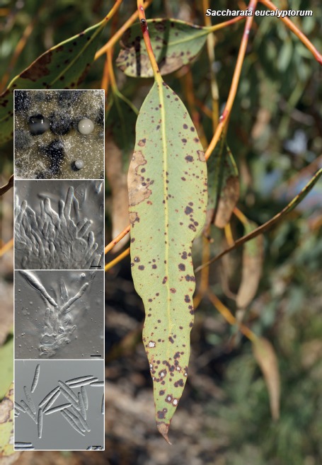
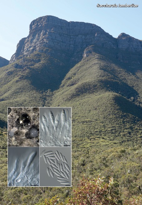
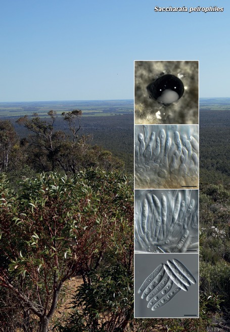
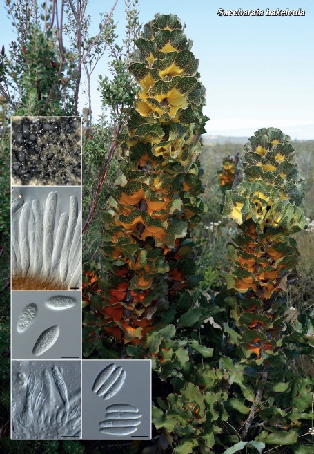
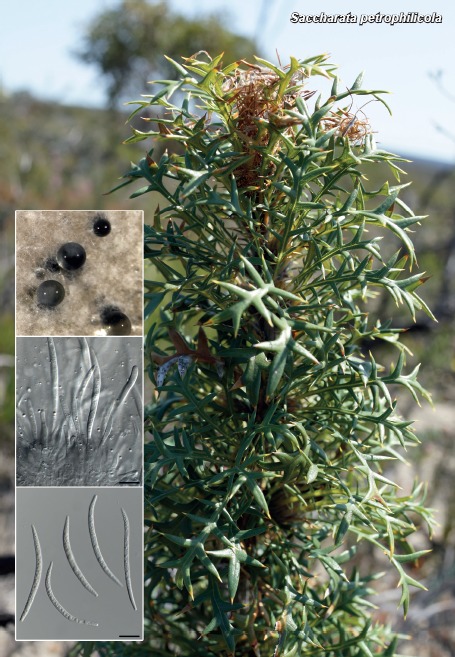
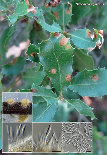
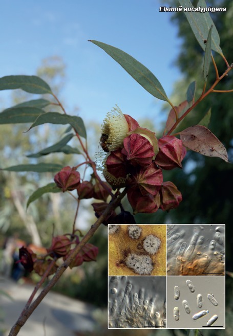
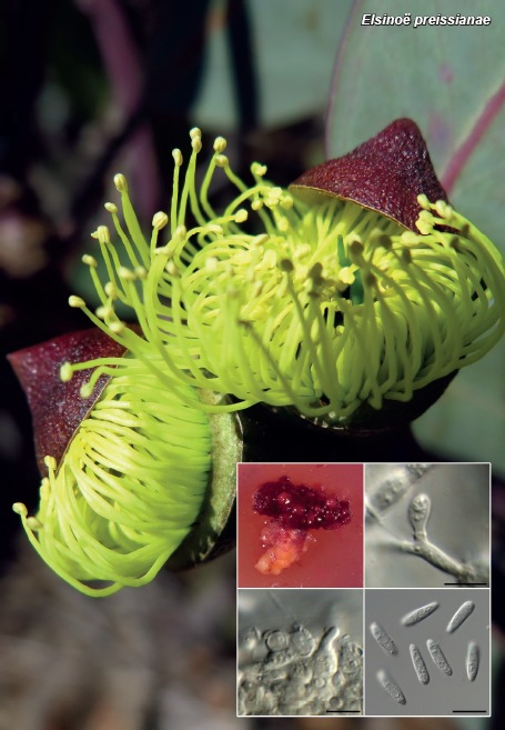
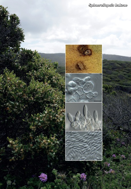
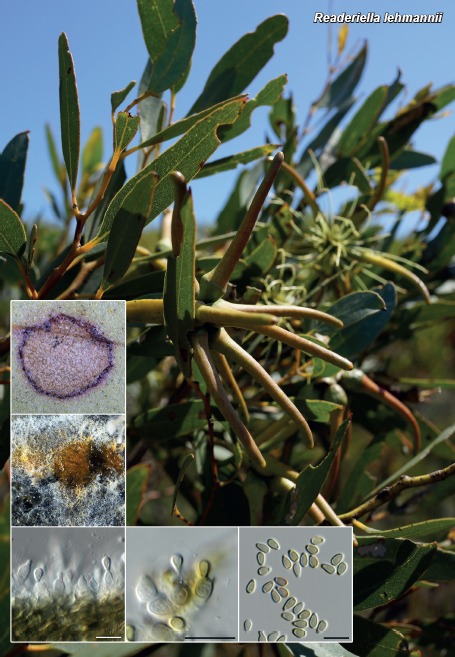
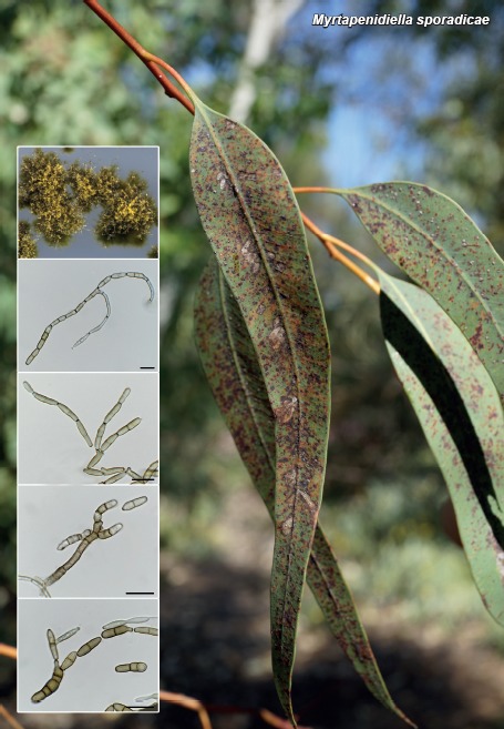
Novel species of fungi described in this study include those from various countries as follows: Australia: Apiognomonia lasiopetali on Lasiopetalum sp., Blastacervulus eucalyptorum on Eucalyptus adesmophloia, Bullanockia australis (incl. Bullanockia gen. nov.) on Kingia australis, Caliciopsis eucalypti on Eucalyptus marginata, Celerioriella petrophiles on Petrophile teretifolia, Coleophoma xanthosiae on Xanthosia rotundifolia, Coniothyrium hakeae on Hakea sp., Diatrypella banksiae on Banksia formosa, Disculoides corymbiae on Corymbia calophylla, Elsinoë eelemani on Melaleuca alternifolia, Elsinoë eucalyptigena on Eucalyptus kingsmillii, Elsinoë preissianae on Eucalyptus preissiana, Eucasphaeria rustici on Eucalyptus creta, Hyweljonesia queenslandica (incl. Hyweljonesia gen. nov.) on the cocoon of an unidentified microlepidoptera, Mycodiella eucalypti (incl. Mycodiella gen. nov.) on Eucalyptus diversicolor, Myrtapenidiella sporadicae on Eucalyptus sporadica, Neocrinula xanthorrhoeae (incl. Neocrinula gen. nov.) on Xanthorrhoea sp., Ophiocordyceps nooreniae on dead ant, Phaeosphaeriopsis agavacearum on Agave sp., Phlogicylindrium mokarei on Eucalyptus sp., Phyllosticta acaciigena on Acacia suaveolens, Pleurophoma acaciae on Acacia glaucoptera, Pyrenochaeta hakeae on Hakea sp., Readeriella lehmannii on Eucalyptus lehmannii, Saccharata banksiae on Banksia grandis, Saccharata daviesiae on Daviesia pachyphylla, Saccharata eucalyptorum on Eucalyptus bigalerita, Saccharata hakeae on Hakea baxteri, Saccharata hakeicola on Hakea victoria, Saccharata lambertiae on Lambertia ericifolia, Saccharata petrophiles on Petrophile sp., Saccharata petrophilicola on Petrophile fastigiata, Sphaerellopsis hakeae on Hakea sp., and Teichospora kingiae on Kingia australis. Brazil: Adautomilanezia caesalpiniae (incl. Adautomilanezia gen. nov.) on Caesalpina echinata, Arthrophiala arthrospora (incl. Arthrophiala gen. nov.) on Sagittaria montevidensis, Diaporthe caatingaensis (endophyte from Tacinga inamoena), Geastrum ishikawae on sandy soil, Geastrum pusillipilosum on soil, Gymnopus pygmaeus on dead leaves and sticks, Inonotus hymenonitens on decayed angiosperm trunk, Pyricularia urashimae on Urochloa brizantha, and Synnemellisia aurantia on Passiflora edulis. Chile: Tubulicrinis australis on Lophosoria quadripinnata. France: Cercophora squamulosa from submerged wood, and Scedosporium cereisporum from fluids of a wastewater treatment plant. Hawaii: Beltraniella acaciae, Dactylaria acaciae, Rhexodenticula acaciae, Rubikia evansii and Torula acaciae (all on Acacia koa). India: Lepidoderma echinosporum on dead semi-woody stems, and Rhodocybe rubrobrunnea from soil. Iran: Talaromyces kabodanensis from hypersaline soil. La Réunion: Neocordana musarum from leaves of Musa sp. Malaysia: Anungitea eucalyptigena on Eucalyptus grandis × pellita, Camptomeriphila leucaenae (incl. Camptomeriphila gen. nov.) on Leucaena leucocephala, Castanediella communis on Eucalyptus pellita, Eucalyptostroma eucalypti (incl. Eucalyptostroma gen. nov.) on Eucalyptus pellita, Melanconiella syzygii on Syzygium sp., Mycophilomyces periconiae (incl. Mycophilomyces gen. nov.) as hyperparasite on Periconia on leaves of Albizia falcataria, Synnemadiella eucalypti (incl. Synnemadiella gen. nov.) on Eucalyptus pellita, and Teichospora nephelii on Nephelium lappaceum. Mexico: Aspergillus bicephalus from soil. New Zealand: Aplosporella sophorae on Sophora microphylla, Libertasomyces platani on Platanus sp., Neothyronectria sophorae (incl. Neothyronectria gen. nov.) on Sophora microphylla, Parastagonospora phoenicicola on Phoenix canariensis, Phaeoacremonium pseudopanacis on Pseudopanax crassifolius, Phlyctema phoenicis on Phoenix canariensis, and Pseudoascochyta novae-zelandiae on Cordyline australis. Panama: Chalara panamensis from needle litter of Pinus cf. caribaea. South Africa: Exophiala eucalypti on leaves of Eucalyptus sp., Fantasmomyces hyalinus (incl. Fantasmomyces gen. nov.) on Acacia exuvialis, Paracladophialophora carceris (incl. Paracladophialophora gen. nov.) on Aloe sp., and Umthunziomyces hagahagensis (incl. Umthunziomyces gen. nov.) on Mimusops caffra. Spain: Clavaria griseobrunnea on bare ground in Pteridium aquilinum field, Cyathus ibericus on small fallen branches of Pinus halepensis, Gyroporus pseudolacteus in humus of Pinus pinaster, and Pseudoascochyta pratensis (incl. Pseudoascochyta gen. nov.) from soil. Thailand: Neoascochyta adenii on Adenium obesum, and Ochroconis capsici on Capsicum annuum. UK: Fusicolla melogrammae from dead stromata of Melogramma campylosporum on bark of Carpinus betulus. Uruguay: Myrmecridium pulvericola from house dust. USA: Neoscolecobasidium agapanthi (incl. Neoscolecobasidium gen. nov.) on Agapanthus sp., Polyscytalum purgamentum on leaf litter, Pseudopithomyces diversisporus from human toenail, Saksenaea trapezispora from knee wound of a soldier, and Sirococcus quercus from Quercus sp. Morphological and culture characteristics along with DNA barcodes are provided.

Keywords: ITS nrDNA barcodes, LSU, novel fungal species, systematics


graphic file with name per-37-218-g001.jpg

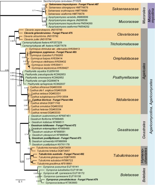
Overview Mucoromycotina and Agaricomycotina phylogeny

Consensus phylogram (50 % majority rule) of 2 394 trees resulting from a Bayesian analysis of the LSU sequence alignment (58 taxa including outgroup; 874 aligned positions; 507 unique site patterns) using MrBayes v. 3.2.6 (Ronquist et al. 2012). Bayesian posterior probabilities (PP) are shown at the nodes and thickened lines represent nodes with PP = 1.00. The scale bar represents the expected changes per site. Families, orders and classes are indicated with coloured blocks to the right of the tree. GenBank accession or Fungal Planet numbers are indicated behind the species names. The tree was rooted to Saccharomyces cerevisiae (GenBank Z73326) and the taxonomic novelties described in this study for which LSU sequence data were available are indicated in bold face. The alignment and tree were deposited in TreeBASE (Submission ID S20202).

graphic file with name per-37-218-g002.jpg

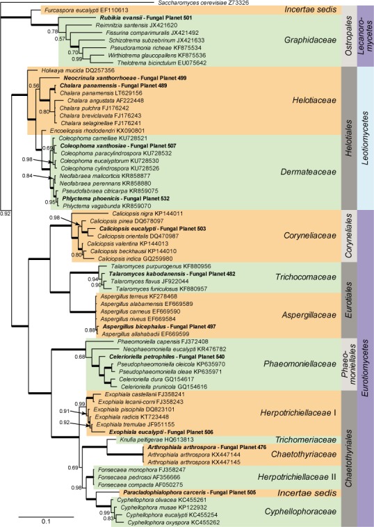
Overview Lecanoromycetes, Leotiomycetes and Eurotiomycetes, phylogeny

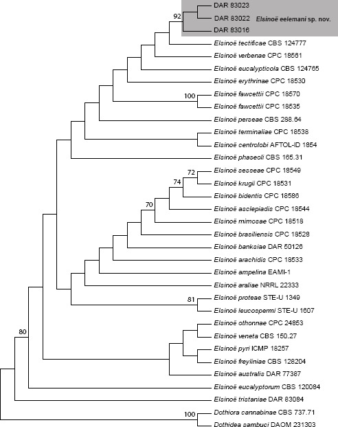
Consensus phylogram (50 % majority rule) of 790 trees resulting from a Bayesian analysis of the LSU sequence alignment (70 taxa including outgroup; 808 aligned positions; 386 unique site patterns) using MrBayes v. 3.2.6 (Ronquist et al. 2012). Bayesian posterior probabilities (PP) are shown at the nodes and thickened lines represent nodes with PP = 1.00. The scale bar represents the expected changes per site. Families, orders and classes are indicated with coloured blocks to the right of the tree. GenBank accession or Fungal Planet numbers are indicated behind the species names. The tree was rooted to Saccharomyces cerevisiae (GenBank Z73326) and the taxonomic novelties described in this study for which LSU sequence data were available are indicated in bold face. The alignment and tree were deposited in TreeBASE (Submission ID S20202).

graphic file with name per-37-218-g003.jpg

graphic file with name per-37-218-g004.jpg

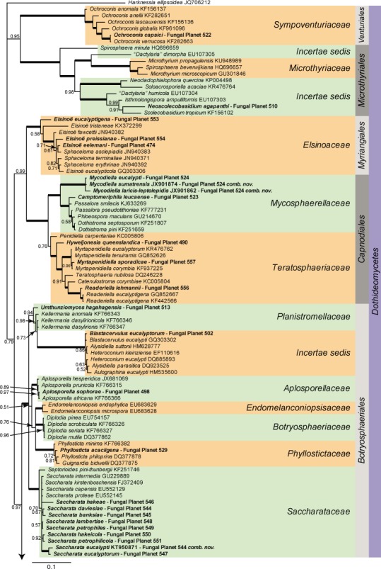
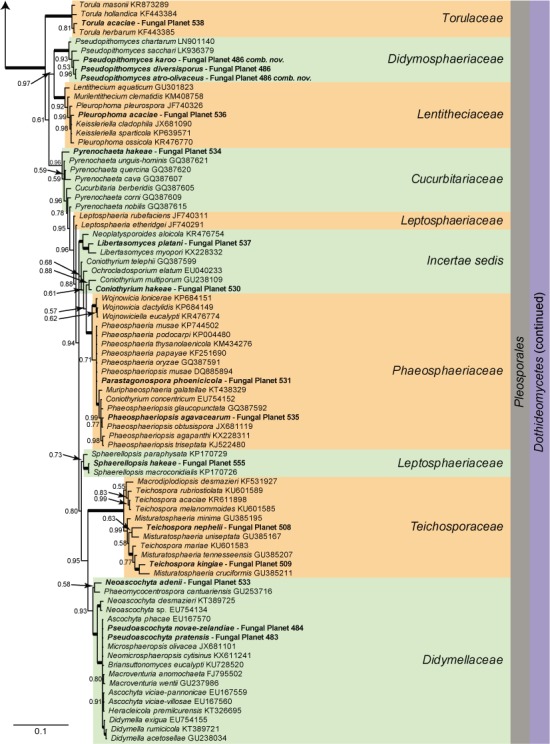
Overview Dothideomycetes phylogeny

Consensus phylogram (50 % majority rule) of 10 030 trees resulting from a Bayesian analysis of the LSU sequence alignment (167 taxa including outgroup; 806 aligned positions; 453 unique site patterns) using MrBayes v. 3.2.6 (Ronquist et al. 2012). Bayesian posterior probabilities (PP) are shown at the nodes and thickened lines represent nodes with PP = 1.00. The scale bar represents the expected changes per site. Families, orders and classes are indicated with coloured blocks to the right of the tree. GenBank accession or Fungal Planet numbers are indicated behind the species names. The tree was rooted to Harknessia ellipsoidea (GenBank JQ706212) and the taxonomic novelties described in this study for which LSU sequence data were available are indicated in bold face. The alignment and tree were deposited in TreeBASE (Submission ID S20202).

graphic file with name per-37-218-g005.jpg

graphic file with name per-37-218-g006.jpg

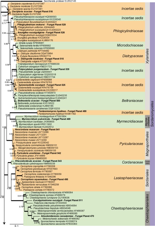
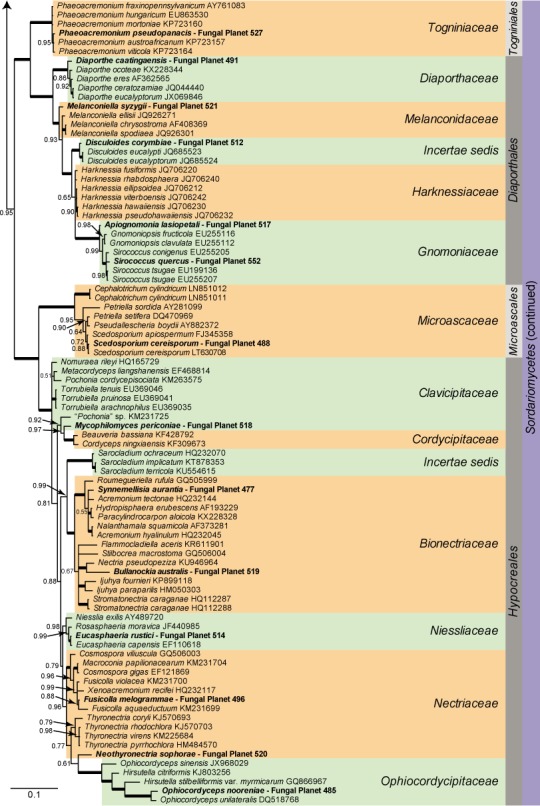
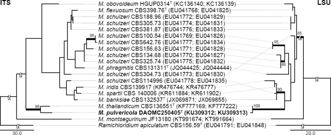
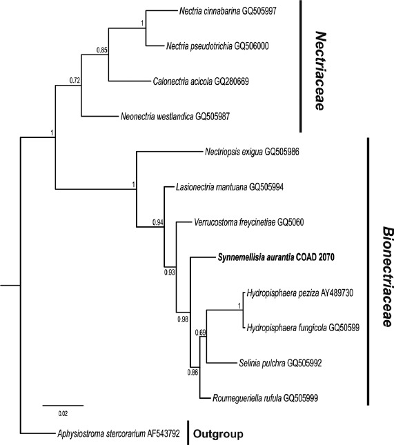
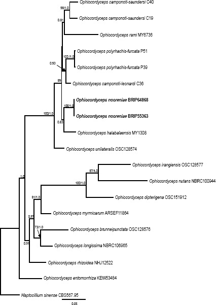
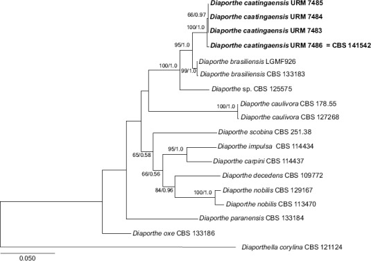
Overview Sordariomycetes phylogeny

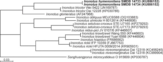
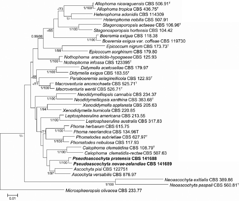
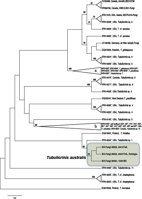
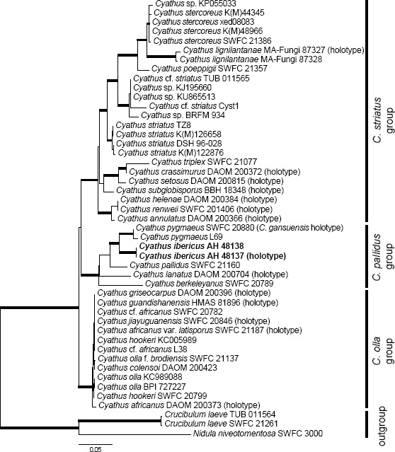
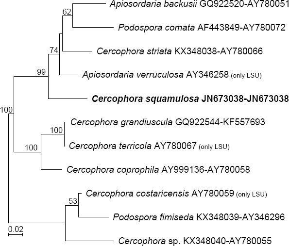
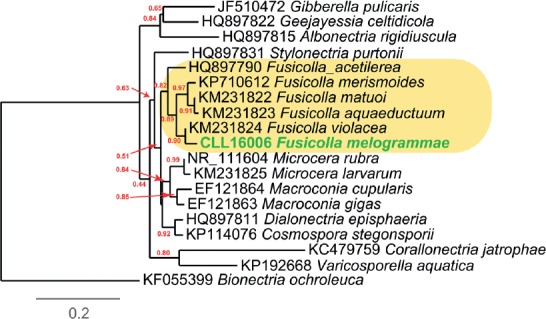
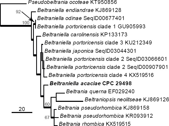
Consensus phylogram (50 % majority rule) of 8 418 trees resulting from a Bayesian analysis of the LSU sequence alignment (174 taxa including outgroup; 788 aligned positions; 393 unique site patterns) using MrBayes v. 3.2.6 (Ronquist et al. 2012). Bayesian posterior probabilities (PP) are shown at the nodes and thickened lines represent nodes with PP = 1.00. The scale bar represents the expected changes per site. Families, orders and classes are indicated with coloured blocks to the right of the tree. GenBank accession or Fungal Planet numbers are indicated behind the species names. The tree was rooted to Saccharata proteae (GenBank EU552145) and the taxonomic novelties described in this study for which LSU sequence data were available are indicated in bold face. The alignment and tree were deposited in TreeBASE (Submission ID S20202).

Acknowledgments

Paulo C. Ceresini acknowledges permission for scientific activities # 39131-3 from the Brazilian Ministry of Environment (MMA) / ‘Chico Mendes’ Institute for Conservation of Biodiversity (ICMBIO). Vanina L. Castroagudín is supported by a Post-Doctoral research fellowship from São Paulo Research Foundation – FAPESP / Higher Education Personnel Improvement Coordination – CAPES, Brazil (PDJ 2014/25904-2, from 2015–2016). Paulo C. Ceresini is supported by a research grant from FAPESP (2015/10453-8) and a fellowship grant from the Brazilian National Council for Scientific and Technological Development – CNPq (307295/2015-0). Santiago Català and Carlos Rojo helped by providing the DNA sequences of Cyathus ibericus used in this study. The research was on-going while J.C. Zamora was a recipient of funding from the Ministerio de Economía y Competitividad (Juan de la Cierva-formación program, FJCI-2014-19801, Spain). Christian Lechat and Nick Aplin acknowledge Amy Y. Rossman (Oregon State University, Corvallis, USA) for her advice and scientific assistance. Jacques Fournier, Las Muros, 09320 Rimont, France, is thanked for the material he collected. Thiago Accioly and co-workers acknowledge Marian Glenn (Seton Hall University, New Jersey) for the English revision of the Geastrum text. Gabriel Moreno and co-workers express their gratitude to Antonio Sánchez and Jaime de Frutos (Mycological Society of Segovia) and Celestino Gelpi (Mycological Society of Extremadura), for sending collections of Gyroporus pseudolacteus; to L. Monje and A. Pueblas of the Department of Drawing and Scientific Photography at the University of Alcalá for their help in preparing the digital photographs; to J. Rejos, curator of the AH herbarium for his assistance with the specimens examined in the present study. The survey which yielded the material considered herein was supported in part by a grant (DEB-0316284) from the National Science Foundation to the University of Arkansas. The assistance of Lal Singh in carrying out the fieldwork in India is gratefully acknowledged. K.N. Anil Raj acknowledges support from the University Grants Commission (UGC), India, in the form of a Rajiv Gandhi National Fellowship (Grant No. F. 14-2(SC)/2009 (SA-III)). K.P. Deepna Latha acknowledges support from the Kerala State Council for Science, Technology and Environment (KSCSTE) in the form of a PhD fellowship (Grant No. 001/FSHP/2011/CSTE), and is also grateful to the Principal Chief Conservator of forests, Kerala State, for granting permission (No. WL10-4937/2012, dated 03-10-2013) to collect agarics from the forests of Kerala. Margarita Dueñas and co-workers acknowledge financial support from the Agreement Endesa and San Ignacio de Huinay Foundations and Consejo Superior de Investigaciones Científicas, CSIC, 2013CL0012 and Plan Nacional I+D+I project n CGL2015-67459P, and thank Marian Glenn (Seton Hall University, USA) for revising the Tubulicrinis australis text. Gordon F. Claridge and co-workers thank Alistair McTaggart for providing the LSU sequence of Ophiocordyceps nooreniae, and Thomas Marney for his valuable advice. Josep Guarro and co-workers acknowledge financial support from the Ministerio de Economía y Competitividad, grant CGL2013-43789P. D.H. Lee acknowledges the Department of Science and Technology (DST)-National Research Foundation (NRF) Centre of Excellence in Tree Health Biotechnology, the National Research Foundation (NRF) for financial support and South African National Parks (SanParks) scientific services at Skukuza for technical and logistic assistance. Work associated with the description of Elsinoe eelemani was funded by the Rural Industries Research and Development Corporation, and the Australian Tea Tree Industry Association. Victor R.M. Coimbra acknowledges CNPq – Conselho Nacional de Desenvolvimento Científico e Tecnológico (SWE 232695/2014-8, Sisbiota 563342/2010-2, PROTAX 562106/2010-3) and FACEPE – Fundação de Amparo à Ciência e Tecnologia do Estado de Pernambuco (IBPG 0827-2.03/12, AMD 0218-2.00/13, APQ 0788-2.03/12) for the PhD scholarships and partially funding this research. Pedro W. Crous and Michael J. Wingfield acknowledge Murdoch University for support via the Sir Walter Murdoch Professorial appointments.

REFERENCES

  1. Alvarez E, Garcia-Hermoso D, Sutton DA, et al. 2010. Molecular phylogeny and proposal of two new species of the emerging pathogenic fungus Saksenaea. Journal of Clinical Microbiology 48: 4410–4416. [DOI] [PMC free article] [PubMed] [Google Scholar]
  2. Antonín V, Noordeloos ME. 2010. A monograph of marasmioid and collybioid fungi in Europe. IHW-Verlag, Eching. [Google Scholar]
  3. Araújo J, Evans H, Geiser DM, et al. 2015. Unravelling the diversity behind the Ophiocordyceps unilateralis (Ophiocordycipitaceae) complex: Three new species of zombie-ant fungi from the Brazilian Amazon. Phytotaxa 220: 224–238. [Google Scholar]
  4. Ariyawansa HA, Hyde KD, Jayasiri SC, et al. 2015. Fungal diversity notes 111–252 – taxonomic and phylogenetic contributions to fungal taxa. Fungal Diversity 75: 27–274. [Google Scholar]
  5. Arzanlou M, Groenewald JZ, Gams W, et al. 2007. Phylogenetic and morphotaxonomic revision of Ramichloridium and allied genera. Studies in Mycology 58: 57–93. [DOI] [PMC free article] [PubMed] [Google Scholar]
  6. Arzanlou M, Samadi R, Frisvad JC, et al. 2016. Two novel Aspergillus species from hypersaline soils of The National Park of Lake Urmia, Iran. Mycological Progress: doi 10.1007/s11557-016-1230-8. [Google Scholar]
  7. Badali H, Gueidan C, Najafzadeh MJ, et al. 2008. Biodiversity of the genus Cladophialophora. Studies in Mycology 61: 175–191. [DOI] [PMC free article] [PubMed] [Google Scholar]
  8. Baroni TJ. 1981. A revision of the genus Rhodocybe Maire (Agaricales). Beihefte zur Nova Hedwigia 67: 1–194. [Google Scholar]
  9. Baroni TJ, Halling RE. 1992. New species of Rhodocybe from South America with a key to species. Mycologia 84: 411–421. [Google Scholar]
  10. Baseia IG, Calonge FD. 2006. Geastrum hirsutum: a new earthstar fungus with a hairy exoperidium. Mycotaxon 95: 301–304. [Google Scholar]
  11. Brodie HJ. 1966. Cyathus pygmaeus from Northwestern United States. Mycologia 58: 973–975. [Google Scholar]
  12. Brodie HJ. 1975. The bird’s nest fungi. University of Toronto Press, Toronto, Canada. [Google Scholar]
  13. Brodie HJ. 1977. A key of the species of Cyathus (Nidulariaceae). Botaniska Notiser 130: 453–459. [Google Scholar]
  14. Brodie HJ. 1984. More bird’s nest fungi (Nidulariaceae) (a supplement to ‘the bird’s nest fungi’). Lejeunia 112: 1–69. [Google Scholar]
  15. Bulliard JBF. 1788. Herbier de la France 8: 337–384. [Google Scholar]
  16. Cabral TS, Silva BDB, Ishikawa NK, et al. 2014. A new species and new records of gasteroid fungi (Basidiomycota) from Central Amazonia, Brazil. Phytotaxa 183: 239–253. [Google Scholar]
  17. Calonge FD. 1998. Gasteromycetes. I. Lycoperdales, Nidulariales, Phallales, Sclerodermatales, Tulostomales. Flora Mycologica Iberica 3: 1–271. [Google Scholar]
  18. Calonge FD, Mata M. 2004. A new species of Geastrum from Costa Rica and Mexico. Boletín de la Sociedad Micológica de Madrid 28: 331–335. [Google Scholar]
  19. Castro ML, Freire L. 1995. Gyroporus ammophilus, a new poisonous bolete from the Iberian Peninsula. Persoonia 16: 123–126. [Google Scholar]
  20. Castroagudín VL, Moreira SI, Pereira DAS, et al. 2016. Pyricularia graminis-tritici, a new Pyricularia species causing wheat blast. Persoonia 37: 199–216. [DOI] [PMC free article] [PubMed] [Google Scholar]
  21. Cheewangkoon R, Groenewald JZ, Hyde KD, et al. 2012. Chocolate spot of Eucalyptus. Mycological Progress 11: 61–69. [Google Scholar]
  22. Cheewangkoon R, Groenewald JZ, Summerell BA, et al. 2009. Myrtaceae, a cache of fungal biodiversity. Persoonia 23: 55–85. [DOI] [PMC free article] [PubMed] [Google Scholar]
  23. Chen C, Verkley GJM, Sun G, et al. 2016. Redefining common endophytes and plant pathogens in Neofabraea, Pezicula, and related genera. Fungal Biology 120: 1291–1322. [DOI] [PubMed] [Google Scholar]
  24. Chen Q, Jiang JR, Zhang GZ, et al. 2015. Resolving the Phoma enigma. Studies in Mycology 82: 137–217. [DOI] [PMC free article] [PubMed] [Google Scholar]
  25. Coimbra VRM, Pinheiro FGB, Wartchow F, et al. 2015. Studies on Gymnopus sect. Impudicae (Omphalotaceae, Agaricales) from Northern Brazil: two new species and notes on G. montagnei. Mycological Progress 14: 110. [Google Scholar]
  26. Condé BD, Pitkethley RN, Smith ESC, et al. 1997. Disease notes or new records: Identification of mango scab caused by Elsinoё mangiferae in Australia. Australasian Plant Pathology 26: 131. [Google Scholar]
  27. Cortez VG, Baseia IG, Silveira RMB. 2008. Gasteromicetos (Basidiomycota) no Parque Estadual de Itapuã, Viamão, Rio Grande do Sul, Brasil. Revista Brasileira de Biociências 6: 291–299. [Google Scholar]
  28. Crane C, Burgess TI. 2013. Luteocirrhus shearii gen. sp. nov. (Diaporthales, Cryphonectriaceae) pathogenic to Proteaceae in the South Western Australian Floristic Region. IMA Fungus 4: 111–122. [DOI] [PMC free article] [PubMed] [Google Scholar]
  29. Crane JL, Miller AN. 2016. Studies in genera similar to Torula: Bahusaganda, Bahusandhika, Pseudotorula, and Simmonsiella gen. nov. IMA Fungus 7: 29–45. [DOI] [PMC free article] [PubMed] [Google Scholar]
  30. Crous PW, Carris LM, Giraldo A, et al. 2015a. The genera of fungi - fixing the application of the type species of generic names – G 2: Allantophomopsis, Latorua, Macrodiplodiopsis, Macrohilum, Milospium, Protostegia, Pyricularia, Robillarda, Rotula, Septoriella, Torula, and Wojnowicia. IMA Fungus 6: 163–198. [DOI] [PMC free article] [PubMed] [Google Scholar]
  31. Crous PW, Groenewald JZ. 2011. Why everlastings don’t last. Persoonia 26: 70–84. [DOI] [PMC free article] [PubMed] [Google Scholar]
  32. Crous PW, Groenewald JZ. 2016. They seldom occur alone. Fungal Biology 120: 1392–1415. [DOI] [PubMed] [Google Scholar]
  33. Crous PW, Groenewald JZ, Smith IW. 2007a. Phlogicylindrium eucalyptorum. Fungal Planet No. 20 Centraalbureau voor Schimmelcultures, Utrecht, The Netherlands. [Google Scholar]
  34. Crous PW, Groenewald JZ, Summerell B. 2007b. Exophiala placitae. Fungal Planet No. 17 Centraalbureau voor Schimmelcultures, Utrecht, The Netherlands. [Google Scholar]
  35. Crous PW, Mohammed C, Glen M, et al. 2007c. Eucalyptus microfungi known from culture. 3. Eucasphaeria and Sympoventuria genera nova, and new species of Furcaspora, Harknessia, Heteroconium and Phacidiella. Fungal Diversity 25: 19–36. [Google Scholar]
  36. Crous PW, Schubert K, Braun U, et al. 2007d. Opportunistic, human-pathogenic species in the Herpotrichiellaceae are phenotypically similar to saprobic or phytopathogenic species in the Venturiaceae. Studies in Mycology 58: 185–217. [DOI] [PMC free article] [PubMed] [Google Scholar]
  37. Crous PW, Schumacher RK, Wingfield MJ, et al. 2015b. Fungal Systematics and Evolution: FUSE 1. Sydowia 67: 81–118. [Google Scholar]
  38. Crous PW, Shivas RG, Quaedvlieg W, et al. 2014. Fungal Planet description sheets: 214–280. Persoonia 32: 184–306. [DOI] [PMC free article] [PubMed] [Google Scholar]
  39. Crous PW, Shivas RG, Wingfield MJ, et al. 2012a. Fungal Planet description sheets: 128–153. Persoonia 29: 146–201. [DOI] [PMC free article] [PubMed] [Google Scholar]
  40. Crous PW, Summerell BA, Alfenas AC, et al. 2012b. Genera of diaporthalean coelomycetes associated with leaf spots of tree hosts. Persoonia 28: 66–75. [DOI] [PMC free article] [PubMed] [Google Scholar]
  41. Crous PW, Summerell BA, Carnegie AJ, et al. 2009. Unravelling Mycosphaerella: do you believe in genera? Persoonia 23: 99–118. [DOI] [PMC free article] [PubMed] [Google Scholar]
  42. Crous PW, Summerell BA, Shivas RG, et al. 2011a. Fungal Planet description sheets: 92–106. Persoonia 27: 130–162. [DOI] [PMC free article] [PubMed] [Google Scholar]
  43. Crous PW, Summerell BA, Shivas RG, et al. 2012c. Fungal Planet description sheets: 107–127. Persoonia 28: 138–182. [DOI] [PMC free article] [PubMed] [Google Scholar]
  44. Crous PW, Summerell BA, Swart L, et al. 2011b. Fungal pathogens of Proteaceae. Persoonia 27: 20–45. [DOI] [PMC free article] [PubMed] [Google Scholar]
  45. Crous PW, Wingfield MJ, Guarro J, et al. 2013. Fungal Planet description sheets: 154–231. Persoonia 31: 188–296. [DOI] [PMC free article] [PubMed] [Google Scholar]
  46. Crous PW, Wingfield MJ, Guarro J, et al. 2015c. Fungal Planet description sheets: 320–370. Persoonia 34: 167–266. [DOI] [PMC free article] [PubMed] [Google Scholar]
  47. Crous PW, Wingfield MJ, Le Roux JJ, et al. 2015d. Fungal Planet description sheets: 371–399. Persoonia 35: 264–327. [DOI] [PMC free article] [PubMed] [Google Scholar]
  48. Crous PW, Wingfield MJ, Mansilla JP, et al. 2006. Phylogenetic reassessment of Mycosphaerella spp. and their anamorphs occurring on Eucalyptus. II. Studies in Mycology 55: 99–131. [DOI] [PMC free article] [PubMed] [Google Scholar]
  49. Crous PW, Wingfield MJ, Richardson DM, et al. 2016. Fungal Planet description sheets: 400–468. Persoonia 36: 316–458. [DOI] [PMC free article] [PubMed] [Google Scholar]
  50. Cunningham GH. 1963. The Thelephoraceae of Australia and New Zealand. Bulletin 145: 1–359. New Zealand, Department of Scientific and Industrial Research. [Google Scholar]
  51. Da Cunha KC, Sutton DA, Gené J, et al. 2014. Pithomyces species (Montagnulaceae) from clinical specimens: identification and antifungal susceptibility profiles. Medical Mycology 52: 748–757. [DOI] [PubMed] [Google Scholar]
  52. Dai YC. 2010. Hymenochaetaceae (Basidiomycota) in China. Fungal Diversity 45: 131–343. [Google Scholar]
  53. Damm U, Fourie PH, Crous PW. 2007. Aplosporella prunicola, a novel species of anamorphic Botryosphaeriaceae. Fungal Diversity 27: 35–43. [Google Scholar]
  54. De Beer ZW, Duong TA, Wingfield MJ. 2016a. The divorce of Sporothrix and Ophiostoma: solution to a problematic relationship. Studies in Mycology 83: 165–191. [DOI] [PMC free article] [PubMed] [Google Scholar]
  55. De Beer ZW, Marincowitz S, Duong TA, et al. 2016b. Hawksworthiomyces gen. nov. (Ophiostomatales), illustrates the urgency for a decision on how to name novel taxa known only from environmental nucleic acid sequences (ENAS). Fungal Biology 120: 1323–1340. [DOI] [PubMed] [Google Scholar]
  56. De Beer ZW, Seifert KA, Wingfield MJ. 2013. The Ophiostomatoid fungi: their dual position in the Sordariomycetes. In: Seifert KA, De Beer ZW, Wingfield MJ. (eds), The Ophiostomatoid fungi: expanding frontiers. CBS Biodiversity Series 12: 1–19. CBS-KNAW Fungal Biodiversity Centre, Utrecht, The Netherlands. [Google Scholar]
  57. De Beer ZW, Wingfield MJ. 2013. Emerging lineages in the Ophiostomatales. In: Seifert KA, De Beer ZW, Wingfield MJ. (eds), The Ophiostomatoid fungi: expanding frontiers. CBS Biodiversity Series 12: 21–46. CBS-KNAW Fungal Biodiversity Centre, Utrecht, The Netherlands. [Google Scholar]
  58. De Gruyter J, Woudenberg JHC, Aveskamp MM, et al. 2013. Redisposition of Phoma-like anamorphs in Pleosporales. Studies in Mycology 75: 1–36. [DOI] [PMC free article] [PubMed] [Google Scholar]
  59. De Hoog GS. 1985. Taxonomy of the Dactylaria complex, IV. Dactylaria, Neta, Subulispora and Scolecobasidium. Studies in Mycology 26: 1–60. [Google Scholar]
  60. De Hoog GS, Guarro J, Gené J, et al. 2000. Atlas of Clinical Fungi, 2nd ed Centraalbureau voor Schimmelcultures / Universitat Rovira i Virgili, Utrecht / Reus. [Google Scholar]
  61. De Hoog GS, Nishikaku AS, Fernández Zeppenfeldt G, et al. 2007. Molecular analysis and pathogenicity of the Cladophialophora carrionii complex, with the description of a novel species. Studies in Mycology 58: 219–234. [DOI] [PMC free article] [PubMed] [Google Scholar]
  62. DeVay JE, Lukezic FL, English WH, et al. 1965. Controlling Ceratocystis canker of stone fruit trees. California Agriculture 19: 2–4. [Google Scholar]
  63. Dulymamode R, Minter DW, Peerally A. 1998. Fungi from Mauritius: Rubikia splendida sp. nov., a coelomycete with unusual features. Mycological Research 102: 1242–1244. [Google Scholar]
  64. Dumortier BC. 1822. Commentationes botanicae. Gasterman-Dieu, Tournay. [Google Scholar]
  65. Ekanayaka AH, Dissanayake AJ, Jayasiri SC, et al. 2016. Aplosporella thailandica; a novel species revealing the sexual-asexual connection in Aplosporellaceae (Botryosphaeriales). Mycosphere 7: 440–447. [Google Scholar]
  66. Ellis MB. 1960. Dematiaceous Hyphomycetes. I. Mycological Papers 76: 1–36. [Google Scholar]
  67. Ellis MB. 1971. Dematiaceous Hyphomycetes. Commonwealth Mycological Institute, Kew, Surrey, UK. [Google Scholar]
  68. Evans HC, Elliot SL, Hughes DP. 2011. Hidden diversity behind the zombie-ant fungus Ophiocordyceps unilateralis: four new species described from carpenter ants in Minas Gerais, Brazil. PLoS ONE 6: e17024. [DOI] [PMC free article] [PubMed] [Google Scholar]
  69. Evans HC, Minter DW. 1985. Two remarkable new fungi on pine from Central America. Transactions of the British Mycological Society 84: 57–78. [Google Scholar]
  70. Evans HC, Samson RA. 1982. Entomogenous fungi from the Galapagos islands. Canadian Journal of Botany 60: 2325–2333. [Google Scholar]
  71. Evans HC, Samson RA. 1984. Cordyceps species and their anamorphs pathogenic on ants (Formicidae) in tropical forest ecosystems II. The Camponotus (Formicinae) complex. Transactions of the British Mycological Society 82: 127–150. [Google Scholar]
  72. Galindo-Leal C, Câmara IG. 2005. Mata Atlântica: biodiversidade, ameaças e perspectivas. Fundação SOS Mata Atlântica – Belo Horizonte, Conservação Internacional. [Google Scholar]
  73. Garrido-Benavent I, Pérez-Ortega S. 2015. Unravelling the diversity of European Caliciopsis (Coryneliaceae, Ascomycota): Caliciopsis valentina sp. nov. and C. beckhausii comb. nov., with a worldwide key to Caliciopsis. Mycological Progress 14: 1–10. [Google Scholar]
  74. Gilgado F, Cano J, Gené J, et al. 2005. Molecular phylogeny of the Pseudallescheria boydii species complex: proposal of two new species. Journal of Clinical Microbiology 43: 4930–4942. [DOI] [PMC free article] [PubMed] [Google Scholar]
  75. Gilgado F, Cano J, Gené J, et al. 2008. Molecular and phenotypic data supporting distinct species statuses for Scedosporium apiospermum and Pseudallescheria boydii and the proposed new species Scedosporium dehoogii. Journal of Clinical Microbiology 46: 766–771. [DOI] [PMC free article] [PubMed] [Google Scholar]
  76. Gomes RR, Glienke C, Videira CIR, et al. 2013. Diaporthe: a genus of endophytic, saprobic and plant pathogenic fungi. Persoonia 31: 1–41. [DOI] [PMC free article] [PubMed] [Google Scholar]
  77. Gouy M, Guindon S, Gascuel O. 2010. SeaView version 4: A multiplatform graphical user interface for sequence alignment and phylogenetic tree building. Molecular Biology and Evolution 27: 221–224. [DOI] [PubMed] [Google Scholar]
  78. Gramaje D, Mostert L, Groenewald JZ, et al. 2015. Phaeoacremonium: From esca disease to phaeohyphomycosis. Fungal Biology 119: 759–783. [DOI] [PubMed] [Google Scholar]
  79. Hashimoto A, Sato G, Matsuda T, et al. 2015. Taxonomic revision of Pseudolachnea and Pseudolachnella and establishment of Neopseudolachnella and Pseudodinemasporium gen. nov. Mycologia 107: 383–408 [DOI] [PubMed] [Google Scholar]
  80. Hernández-Restrepo M, Groenewald JZ, Crous PW. 2015. Neocordana gen. nov., the causal organism of Cordana leaf spot of banana. Phytotaxa 205: 229–238. [Google Scholar]
  81. Hernández-Restrepo M, Groenewald JZ, Crous PW. 2016. Taxonomic and phylogenetic re-evaluation of Microdochium, Monographella and Idriella. Persoonia 36: 57–82. [DOI] [PMC free article] [PubMed] [Google Scholar]
  82. Hirooka Y, Rossman AY, Samuels GJ, et al. 2012. A monograph of Allantonectria, Nectria, and Pleonectria (Nectriaceae, Hypocreales, Ascomycota) and their pycnidial, sporodochial, and synnematous anamorphs. Studies in Mycology 71: 1–210. [DOI] [PMC free article] [PubMed] [Google Scholar]
  83. Hjortstam K, Larsson K-H, Ryvarden L. 1988. The Corticiaceae of North Europe. Vol. 8 Phlebiella, Thanatephorus – Ypsilonidium. Fungiflora, Oslo, Norway. [Google Scholar]
  84. Honrubia M, Calonge FD, Demoulin V, et al. 1982. Aportación al conocimiento de los hongos del SE. de España VI: Esclerodermatales, Licoperdales, Nidulariales, Falales, Himenogasterales, Podaxales (Gasteromicetes, Basidiomicetes). Anales de la Universidad de Murcia - Ciencias 38: 101–132. [Google Scholar]
  85. Hyde KD, Hongsanan S, Jeewon R, et al. 2016. Fungal diversity notes 367–490: taxonomic and phylogenetic contributions to fungal taxa. Fungal Diversity 80: 1–270. [Google Scholar]
  86. Ito K, Sato K, Ota N. 1957. Studies on the needle cast of Japanese larch. I. Life history of the causal fungus, Mycosphaerella larici-leptolepis. Bulletin of the Government Forest Experiment Station, Meguro 96: 69–88. [Google Scholar]
  87. Jaklitsch WM, Olariaga I, Voglmayr H. 2016. Teichospora and Teichosporaceae. Mycological Progress 15: 31. [DOI] [PMC free article] [PubMed] [Google Scholar]
  88. Jaklitsch WM, Voglmayr H. 2014. Persistent hamathecial threads in the Nectriaceae, Hypocreales: Thyronectria revisited and re-instated. Persoonia 33: 182–211. [DOI] [PMC free article] [PubMed] [Google Scholar]
  89. Jie C-Y, Zhou Q-X, Zhao W-S, et al. 2013. A new Myrmecridium species from Guizhou, China. Mycotaxon 124: 1–8. [Google Scholar]
  90. Johnston PR, Seifert KA, Stone JK, et al. 2014. Recommendations on generic names competing for use in Leotiomycetes (Ascomycota). IMA Fungus 5: 91–120. [DOI] [PMC free article] [PubMed] [Google Scholar]
  91. Karsten PA. 1876. Symbolae ad mycologiam Fennicam. III. Meddelanden af Societas pro Fauna et Flora Fennica 1: 55–59. [Google Scholar]
  92. Kile GA. 1993. Plant diseases caused by species of Ceratocystis sensu stricto and Chalara. In: Wingfield MJ, Seifert KA, Webber JF. (eds), Ceratocystis and Ophiostoma: taxonomy, ecology and pathogenicity: 173–183. American Phytopathological Society Press, St. Paul, Minnesota, USA. [Google Scholar]
  93. Klaubauf S, Tharreau D, Fournier E, et al. 2014. Resolving the polyphyletic nature of Pyricularia (Pyriculariaceae). Studies in Mycology 79: 85–120. [DOI] [PMC free article] [PubMed] [Google Scholar]
  94. Klich MA. 2002. Identification of common Aspergillus species. 1st ed Centraalbureau voor Schimmelcultures, Utrecht, The Netherlands. [Google Scholar]
  95. Kobmoo N, Mongkolsamrit S, Tasanathai K, et al. 2012. Molecular phylogenies reveal host-specific divergence of Ophiocordyceps unilateralis sensu lato following its host ants. Molecular Ecology 21: 3022–3031. [DOI] [PubMed] [Google Scholar]
  96. Kornerup A, Wanscher JH. 1978. Methuen handbook of colour. 3rd ed Eyre Methuen, London. [Google Scholar]
  97. Kurose D, Evans HC, Djeddour DH, et al. 2009. Systematics of Mycosphaerella species associated with the invasive weed Fallopia japonica, including the potential biological control agent M. polygoni-cuspidati. Mycoscience, 50: 179–189. [Google Scholar]
  98. Lackner M, De Hoog GS, Yang L, et al. 2014. Proposed nomenclature for Pseudallescheria, Scedosporium and related genera. Fungal Diversity 67: 1–10. [Google Scholar]
  99. Lakhanpal TN, Mukerji KG. 1981. Taxonomy of the Indian Myxomycetes. Bibliotheca Mycologica 78: 1–531. [Google Scholar]
  100. Lechat C, Lesaga-Meessen L, Favel A. 2015. A new species of Ijuhya, I. fournieri, from French Guiana. Ascomycete.org 7: 101–104. [Google Scholar]
  101. Léveillé JH. 1848. Fragments mycologiques. Annales des Sciences Naturelles Botanique 9: 119–144. [Google Scholar]
  102. Li DW, Chen JY, Wang YX. 2011. Rhexodenticula zhengii sp. nov. from fallen leaves from China. Mycotaxon 117: 287–290. [Google Scholar]
  103. Li Y, Li HZ, Wang Q, et al. 2007. Flora fungorum sinicorum. Myxomycetes II: 1–131. Science Press, Beijing. [Google Scholar]
  104. Lombard L, Van der Merwe NA, Groenewald JZ, et al. 2015. Generic concepts in Nectriaceae. Studies in Mycology 80: 189–245. [DOI] [PMC free article] [PubMed] [Google Scholar]
  105. Marasas WFO, Schumann IH. 1972. The genus Pithomyces in South Africa. Bothalia 10: 509–516. [Google Scholar]
  106. Marincowitz S, Crous PW, Groenewald JZ, et al. 2008. Microfungi occurring on the Proteaceae in the fynbos. CBS Biodiversity Series 7: 1–166. [Google Scholar]
  107. Mchau GRA, Crous PW, Philips AJL. 1998. Molecular characterization of some Elsinoë isolates from leguminous hosts. Plant Pathology 47: 773–779. [Google Scholar]
  108. McKenzie EHC, Pinnoi A, Wong MKM, et al. 2002. Two new hyaline Chalara species, and a key to species described since 1975. Fungal Diversity 11: 129–139. [Google Scholar]
  109. Mel’nik V, Lee S, Groenewald JZ, et al. 2004. New hyphomycetes from Restionaceae in the fynbos: Parasarcopodium ceratocaryi gen. et sp. nov. and Rhexodenticulata elegiae sp. nov. Mycological Progress 3: 19–28. [Google Scholar]
  110. Melo Santos AM, Cavalcanti DR, Da Silva JMC, et al. 2007. Biogeographical relationships among tropical forests in north-eastern Brazil. Journal of Biogeography 34: 437–446. [Google Scholar]
  111. Miadlikowska J, Kauff F, Högnabba F, et al. 2014. A multigene phylogenetic synthesis for the class Lecanoromycetes (Ascomycota): 1307 fungi representing 1139 infrageneric taxa, 317 genera and 66 families. Molecular Phylogenetics and Evolution 79: 132–168. [DOI] [PMC free article] [PubMed] [Google Scholar]
  112. Miles AK, Tan YP, Shivas RG, et al. 2015. Novel pathotypes of Elsinoë australis associated with Citrus australasica and Simmondsia chinensis in Australia. Tropical Plant Pathology 40: 26–34. [Google Scholar]
  113. Miller AN, Huhndorf SM. 2001. Neotropical Ascomycetes 10. New and interesting Cercophora species. Sydowia 53: 211–226. [Google Scholar]
  114. Milne I, Lindner D, Bayer M, et al. 2009. TOPALi v2: a rich graphical interface for evolutionary analyses of multiple alignments on HPC clusters and multi-core desktops. Bioinformatics 25: 126–127. [DOI] [PMC free article] [PubMed] [Google Scholar]
  115. Minnis AM, Kennedy AH, Grenier DB, et al. 2012. Phylogeny and taxonomic revision of the Planistromellaceae including its coelomycetous anamorphs: contributions towards a monograph of the genus Kellermania. Persoonia 29: 11–28. [DOI] [PMC free article] [PubMed] [Google Scholar]
  116. Mostert L, Groenewald JZ, Summerbell RC, et al. 2005. Species of Phaeoacremonium associated with human infections and environmental reservoirs in infected woody plants. Journal of Clinical Microbiology 43: 1752–1767. [DOI] [PMC free article] [PubMed] [Google Scholar]
  117. Mostert L, Groenewald JZ, Summerbell RC, et al. 2006. Taxonomy and pathology of Togninia (Diaporthales) and its Phaeoacremonium anamorphs. Studies in Mycology 54: 1–115. [Google Scholar]
  118. Mugambi GK, Huhndorf SM. 2009. Molecular phylogenetics of Pleosporales: Melanommataceae and Lophiostomataceae recircumscribed (Pleosporomycetidae, Dothideomycetes, Ascomycota). Studies in Mycology 64: 103–121. [DOI] [PMC free article] [PubMed] [Google Scholar]
  119. Muñoz JA. 2005. Boletus s.l. (excl. Xerocomus). Fungi Europaei 2. Edizioni Candusso, Alassio. [Google Scholar]
  120. Nag Raj TR, Kendrick WB. 1976. A monograph of Chalara and allied genera. Wilfred Laurier University Press, Waterloo. [Google Scholar]
  121. Olariaga I, Salcedo I, Daniëls PP, et al. 2015. Taxonomy and phylogeny of yellow Clavaria species with clamped basidia – Clavaria flavostellifera sp. nov. and the typification of C. argillacea, C. flavipes and C. sphagnicola. Mycologia 107: 104–122. [DOI] [PubMed] [Google Scholar]
  122. Olson DM, Dinerstein E, Wikramanayake ED, et al. 2001. Terrestrial ecoregions of the world: A new map of life on earth. BioScience 51: 933–938. [Google Scholar]
  123. Pascoe I, Crous PW, Groenewald JZ. 2007. Sphaceloma banksiicola. Fungal Planet description sheet no. 14. CBS-KNAW Fungal Biodiversity Centre, Utrecht, The Netherlands. [Google Scholar]
  124. Pereira JM, Barreto RW, Ellison C, et al. 2003. Corynespora cassiicola f. sp. lantanae: a potential biocontrol agent for Lantana camara from Brazil. Biological Control 26: 21–31. [Google Scholar]
  125. Pratibha J, Prabhugaonkar A. 2015. Multi-gene phylogeny of Pithomyces with the sexual morph of P. flavus Berk. & Broome. Phytotaxa 218: 84–90. [Google Scholar]
  126. Quaedvlieg W, Binder M, Groenewald JZ, et al. 2014. Introducing the Consolidated Species Concept to resolve species in the Teratosphaeriaceae. Persoonia 33: 1–40. [DOI] [PMC free article] [PubMed] [Google Scholar]
  127. Quaedvlieg W, Verkley GJM, Shin H-D, et al. 2013. Sizing up Septoria. Studies in Mycology 75: 307–390. [DOI] [PMC free article] [PubMed] [Google Scholar]
  128. Quandt CA, Kepler RM, Gams W, et al. 2014. Phylogenetic-based nomenclatural proposals for Ophiocordycipitaceae (Hypocreales) with new combinations in Tolypocladium. IMA Fungus 5: 121–134. [DOI] [PMC free article] [PubMed] [Google Scholar]
  129. Rajeshkumar KC, Crous PW, Groenewald JZ, et al. 2016. Resolving the phylogenetic placement of Porobeltraniella and allied genera in the Beltraniaceae. Mycological Progress: doi: 10.1007/s11557-016-1234-4. [Google Scholar]
  130. Ramaley AW. 1997. New Paraphaeosphaeria species and their anamorphs. Mycotaxon 61: 347–358. [Google Scholar]
  131. Rao NK, Manoharachary C, Goos RD. 1988. Forest litter Hyphomycetes from Andhra Pradesh, India. III. A new synnematous Hyphomycete. Mycologia 80: 896–899. [Google Scholar]
  132. Rayner RW. 1970. A mycological colour chart. Commonwealth Mycological Institute and British Mycological Society, Kew, Surrey, UK. [Google Scholar]
  133. Réblová M. 2006. Molecular systematics of Ceratostomella sensu lato and morphologically similar fungi. Mycologia 98: 68–93. [DOI] [PubMed] [Google Scholar]
  134. Réblová M. 2013. Two taxonomic novelties in the Sordariomycetidae: Ceratolenta caudata gen. et sp. nov. and Platytrachelan abietis gen. et comb. nov. for Ceratosphaeria abietis. Mycologia 105: 462–475. [DOI] [PubMed] [Google Scholar]
  135. Réblová M, Fournier J, Štěpánek V. 2016. Two new lineages of aquatic ascomycetes: Atractospora gen. nov. and Rubellisphaeria gen. et sp. nov., and a sexual morph of Myrmecridium montsegurinum sp. nov. Mycological Progress 15: 21. [Google Scholar]
  136. Réblová M, Réblová K, Štěpánek V. 2015. Molecular systematics of Barbatosphaeria (Sordariomycetes): multigene phylogeny and secondary ITS structure. Persoonia 35: 21–38. [DOI] [PMC free article] [PubMed] [Google Scholar]
  137. Reeder R, Kelly PL, Hill AA St, et al. 2009. Superelongation disease, caused by Elsinoë brasiliensis, confirmed on cassava in Trinidad and Tobago. Plant Pathology 58: 800. [Google Scholar]
  138. Reges JTL, Negrisoli MM, Dorigan AF, et al. 2016. Pyricularia pennisetigena and P. zingibericola from invasive grasses infect signal grass, barley and wheat. Pesquisa Agropecuaria Tropical 46: 206–214. [Google Scholar]
  139. Rodrigues A, Passarini MRZ, Ferro M, et al. 2014. Fungal communities in the garden chamber soils of leaf-cutting ants. Journal of Basic Microbiology 54: 1186–1196. [DOI] [PubMed] [Google Scholar]
  140. Ronquist F, Teslenko M, Mark P, et al. 2012. MrBayes 3.2: Efficient Bayesian phylogenetic inference and model choice across a large model space. Systematic Biology 61: 539–542. [DOI] [PMC free article] [PubMed] [Google Scholar]
  141. Rossman AY, Adams GC, Cannon PF, et al. 2015. Recommendations of generic names in Diaporthales competing for protection or use. IMA Fungus 6: 145–154. [DOI] [PMC free article] [PubMed] [Google Scholar]
  142. Rossman AY, Castlebury LA, Farr DF, et al. 2008. Sirococcus conigenus, Sirococcus piceicola sp. nov. and Sirococcus tsugae sp. nov. on conifers: anamorphic fungi in the Gnomoniaceae, Diaporthales. Forest Pathology 38: 47–60. [Google Scholar]
  143. Rossman AY, Samuels GJ, Rogerson CT, et al. 1999. Genera of Bionectriaceae, Hypocreaceae and Nectriaceae (Hypocreales, Ascomycetes). Studies in Mycology 42: 1–248. [Google Scholar]
  144. Roumeguère C. 1880. Publication des ‘Reliquiae Libertianae’. Pars 1. Revue Mycologique Toulouse 2: 7–24. [Google Scholar]
  145. Ryvarden L. 2002. Studies in neotropical polypores 17. New neotropical Inonotus species. Synopsis Fungorum 15: 70–80. [Google Scholar]
  146. Ryvarden L. 2005. The Genus Inonotus, a synopsis. Synopsis Fungorum 21: 1–149. [Google Scholar]
  147. Samerpitak K, Van der Linde E, Choi H-J, et al. 2014. Taxonomy of Ochroconis, genus including opportunistic pathogens on humans and animals. Fungal Diversity 65: 89–126. [Google Scholar]
  148. Samson RA, Peterson SW, Frisvad JC, et al. 2011. New species in Aspergillus section Terrei. Studies in Mycology 69: 39–55. [DOI] [PMC free article] [PubMed] [Google Scholar]
  149. Seifert K, Morgan-Jones G, Gams W, et al. 2011. The genera of Hyphomycetes. CBS Biodiversity Series 9. CBS-KNAW Fungal Biodiversity Centre, Utrecht, The Netherlands. [Google Scholar]
  150. Seifert KA, De Beer ZW, Wingfield MJ. 2013. The Ophiostomatoid fungi: expanding frontiers. In: Seifert KA, De Beer ZW, Wingfield MJ. (eds), The Ophiostomatoid fungi: expanding frontiers. CBS Biodiversity Series 12: 21–46. CBS-KNAW Fungal Biodiversity Centre, Utrecht, The Netherlands. [Google Scholar]
  151. Seifert KA, Okada G. 1990. Taxonomic implications of conidiomatal anatomy in synnematous Hyphomycetes. Studies in Mycology 32: 29–40. [Google Scholar]
  152. Simmons DR, Kepler RM, Rehner SA, et al. 2015a. Phylogeny of Hirsutella species (Ophiocordycipitaceae) from the USA: remedying the paucity of Hirsutella sequence data. IMA Fungus 6: 345–356. [DOI] [PMC free article] [PubMed] [Google Scholar]
  153. Simmons DR, Lund J, Levitsky T, et al. 2015b. Ophiocordyceps myrmicarum, a new species infecting invasive Myrmica rubra in Maine. Journal of Invertebrate Pathology 125: 23–30. [DOI] [PubMed] [Google Scholar]
  154. Slippers B, Boissin E, Phillips AJL, et al. 2013. Phylogenetic lineages in the Botryosphaeriales: A systematic and evolutionary framework. Studies in Mycology 76: 31–49. [DOI] [PMC free article] [PubMed] [Google Scholar]
  155. Slippers B, Roux J, Wingfield MJ, et al. 2014. Confronting the constraints of morphological taxonomy in the Botryosphaeriales. Persoonia 33: 155–168. [DOI] [PMC free article] [PubMed] [Google Scholar]
  156. Soares DJ, Barreto RW, Braun U. 2009. Brazilian mycobiota of the aquatic weed Sagittaria montevidensis. Mycologia 101: 401–416. [DOI] [PubMed] [Google Scholar]
  157. Sousa JO, Silva BDB, Alfredo DS, et al. 2014. New records of Geastraceae (Basidiomycota: Phallomycetidae) from Atlantic rainforest remnants and relicts of northeastern Brazil. Darwiniana, Nueva Serie 2: 207–221. [Google Scholar]
  158. Stamatakis A, Alchiotis N. 2010. Time and memory efficient likelihood-based tree searches on phylogenomic alignments with missing data. Bioinformatics 26: i132–i139. [DOI] [PMC free article] [PubMed] [Google Scholar]
  159. Summerell BA, Groenewald JZ, Carnegie AJ, et al. 2006. Eucalyptus microfungi known from culture. 2. Alysidiella, Fusculina and Phlogicylindrium genera nova, with notes on some other poorly known taxa. Fungal Diversity 23: 323–350. [Google Scholar]
  160. Sunhede S. 1989. Geastraceae (Basidiomycotina). Morphology, ecology and systematics with special emphasis on the North European species. Synopsis Fungorum 1: 1–534. [Google Scholar]
  161. Sutton BC. 1980. The Coelomycetes. Fungi imperfecti with pycnidia, acervuli and stromata. CMI, Kew, UK. [Google Scholar]
  162. Swart HJ. 1988. Australian leaf-inhabiting fungi. XXVI. Some noteworthy Coelomycetes on Eucalyptus. Transactions of the British Mycological Society 90: 279–291. [Google Scholar]
  163. Swart L, Crous PW, Kang JC, et al. 2001. Differentiation of species of Elsinoë associated with scab disease of Proteaceae based on morphology, symptomatology, and ITS sequence phylogeny. Mycologia 93: 366–379. [Google Scholar]
  164. Swofford DL. 2003. PAUP*. Phylogenetic Analysis Using Parsimony (*and other methods). Sinauer Associates, Sunderland, Massachusetts. [Google Scholar]
  165. Tamura K, Stecher G, Peterson D, et al. 2013. MEGA 6: Molecular Evolutionary Genetics Analysis version 6.0. Molecular Biology and Evolution 30: 2725–2729. [DOI] [PMC free article] [PubMed] [Google Scholar]
  166. Thind KS. 1977. The Myxomycetes of India. Indian Council of Agricultural Research, New Delhi. [Google Scholar]
  167. Trakunyingcharoen T, Lombard L, Groenewald JZ, et al. 2014. Mycoparasitic species of Sphaerellopsis, and allied lichenicolous and other genera. IMA Fungus 5: 391–414. [DOI] [PMC free article] [PubMed] [Google Scholar]
  168. Trakunyingcharoen T, Lombard L, Groenewald JZ, et al. 2015. Caulicolous Botryosphaeriales from Thailand. Persoonia 34: 87–99. [DOI] [PMC free article] [PubMed] [Google Scholar]
  169. Trouillas FP, Pitt WM, Sosnowski MR, et al. 2011. Taxonomy and DNA phylogeny of Diatrypaceae associated with Vitis vinifera and other woody plants in Australia. Fungal Diversity 49: 203–223. [Google Scholar]
  170. Untereiner WA, Gueidan C, Orr MJ, et al. 2011. The phylogenetic position of the lichenicolous ascomycete Capronia peltigerae. Fungal Diversity 49: 225–233. [Google Scholar]
  171. Van der Aa HA, Vanev S. 2002. A revision of the species described in Phyllosticta. Centraalbureau voor Schimmelcultures, Utrecht, The Netherlands. [Google Scholar]
  172. Videira SIR, Groenewald JZ, Braun U, et al. 2016. All that glitters is not Ramularia. Studies in Mycology 83: 49–163. [DOI] [PMC free article] [PubMed] [Google Scholar]
  173. Videira SIR, Groenewald JZ, Kolecka A, et al. 2015a. Elucidating the Ramularia eucalypti species complex. Persoonia 34: 50–64. [DOI] [PMC free article] [PubMed] [Google Scholar]
  174. Videira SIR, Groenewald JZ, Verkley GJM, et al. 2015b. The rise of Ramularia from the Mycosphaerella labyrinth. Fungal Biology 119: 823–843. [DOI] [PubMed] [Google Scholar]
  175. Vizzini A, Angelini C, Ercole E. 2015. Molecular confirmation of Gyroporus lacteus and typification of Boletus cyanescens. Phytotaxa 226: 27–38. [Google Scholar]
  176. Voglmayr H, Rossman AY, Castlebury LA, et al. 2012. Multigene phylogeny and taxonomy of the genus Melanconiella (Diaporthales). Fungal Diversity 57: 1–44. [Google Scholar]
  177. Wikee S, Lombard L, Crous PW, et al. 2013a. Phyllosticta capitalensis, a widespread endophyte of plants. Fungal Diversity 60: 91–105. [Google Scholar]
  178. Wikee S, Lombard L, Nakashima C, et al. 2013b. A phylogenetic re-evaluation of Phyllosticta (Botryosphaeriales). Studies in Mycology 76: 1–29. [DOI] [PMC free article] [PubMed] [Google Scholar]
  179. Wingfield MJ, De Beer C, Visser C, et al. 1996. A new Ceratocystis species defined using morphological and ribosomal DNA sequence comparisons. Systematic and Applied Microbiology 19: 191–202. [Google Scholar]
  180. Wingfield MJ, Seifert KA, Webber J. 1993. Ceratocystis and Ophiostoma: Taxonomy, Ecology and Pathogenicity. APS Press, St. Paul, Minnesota, USA. [Google Scholar]
  181. Wood AR, Damm U, Van der Linde EJ, et al. 2016. Finding the missing link: Resolving the Coryneliomycetidae within Eurotiomycetes. Persoonia 37: 37–56. [DOI] [PMC free article] [PubMed] [Google Scholar]
  182. Yamamoto Y. 1998. The Myxomycete biota of Japan. Toyo Shorin Publishing Co., Ltd; Japan. [Google Scholar]
  183. Yilmaz N, Visagie CM, Houbraken J, et al. 2014. Polyphasic taxonomy of the genus Talaromyces. Studies in Mycology 78: 175–341. [DOI] [PMC free article] [PubMed] [Google Scholar]
  184. Zamora JC, Calonge FD, Hosaka K, et al. 2014. Systematics of the genus Geastrum (Fungi: Basidiomycota) revisited. Taxon 63: 477–497. [Google Scholar]
  185. Zamora JC, Calonge FD, Martín MP. 2015. Integrative taxonomy reveals an unexpected diversity in Geastrum section Geastrum (Geastrales, Basidiomycota). Persoonia 34: 130–165. [DOI] [PMC free article] [PubMed] [Google Scholar]
  186. Zamora JC, Kuhar F, Castiglia V, et al. 2014. On Geastrum argentinum, a forgotten species. Mycoscience 55: 177–182. [Google Scholar]
  187. Zhao R-L, Desjardin DE, Soytong D, et al. 2006. Proposed synonyms in Cyathus. Mycotaxon 97: 327–335. [Google Scholar]
  188. Zhao R-L, Jeewon R, Desjardin DE, et al. 2007. Ribosomal DNA phylogenies: Is the current infrageneric classification accurate? Mycologia 99: 385–395. [DOI] [PubMed] [Google Scholar]
Persoonia. 2016 Dec 21;37:226–227.

Fungal Planet 469 – 21 December 2016

Myrmecridium pulvericola Koppel, Visagie, Hirooka & Seifert, sp. nov.

Koppel Olga 1,2, Cobus M Visagie 1,2, Keith A Seifert 1,2, Hirooka Yuuri 3,4

Etymology. Latin, pulvericola, meaning living in dust.

Classification — Myrmecridiaceae, Myrmecridiales, Sordariomycetes.

Mycelium consisting of hyaline, thin-walled, smooth, 2–3 μm diam hyphae. Conidiophores erect, unbranched, straight, pigmented brown, thick-walled, septate, 22–77 × 2.5–3.5 μm, after 1 mo of incubation up to 350 μm tall, basal cell sometimes inflated, 3–4 μm wide. Conidiogenous cells integrated, cylindrical, 10–32 μm long, brownish, becoming reddish brown with age, fertile region forming a rachis with pimple-like denticles arranged in whorls along the conidiogenous axis, less than 0.5 × 0.5 μm, with unpigmented, slightly thickened scars. Conidia solitary, hyaline to somewhat pale brown, thin-walled, smooth to finely roughened, wing-like gelatinous sheaths present 0.5–1 μm, ellipsoidal to obovoid or fusiform, 5.5–7(–8) × 2.5–4 μm (6.7 ± 0.7 × 3.5 ± 0.4); tapering from obtuse apex to subtruncate base; hilum unthickened.

Culture characteristics — Colonies on MEA 30 mm diam after 14 d, lightly sulcate; margins entire; sporulation moderately dense, olive-brown (4E5–8) to greyish brown (5D3) near centre; reverse greyish yellow (4C7–4C6). Colonies on OA 36 mm after 14 d.

Typus. URUGUAY, Santiago de Auca, Montevideo Dept., from house dust, 2008, coll. Z. Torrano, isol. E. Whitfield & K. Mwange as ZT01UY-27 (holotype DAOM 675861, cultures ex-type DAOMC 250405, ITS sequence GenBank KU309312, LSU sequence GenBank KU309313, MycoBank MB815486).

Notes — Here we introduce the new species M. pulvericola, isolated from house dust collected in Uruguay. Myrmecridium pulvericola is distinct from other species and closely related to M. thailandicum. Morphologically the mostly smooth-walled conidia of M. pulvericola differ from the finely verruculose conidia of M. thailandicum. In addition, M. thailandicum grows significantly faster than M. pulvericola (60 mm vs 30 mm after 2 wk on MEA). Sequence divergence between M. pulvericola and M. thailandicum is comparable to variation between recently described Myrmecridium species. Based on a MegaBLAST search of GenBank’s nucleotide database, ITS of M. pulvericola is 100 % and 99 % similar to two environmental clones (GenBank JQ081655, JQ081571) from soil collected near a nest of leaf-cutting ants (Atta bisphaerica) in Brazil (Rodrigues et al. 2014). A separate analysis (not shown) confirmed the identity of these clones as M. pulvericola, suggesting this is a broadly distributed South American species.

Myrmecridium was introduced for Ramichloridium schulzeri, when it was found to be phylogenetically distinct from other Ramichloridium species (Arzanlou et al. 2007). Based on morphological differences, new combinations were introduced as M. schulzeri var. schulzeri, M. schulzeri var. tritici and M. flexuosum. In their ITS phylogeny, the taxonomic status of the varieties of M. schulzeri was not finalised, mainly because of the lack of ex-type sequences. Although these clades remain unresolved (up to six clades in our phylogeny), several new species were described in recent years (Arzanlou et al. 2007, Crous et al. 2011a, 2012c, 2013, 2015b, c, Jie et al. 2013), including M. montsegurinum producing an ascomatal sexual state (Réblová et al. 2016). The genus now includes 10 species that vary in conidial and colony characteristics. For morphological identification, we include a dichotomous key adapted from Jie et al. (2013).

Key to Myrmecridium species

1. Ascomata present, immersed in wood under a clypeus . . . . . . . . . . . . . . . . . . M. montsegurinum

1. Sexual state not present . . . . . . . . . . . . . . . . . . 2

2. Conidial hilum pigmented . . . . . . . . . . . . . . . . . . 3

2. Conidial hilum unpigmented . . . . . . . . . . . . . . . . . . 4

3. Conidia < 10 μm long . . . . . . . . . . . . . . . . . . M. obovoideum

3. Conidia 10–20 μm long . . . . . . . . . . . . . . . . . . M. iridis

4. Conidia typically < 10 μm long . . . . . . . . . . . . . . . . . . 5

4. Conidia at least 10 μm long . . . . . . . . . . . . . . . . . . 9

5. Conidia mostly smooth . . . . . . . . . . . . . . . . . . 6

5. Conidia textured, finely verruculose or vein-like . . . . . . . . . . . . . . . . . . 7

6. Conidia septate . . . . . . . . . . . . . . . . . . M. phragmatis

6. Conidia aseptate . . . . . . . . . . . . . . . . . . M. pulvericola

7. Colonies reaching 60 mm in 2 wk . . . . . . . . . . . . . . . . . . M. thailandicum

7. Colonies not reaching 60 mm in 2 wk . . . . . . . . . . . . . . . . . . 8

8. Conidia ellipsoid to obovoid, (5–)6–7(–9) μm long . . . . . . . . . . . . . . . . . . M. flexuosum

8. Conidia obovoid to fusiform (6.5–)8–9(–10) μm long . . . . . . . . . . . . . . . . . . M. spartii

9. Conidia septate . . . . . . . . . . . . . . . . . . M. schulzeri var. tritici

9. Conidia aseptate . . . . . . . . . . . . . . . . . . . . . . . . . . . . . . . . . 10

10. Colony surface colour grey olivaceous, conidia up to 14 μm long . . . . . . . . . . . . . . . . . . M. banksiae

10. Colony surface colour pale orange to orange, conidia up to 12 μm long . . . . . . . . . . . . . . . . . . M. schulzeri var. schulzeri

Phylogenetic trees of ITS and LSU. MAFFT aligned datasets were analysed using maximum parsimony. A heuristic search was performed in PAUP* 4.0b.10 with support in nodes calculated using a bootstrap analysis with 1 000 replicates. The new species is indicated by bold text, T = ex-type strain.

graphic file with name per-37-218-i001.jpg

Colour illustrations. House dust; colonies on MEA and OA, conidiophores and conidia. Scale bars = 10 μm.

graphic file with name per-37-218-g007.jpg

Myrmecridium pulvericola

Persoonia. 2016 Dec 21;37:228–229.

Fungal Planet 470 – 21 December 2016

Adautomilanezia Gusmão, S.S. Silva, Fiuza, L.A. Costa, T.A.B. Santos, gen. nov.

Luís FP Gusmão 1, Silvana S da Silva 1, Patrícia O Fiuza 1, Loise A Costa 1, Tiago AB Santos 1

Etymology. Named Latinised, in honour to the Brazilian mycologist, Dr Adauto Ivo Milanez, who dedicated his life to study the zoosporic fungi.

Classification — Chaetosphaeriaceae, Chaetosphaeriales, Sordariomycetes.

Colonies on natural substrata effuse, black. Stromata absent. Conidiomata sporodochial, determinate, solitary or in groups, black. Setae arising intermixed with conidiogenous cells, straight to curved, acute apex, smooth, septate, inflated at the base, dark brown to black. Conidiophores reduced to conidiogenous cells. Conidiogenous cells enteroblastic, unilocal, conidiogenous loci lacerate, arising from superficial hyphae, thick-walled, sessile, grouped, globose to subglobose, pale brown. Conidia solitary, exogenously produced, dry, smooth, oblong to clavate, multi-septate, pale brown to brown, secession schizolytic.

Type species. Adautomilanezia caesalpiniae Gusmão, S.S. Silva, Fiuza, L.A. Costa, T.A.B. Santos.

MycoBank MB815142.

Persoonia. 2016 Dec 21;37:228–229.

Fungal Planet 470 – 21 December 2016

Adautomilanezia caesalpiniae Gusmão, S.S. Silva, Fiuza, L.A. Costa, T.A.B. Santos, sp. nov.

Luís FP Gusmão 1, Silvana S da Silva 1, Patrícia O Fiuza 1, Loise A Costa 1, Tiago AB Santos 1

Etymology. Name reflects the host genus Caesalpinia, from which the species was isolated.

Colonies on the natural substrate effuse, black. Mycelium mostly immersed in substrate. Stromata absent. Conidiomata sporodochial, determinate, solitary or in groups, black. Setae arising from globose basal cells intermixed with conidiogenous cells, straight to curved, acute apex, smooth, septate, dark brown to black, 75–217.5 × 5–10 μm; basal cells 7.5–12.5 μm. Conidiophores absent. Conidiogenous cells enteroblastic, unilocal, with a large lacerate aperture on conidiogenous loci, 3.8–6.3 μm wide, arising from superficial hyphae, thick-walled, sessile, grouped, globose to subglobose, pale brown, 15–18.8 × 10.5–20 μm. Conidia solitary, exogenous, dry, smooth, oblong to clavate, (1–)3-septate, brown, 27.5–42.5 × 12.5–22.5 μm; thick and darkly pigmented at the septa, 2.5–3.7 μm wide.

Culture characteristics — Colonies on AWA (Antibiotic Water Agar), reaching 50 mm diam after 60 d at 25 °C, immersed mycelium, circular, entire edges, dark brown. Reproductive structures and setae present on the surface of the culture medium, and abundant in the centre and the edge of the colony. Colonies on CMA (Corn Meal Agar) attaining 52 mm diam after 15 d at 25 °C, moderate or abundant aerial mycelium, circular, umbonate, entire edges, pale brown.

Typus. BRAZIL, Bahia, Feira de Santana, Universidade Estadual de Feira de Santana, on decaying twigs of Caesalpina echinata (Caesalpiniaceae), 23 Nov. 2011, S.S. Silva (holotype HUEFS216632, cultures ex-type CCLAMIC 102/12, ITS sequence GenBank KX821777, LSU sequence GenBank KU170671, TreeBASE ID19855, MycoBank MB815303).

Notes — Based on a megablast search of NCBIs GenBank nucleotide database using the LSU sequence, the closest hits are Melanochaeta hemipsila (GenBank AF466084) M. aotearoae (GenBank AF466082), Sporoschisma saccardoi (GenBank KF833363) and S. hemipsila (GenBank KX358074). No data for ITS was deposited in GenBank for either Melanochaeta or Sporoschisma. Morphologically, Adautomilanezia can be compared with Sporoschisma (Melanochaeta-Chaetosphaeriaceae) and Endogenospora in conidial ontogeny; in both genera conidia are formed endogenously and successively after schizolytic secession.

Maximum likelihood (ML) tree based on partial sequences of LSU. The Bayesian analysis (MrBayes v. 3.3) was performed under a GTR+G+I model for 2 M generations. The values of maximum parsimony and maximum likelihood bootstrap (BP-MP and BP-ML) and posterior probabilities (PP-BI) were plotted at the nodes for which threshold values (BP-MP: > 50 % / BP-ML: > 70 % / PP: > 0.95) were achieved.

graphic file with name per-37-218-i002.jpg

Colour illustrations. Caesalpinia echinata grove at Universidade Estadual de Feira de Santana; general aspect, conidiogenous cells, conidial development, and mature conidia. Scale bars = 10 μm.

graphic file with name per-37-218-g008.jpg

Adautomilanezia caesalpiniae

Persoonia. 2016 Dec 21;37:230–231.

Fungal Planet 471 – 21 December 2016

Lepidoderma echinosporum G. Moreno, López-Villalba & S.L. Stephenson, sp. nov.

Gabriel Moreno 1, Ángela López-Villalba 1, Aurelio Castillo 1, Steven L Stephenson 2

Etymology. Echinosporum (from Latin echinos = spine and spora = spore) refers to the conspicuous spines on the spores.

Classification — Didymiaceae, Physarales, Myxomycetes.

Sporocarps sessile, 1–2 mm diam and 1–1.2 mm high. Peridium double with apical longitudinal dehiscence, fissures forming stellate radial plates. Outer layer membranous, dark brown, the inner layer formed by small white flat and prismatic plates of calcium carbonate, which are projected as small lumps on the outer layer, these falling off after dehiscence of the sporocarps. Columella well developed, cylindrical, straw-coloured to brown-dark, formed by small plates of calcium carbonate, the latter similar to those of the inner layer, which occupies almost the entire interior of the sporotheca. Hypothallus light brown, membranous. Capillitium branched and arising along the columella and extending towards the inner peridium, formed by dark brown filaments, 1.5–2 mm diam. Spores globose to subglobose, 15–17.7(–18) × 14–17 μm, av. 16.3 × 15.4 μm, Qav = 1.06, brown blackish in transmitted light, with numerous spines, the spines very sharp, curved, sometimes dichotomously branched at the apex. Under SEM the spore ornamentation is formed by large baculae, these sometimes fused into short ridges and the apices with more or less coralloid branches.

Habitat & Distribution — This is a nivicolous species occurring gregariously in groups of 5–30 sporocarps. It is apparently very rare in the study area.

Typus. INDIA, Himachal Pradesh Province, 50 km N of Manali and S of Rohtang Pass, N32°20’23.9" and E77°13’08.2", elevation 3240 m, on dead semi-woody stems near the margin of a melting snowbank along National Highway 21, 19 May 2006, S.L. Stephenson 21862 (holotype AH 46061; isotype in BPI, MycoBank MB818415).

Additional specimens examined. Lepidoderma peyerimhoffii: SPAIN, Granada, Sierra Nevada, Cauchiles, in Carduus carlinoides subsp. hispanicus (Asteraceae), 14 Aug. 2001, A. Sánchez & M. Sánchez, AH 29293. Lepidoderma trevelyanii: SPAIN, Ávila, Aveinte, in bark of Populus sp. (Salicaceae), 26 Mar. 1977, G. Moreno, AH 45471.

Notes — Lepidoderma echinosporum is characterised by having sporocarps with stellate dehiscence, a cylindrical columella which occupies almost the entire interior of the sporotheca, an inner layer of the peridium formed by prismatic thick plates of calcium carbonate and spores 16.3 × 15.4 μm on average, with very dark and marked prominent spines. Lepidoderma trevelyanii shares a similar morphology but differs in having a smaller and often globose but non-cylindrical columella and warted spores that range in diam from 11–12.5 μm. Lepidoderma peyerimhoffii differs by having sporocarps with a tesserae, well-marked, dehiscence with prismatic plates, the latter with whitish edges and smaller (12.0–13.5 μm diam) warted spores. Lepidoderma takahashii can be distinguished by its warted spores which are only 9–11.7 × 7.8–9.6 μm diam and its very small globose columella at the base of the sporotheca (Yamamoto 1998).

In order to determine if there were any morphologically similar species in Asia, the monographs of Japan (Yamamoto 1998), India (Thind 1977, Lakhanpal & Mukerji 1981) and China (Li et al. 2007) were reviewed, but no such species were noted.

Colour illustrations. India, 50 km N of Manali and S of Rohtang Pass, 3240 m, on dead semi-woody stems near the margin of a melting snowbank along National Highway 21, where the holotype was collected; sporocarps with a double peridium, cylindrical columella, prismatic plates of calcium carbonate, prismatic plates of calcium carbonate as observed under scanning electron microscope (SEM), detail of a prismatic plate; capillitium; spores and capillitium, capillitium observed under SEM, spore, details of the spore ornamentation (holotype AH 46061). Scale bars = 1 mm (sporocarps), 100 μm (prismatic plates under light microscope), 50 μm (prismatic plates under SEM), 5 μm (detail of prismatic plate under SEM), 100 μm (capillitium), 10 μm (spores and capillitium), 5 μm (capillitium under SEM), 5 μm (spore under SEM), 2 μm (detail of spore ornamentation under SEM).

graphic file with name per-37-218-g009.jpg

Lepidoderma echinosporum

Persoonia. 2016 Dec 21;37:232–233.

Fungal Planet 472 – 21 December 2016

Geastrum ishikawae Accioly, J.O. Sousa, Baseia & M.P. Martín, sp. nov.

Thiago Accioly 1, Julieth O Sousa 1, Iuri G Baseia 1, María P Martín 2

Etymology. In the honour of Noemia Kazue Ishikawa, scientific researcher at INPA for her great contribution to the study of fungi in the Amazon.

Classification — Geastraceae, Geastrales, Agaricomycetes.

Basidiomata 12–25 mm diam when mature, rhizomorphs present, white (KW1A1, Kornerup & Wanscher 1978). Exoperidium saccate, rays revolute, non-hygroscopic. Mycelial layer detaching in patches, cottony to woolly, incrusted with sand debris, white (KW1A1), hyphae 1.2–2.1 μm diam, encrusted, rarely branched, sinuous, hyaline in 5 % KOH. Fibrous layer papery to coriaceous, greyish orange (KW5B3), hyphae 2.7–6.4 μm diam, incrusted, not branching, almost hyaline in 5 % KOH. Pseudoparenchymatous layer smooth to rimulose, persistent, cracking radially around the endoperidium, dark blond (KW 5D4). Endoperidium subglobose, 4–9 mm diam, sessile, felted, pruinose, greyish brown/mouse grey (KW5E3), composed by interwoven, some protruding hyphae. Peristome mammiform to flattened, folded, not delimited, concolorous with the endoperidium, mouth fibrillose. Gleba greyish brown (KW5F3). Basidiospores globose, 4.5–7 μm diam, surface covered with conspicuous columnar warts with truncated tips, some confluent, yellowish brown to brown in 5 % KOH. Capillitia 3–5.5 μm diam, verrucose, thick walled (< 4 μm), yellowish brown in 5 % KOH.

Typus. BRAZIL, Amazonas, Manaus, Instituto Nacional de Pesquisas da Amazônia (INPA), Campus I, alt. 64 m, S3°5’31" W59°59’40", on sandy soil close to palm trees, 12 Jan. 2013, T. Accioly & N.K. Ishikawa (holotype UFRN-Fungos 2785; ITS sequence GenBank KX753671, LSU sequence GenBank KX765817, MycoBank MB817951).

Notes — Geastrum ishikawae was found growing in an anthropised secondary Amazon Forest, within an urban area. Many other Geastrum species also present tiny and non-subiculose basidiomata (G. kotlabae, G. minimum, G. schmidelii, G. floriforme and G. hungaricum). Although G. floriforme and G. hungaricum both have very similar mycelial layers to those of G. ishikawae, they clearly differ by presenting strongly hygroscopic rays and non-folded peristome. Geastrum minimum and G. schmidelii also present compatible basidiospore dimensions (up to 7 μm diam) with G. ishikawae, but they differ on their stalked endoperidium, and delimited peristome. In the case of G. kotlabae, the mycelial layer and basidiospore dimensions (up to 7 μm diam) bring it morphologically close to G. ishikawae, but its plicate peristome and strongly hygroscopic rays separate them. An irregularly plicated or folded peristome is also found in G. rufescens and G. morganii, but they contrast with G. ishikawae by exhibiting a brownish coloured and non-woolly mycelial layer, and stalked endoperidium (G. rufescens). In the ITS analyses, G. ishikawae clustered together with G. albonigrum, G. inpaense and G. argentinum. However, G. albonigrum and G. inpaense exhibit smaller basidiospores (3–5 μm and 2.6–3.8 μm, respectively) than those of G. ishikawae; and G. argentinum presents a non-folded fibrillose peristome, and much more regular basidiospore ornamentation. The woolly mycelial layer plus folded peristome are unique characteristics of G. ishikawae (Sunhede 1989, Calonge & Mata 2004, Cabral et al. 2014, Zamora et al. 2014).

One of the 19 equally most parsimonious trees of ITS nrDNA sequences obtained after a heuristic search using SeaView v. 4.6 (Gouy et al. 2010). The two new Geastrum species described in this issue are marked with rectangles: G. ishikawae and G. pusillipilosum (see Fungal Planet 473 in this manuscript). The accession numbers from EMBL/GenBank databases are indicated on the tree. Bootstrap support values greater than 50 % are indicated on the branches, as well as posterior probabilities obtained after Bayesian analyses. Geastrum elegans was included as outgroup.

graphic file with name per-37-218-i003.jpg

Colour illustrations. Brazil, Instituto Nacional de Pesquisas da Amazônia (INPA), Campus I, ‘Buriti’ palm trees growing at the locality where the type specimens were collected; mature basidiome (UFRN-Fungos 2785, holotype) detailing folded peristome, Scanning Electron Microscopy (SEM) of basidiospores detailing ornamentation (UFRN-Fungos 2785), SEM of capillitium detailing surface (UFRN-Fungos 2785). Scale bars = 5 mm (mature basidiome), 2 μm (basidiospores), 5 μm (capillitium).

graphic file with name per-37-218-g010.jpg

Geastrum ishikawae

Persoonia. 2016 Dec 21;37:234–235.

Fungal Planet 473 – 21 December 2016

Geastrum pusillipilosumJ.O. Sousa, Alfredo, R.J. Ferreira, M.P. Martín & Baseia, sp. nov.

Julieth O Sousa 1, Donis S Alfredo 1, Renato J Ferreira 2, María P Martín 3, Iuri G Baseia 4

Etymology. Named in reference to reduced size of basidiomata and the hirsute exoperidium.

Classification — Geastraceae, Geastrales, Agaricomycetes.

Unexpanded basidiomata epigeous, globose to subglobose, 3–10 mm wide, surface densely hairy, short hair (< 1 mm high), presence of subiculum under some basidiomata, yellowish brown (4C8, 5D5, Kornerup & Wanscher 1978). Expanded basidiomata saccate, 4–10 mm high (including peristome) × 6–17 mm wide. Exoperidium splitting into 5–9 revolute, triangular rays, non-hygroscopic. Mycelial layer not encrusted with debris, persistent, pale yellow (colour 3A3), densely hairy, formed of hyaline to greenish hyphae, 2.5–7 μm diam, lumen evident, hairs formed by brownish, interlaced hyphae, 5.5–9 μm diam, thick-walled (0.5–1.5 μm), lumen evident. Fibrous layer papery, yellowish white (colour 4A2), formed of brownish, sinuous hyphae, 3.5–7.5 μm diam, thin-walled (< 1 μm), lumen evident. Pseudoparenchymatous layer persistent, glabrous, brown (colour 5E6), formed by brownish hyphal cells, subglobose, oval to pyriform, 15–34 × 15–31.5 μm, thin-walled (< 1 μm). Endoperidial body globose, 1–7 mm wide, sessile, glabrous, brown (colour 5E4) to greyish brown (colour 5D3). Peristome finally fibrillose, delimited, delimitation dark brown (colour 6F4), concolorous or lighter than endoperidium, up to 1 mm high. Columella elliptical, central, inconspicuous, white (colour 4A1). Mature gleba greyish brown (colour 5F3). Basidiospores globose, 5–6.5 μm diam, dark brown in 5 % KOH, ornamentation densely verrucose, warts columnar, slightly truncated, with planar tips, apiculus reduced. Basidia clavate, pyriform to lageniform, 10.5–16.5 × 5–7.5 μm, 3–4 sterigmata, yellowish in 5 % KOH. Eucapillitium 2–4.5 μm diam, thin-walled (< 1 μm diam), sinuous, unbranched, surface slightly verrucose, encrusted, lumen not evident, brownish in 5 % KOH.

Ecology & Distribution — The specimens present a gregarious habit and colonise two different types of substrates: leaf-litter and decaying wood. Until now, the distribution of G. pusillipilosum is restricted to Brazil and Argentina. In Brazil, this species occurs in the North-east (Paraíba and Ceará States) and South-east (Minas Gerais State) regions. Specimens were found in three Conversation Units of the Brazilian Atlantic Rainforest domain (Reserva Biológica Guaribas and Reserva Biológica Mitzi Brandão), and Caatinga domain (Floresta Nacional do Araripe), in different phytophysiognomies: tropical montane forest low broadleaf; upland, wet forest enclaves and coastal tableland. The Atlantic Rainforest is a ‘hotspot’ of biodiversity and Caatinga is an endemic vegetation formation of Brazil; however these areas are extremely degraded, and these Conservation Units represent a few remaining of this domain (Galindo-Leal & Câmara 2005, Melo Santos et al. 2007).

Typus. BRAZIL, Paraíba, Mamanguape, Reserva Biológica Guaribas, alt. 150 m, S06°44’28.0" W35°08’23.8", on soil covered by leaf-litter or decaying wood, 26 June 2014, J.O. Sousa et al. (holotype UFRN-Fungos 2315, ITS sequence GenBank KX761175, LSU sequence GenBank KX761176, isotype UFRN-Fungos 2316, ITS sequence GenBank KX761179, MycoBank MB812875).

Notes — Geastrum pusillipilosum is recognised by its small basidiomata (up to 17 mm wide), fibrillose, delimited peristome, exoperidium totally covered by short hairs (up to 1 mm in length) and basidiospores 5.0–6.8 μm diam, with columnar warts. The presence of hairs on the exoperidium is a rare feature in the genus Geastrum. One species with this characteristic is G. hirsutum, which has a morphology closely related to G. pusillipilosum. However, it is easily distinguished by its larger basidiomata (25–20 mm wide), subicular base, long, dark-brown hairs (1.5–3 mm in length) and smaller basidiospores (2.5–3 μm diam) (Baseia & Calonge 2006). Other species in the genus Geastrum that present an exoperidium with hairs are G. inpaense and G. albonigrun. However, these species are distinguished by non-delimited peristome, dark brown endoperidium and larger basidiomata, 25–40 mm and 32–40 mm wide, respectively (Calonge & Mata 2004, Cabral et al. 2014). Geastrum schweinitzii is another species similar to G. pusillipilosum, both having small basidiomata, subiculum, and lignicolous habit, but G. schweinitzii differs by not having hairs on the exoperidium and having smaller basidiospores, up to 5 μm diam (Cortez et al. 2008, Sousa et al. 2014). The ITS nrDNA (see tree figure in G. ishikawae (FP472)) and LSU sequences of G. pusillipilosum show that it is a well-delimited species.

Colour illustrations. Brazil, Paraíba, Reserva Biológica Guaribas, field track where the type species was collected; a. Basidiomata in situ (UFRN-Fungos 2316, isotype); b. detail of hairy exoperidium (UFRN-Fungos 2315, holotype); c. basidiospores under the light microscope (UFRN-Fungos 2314); d. verrucose basidiospore with columnar warts (UFRN-Fungos 2314). Scale bars: a = 2.5 mm; b = 0.5 mm; c = 10μm; d = 1 μm.

graphic file with name per-37-218-g011.jpg

Geastrum pusillipilosum

Persoonia. 2016 Dec 21;37:236–237.

Fungal Planet 474 – 21 December 2016

Elsinoë eelemaniL.A. Shuttlew., K. Scarlett, Entwistle & R. Daniel, sp. nov.

Lucas Shuttleworth 1, Rosalie Daniel 1, Kelly Scarlett 2, Peter Entwistle 3

Etymology. Named after the legendary Princess Eelemani of the Bundjalung people. The Bundjalung are Indigenous Australians, the original custodians of coastal areas of northern New South Wales.

Classification — Elsinoaceae, Myriangiales, Dothideomycetes.

Leaf spots observed on edges and centre of leaves, circular to subcircular, starting as yellow to red coloured spots, progressing to dark purple to sepia before developing a buff coloured centre surrounded by dark-purple to sepia coloured border, 0.5–1.5 mm diam. Lesions on stems circular to ovate, cream, red to pale-brown centre with raised, scabby margin. Conidiomatal walls composed of luteous to orange textura angularis. Conidiophores hyaline, luteous to orange, smooth, cylindrical to doliiform, unbranched, (11–)15.5(–22.5) × (2.5–)3.5(–4.5) mm. Conidiogenous cells hyaline, cylindrical, slight tapering at terminal end, polyphialidic with 1–2 apical loci, (5–)10(–19.5) × (2–)2.5(–4) mm. Conidia formed terminally on conidiogenous cells, hyaline, smooth, ovate to subcylindrical, rounded at terminal end, sides tapering toward the base, truncate at base, containing 0–6 guttules, (4.5–)5.5(–8) × (2–)2.5(–3.5) mm. Chlamydospores hyaline, luteous to orange, smooth, terminal, intercalary, unbranched or branched, up to 25-septate, produced on PDA after 4–6 wk.

Culture characteristics — Colony colours were determined with Rayner (1970). Colonies after 1 mo in the dark at 25 °C. On PDA: 11 × 17 mm diam, erumpent, subcircular, margins lobed, top surface of colony folded, centre rust to chestnut coloured overlain with dense flesh coloured mycelia with firm, velvet texture, edges luteous and orange overlain with flat velvety flesh coloured mycelia, reverse centre chestnut coloured, edge rust with luteous edge. On MEA: 12.5 × 13 mm diam, top surface velvety and rosy buff in centre, extending to smooth, folded and brick coloured, edge of colony cinnamon, a hyaline gummy substance was produced 1–2 mm around the edge of the colony, reverse centre scarlet and orange, edge cinnamon.

Typus. AUSTRALIA, New South Wales, Coraki, on leaves and stems of Melaleuca alternifolia (Myrtaceae), 2014, R. Daniel & P. Entwistle (holotype specimen and ex-type culture DAR 83016, ITS sequence GenBank KX372292, LSU sequence GenBank KX372296, tef1 sequence GenBank KX398203, rpb2 sequence GenBank KX398204, MycoBank MB817349); Dec. 2015, R. Daniel & P. Entwistle (paratype specimen and paratype culture DAR 83022, ITS sequence GenBank KX372293, LSU sequence GenBank KX372297).

Additional specimen examined. AUSTRALIA, New South Wales, Ruthven, on leaves and stems of Melaleuca alternifolia, Dec. 2015, R. Daniel & P. Entwistle, culture DAR 83023, ITS sequence GenBank KX372294, LSU sequence GenBank KX372298.

Notes — Species of Elsinoë are known as pathogens causing scab diseases and are associated with various hosts including citrus (Miles et al. 2015), cassava (Reeder et al. 2009), legumes (Mchau et al. 1998), Othonna quinquedentata (Crous et al. 2015b), mango (Condé et al. 1997), Eucalyptus (Cheewangkoon et al. 2009), and Proteaceae (Swart et al. 2001, Pascoe et al. 2007). The combined LSU-ITS phylogeny showed that E. eelemani forms a distinct clade with 92 % maximum parsimony bootstrap support (MPBS). On ITS, Elsinoë eelemani is phylogenetically most closely related to E. tectificae from Eucalyptus tectifica and E. eucalypticola from Eucalyptus sp. (Cheewangkoon et al. 2009). However, E. eelemani is supported as distinct with 100 % MPBS. On LSU, E. eelemani was most closely related to E. centrolobii from Centrolobium robustum and was distinct with 85 % MPBS. The ITS sequence was not available for E. centrolobii on GenBank so could not be compared, however on tef1 E. eelemani had 17 polymorphisms when compared to E. centrolobii. Elsinoë tristaniae DAR 83084 from leaf spots of Lophostemon confertus (Myrtaceae) collected in Eveleigh, New South Wales, was sequenced in the current study for phylogenetic comparison (ITS sequence GenBank KX372295, LSU sequence GenBank KX372299). Elsinoë tristaniae was basal in the LSU-ITS phylogeny to all known Elsinoë species, being most closely related to Elsinoë eucalyptorum from Eucalyptus propinqua from the North Coast, New South Wales, a similar geographic area to Elsinoë eelemani. Morphologically, E. eelemani has longer conidia than E. tectificae. The Sphaceloma morph of E. eucalypticola was not reported so could not be compared.

graphic file with name per-37-218-i004.jpg

Most parsimonious tree obtained in MEGA v. 7 using a concatenated LSU-ITS dataset with maximum parsimony bootstrap support values ≥ 70 % plotted at the nodes.

Colour illustrations. Melaleuca alternifolia infected with E. eelemani growing in a plantation, Coraki, New South Wales, Australia; close up of leaf lesion, colony on PDA, conidia. Scale bars = 0.5 mm (lesion), 10 mm (colony), and 20 μm (conidia).

graphic file with name per-37-218-g012.jpg

Elsinoë eelemani

Persoonia. 2016 Dec 21;37:238–239.

Fungal Planet 475 – 21 December 2016

Clavaria griseobrunnea Olariaga, Salcedo, Albizu & Kautmanová, sp. nov.

Ibai Olariaga 1, Isabel Salcedo 1, José Luis Albizu 2, Ivona Kautmanová 3

Etymology. The epithet refers to the combination of a pale grey clavula and a brown stipe. From Latin ‘griseus’ (grey) and ‘brunneus’ (brown).

Classification — Clavariaceae, Agaricales, Agaricomycetes.

Basidiomata gregarious or fascicled (2–3 basidiomata), 1.5–6 mm long, simple, rarely bifid at apex, stipe well-delimited. Clavula 1.1–3.5 × 0.15–0.25 mm, cylindrical to narrowly claviform, often flattened, pale grey (paler than 195A), grey (2.5YR 6/2, 6/3) when dried. Apex obtuse, concolorous. Stipe 1.3–2.5 × 0.15–0.25 mm, cylindrical, reddish brown (5YR 4/6). Context white (155A), taste mild; smell none. Basidiospores ellipsoidal to narrowly ellipsoidal in side view, rounded in face view, thin-walled, smooth, with refringent content, non-amyloid, often aggregated in tetrads, with cubic apiculus, 4.5–6(–7) × 2.5(–3) μm (Lm = 5.4; Wm = 2.7; Qm = 1.99). Basidia claviform, 4-spored, clampless, 26–30 × 4.5–6 μm. Cystidia absent. Subhymenium formed by densely interwoven hyphae, cylindrical to inflated, thin-walled, clampless, 2.5–4.5 μm. Context hyphae parallel-arranged, inflated, thin-walled, not closely septate, hyaline, smooth, clampless, 4–12 μm. Basal mycelium white, composed of woven hyphae, cylindrical, thick-walled, scarcely septate, hyaline, clampless, 1–2 μm wide.

Distribution — Currently only known from the type location.

Phylogeny — A sequence of the holotype of C. griseobrunnea nests in Clavaria in the LSU analyses by Olariaga et al. (2015, as C. guillemini), holding an isolated position within a supported clade encompassing also C. asperulospora and C. atrofusca. A megablast search of the NCBIs GenBank nucleotide sequence database using the LSU sequence of C. griseobrunnea retrieves as closest hits C. asperulospora (GenBank JN315790; Identities = 1295/1381 (94 %), Gaps = 31/1381 (2 %)) and C. atrofusca (GenBank HQ606080; Identities = 426/526 (81 %), Gaps = 34/526 (6 %)) when the ITS region is employed.

Typus. SPAIN, Basque Country, Gipuzkoa, Berastegi, Artaleku, 43.102542, -1.956509, 520 m a.s.l., on bare ground in Pteridium aquilinum field, 12 Oct. 2007, J.L. Albizu & I. Olariaga (holotype BIO-Fungi 12566, ITS sequence GenBank KY091644, LSU sequence GenBank JQ415939, MycoBank MB817883).

Notes — Clavaria griseobrunnea is characterised by producing small-sized basidiomata, its pale grey clavula and a distinct reddish brown stipe. Its clampless basidia, characteristic of Clavaria subg. Clavaria, and its rather small spores are also distinctive. Clavaria atrofusca and C. asperulospora differ from C. griseobrunnea in having larger dark-coloured basidiomata and ornamented spores. Clavaria crosslandii, C. guilleminii and C. corbierei also have small basidiomata and small spores like C. griseobrunnea. Clavaria crosslandii differs in having a white stipe and clamped basidia (syntype K(M) 120243!). Clavaria guilleminii has basidiomata with truncate apex, a white stipe, clamped basidia and scattered cystidia (holotype PC0094980!), which makes it close to C. taxophila. We also attribute C. corbierei to C. taxophila due to its truncate apex and after observing clamp connections in an authentic specimen from the type locality (leg. Corbière, November 1925, UPS).

Colour illustrations. Pteridium aquilinum field where the holotype of Clavaria griseobrunnea was encountered; basidiomata growing in situ under Pteridium aquilinum, basidia and spores aggregated sometimes in tetrads. Scale bars = 10 μm.

graphic file with name per-37-218-g013.jpg

Clavaria griseobrunnea

Persoonia. 2016 Dec 21;37:240–241.

Fungal Planet 476 – 21 December 2016

Arthrophiala W.S. Lisboa, Meir. Silva & R.W. Barreto, gen. nov.

Willyane S Lisboa 1, Meiriele Silva 1, Robert W Barreto 1

Etymology. Referring to the combination of arthric conidia (from primary conidiophores) and phialoconidia (from secondary conidiophores).

Classification — Chaetothyriaceae, Chaetothyriales, Eurotiomycetes.

Internal mycelium, loosely branched, septate, hyaline to pale olivaceous. Conidiophores dimorphic: a) primary – cercosporoid-like, forming dense fascicles on host tissue, epiphyllous, subcylindrical, straight to slightly curved, geniculate, septate, not branched, subhyaline to pale olivaceous or olivaceous brown, paler towards the apex, smooth; b) secondary – restricted to phialidic conidiogenous cells formed on mycelium (only seen in culture), solitary, oblong to subclavate, somewhat narrowed at base and slightly papillate at apical conidiogenous locus, straight or slightly curved, hyaline to subhyaline, smooth. Conidia dimorphic: a) on primary conidiophores – mostly entire, or fragmenting as arthroconidia, obclavate to obclavate-cylindrical, thick-walled, straight to curved, septate, septation sometimes almost indistinct, hyaline or subhyaline, smooth, guttulate or eguttulate, hila unthickened, not darkened; b) on secondary conidiophores – phialoconidia formed singly or aggregated in slimy heads, subcylindrical, oval to subclavate, apex rounded, base acute, smooth, hyaline.

Type species: Arthrophiala arthrospora (D.J. Soares et al.) W.S. Lisboa, Meir. Silva & R.W. Barreto.

MycoBank MB817842.

Persoonia. 2016 Dec 21;37:240–241.

Fungal Planet 476 – 21 December 2016

Arthrophiala arthrospora (D.J. Soares et al.) W.S. Lisboa, Meir. Silva & R.W. Barreto, comb. nov. emend.

Willyane S Lisboa 1, Meiriele Silva 1, Robert W Barreto 1

Basionym. Pseudocercospora arthrospora D.J. Soares et al., Mycologia 101: 409. 2009.

Internal mycelium 2–3 μm diam, loosely branched, septate, hyaline to pale olivaceous. Conidiophores dimorphic. Primary conidiophores epiphyllous, cercosporoid-like, forming dense fascicles on host tissue, subcylindrical, straight to slightly curved, geniculate, 20–39 × 2–4 μm, unbranched, 1–3-septate, subhyaline to pale olivaceous or olivaceous-brown, paler towards the apex, smooth. Conidiogenous cells terminal, integrated, subcylindrical, 11–21 × 2–3.5 μm, conidiogenous loci almost indistinct to subdenticulate, unthickened or only slightly thickened, somewhat refractive, not darkened, subhyaline, smooth. Secondary conidiophores (only seen in culture) restricted to phialidic conidiogenous cells formed on mycelium, solitary, oblong to subclavate, somewhat narrowed at base and slightly papillate at apical conidiogenous locus, straight or slightly curved, narrowed at the base, 3–11 × 1–2.5 μm, hyaline to subhyaline, smooth. Conidia dimorphic: a) on primary conidiophores – either entire or as fragment arthroconidia, obclavate to obclavate-cylindrical, straight to curved, 20–190 × 1.5–3.5 μm, thick-walled, apex obtuse, base truncate to obconically truncate, 1–2.5 μm wide, (1–)3–16(–30)-septate, septation almost indistinct, hyaline or subhyaline, smooth, guttulate or eguttulate; hila unthickened, not darkened; b) on secondary conidiophores – phialoconidia formed single or aggregated in slimy heads, subcylindrical, oval to subclavate, 2.5–5 × 1–1.5 μm, apex rounded, base subacute, 0.5–1 μm wide, smooth, hyaline.

Cultural characteristicsOn PDA slow-growing, reaching 2 cm diam after 10 d, grey, powdery, sporulating abundantly, velvety to chamois-like, reverse black at centre, dark grey to pale grey towards the edges; on OA aerial mycelium sparse, flat, with irregular edges, pale grey, grey reverse; sporulating abundantly.

Typus. BRAZIL, Santa Catarina, Forquilhinha, rice field at roadside, on leaves of Sagittaria montevidensis (Alismataceae), 21 Apr. 2005, D.J. Soares 172 (holotype VIC 30505, isotype HAL 1884 F, culture ex-type COAD 658 = DJS 172 = CPC 18514); ITS sequence GenBank KY173473, LSU sequence GenBank KX447143, MycoBank MB817842).

Additional specimens examined. All on leaves of S. montevidensis. BRAZIL, Rio de Janeiro, Nova Friburgo, Alto dos Micheis, 30 Oct. 2007, R.W. Barreto 814, culture COAD 367 = CPC 19480, ITS sequence GenBank KY173474, LSU sequence GenBank KX447144; Santa Catarina, Itajaí, experimental rice field of EPAGRI, 10 July 2008, R.W. Barreto 969 = VIC 30704, culture COAD 368 = CPC 19482; ITS sequence GenBank KY173475, LSU sequence GenBank KX447145.

Notes — Arthrophiala arthrospora was previously described as Pseudocercospora arthrospora (Soares et al. 2009). At the time of its description there were doubts on its adequate placement and the possibility of describing it as a new species of Thedgonia was contemplated. Nevertheless, in the absence of molecular information on this isolate and considering its morphological similarity with Pseudocercospora it was placed in this genus. More recently it was observed that the fungus formed phialides in culture and hence its placement in Pseudocercospora was regarded to be clearly inadequate. Additionally LSU sequences obtained from COAD 368 were compared in a BLASTn search with GenBank sequences and the highest homology found was with an LSU sequence of Capronia peltigerae UAMH 11090 (GenBank HQ613813) – identities 94 %. Capronia belongs to the fungal group known as ‘black yeasts (Chaetothyriales) which includes species of opportunistic vertebrate pathogens (De Hoog et al. 2000, Untereiner et al. 2011). The phylogenetic analysis based on the LSU sequence placed Arthrophiala in the Chaetothyriaceae close to another genus with phialidic conidial states, Exophiala. Nevertheless, fungi in Exophiala (Ellis 1971) lack the cercosporoid-like morph which predominates under natural conditions for Arthrophiala and the newly described genus is a demonstrated plant pathogenic fungus, hence occupying a different niche from other Chaetothyriaceae. For phylogenetic tree see MycoBank.

Colour illustrations. Brazil, Santa Catarina, Garuva, rice plantation invaded by Sagittaria montevidensis bearing leaf spots; COAD 678: primary conidium, primary conidia showing arthric fragmentation, secondary conidia and phialides. Scale bars = 5, 10, 5 μm from left to right.

graphic file with name per-37-218-g014.jpg

Arthrophiala arthrospora

Persoonia. 2016 Dec 21;37:242–243.

Fungal Planet 477 – 21 December 2016

Synnemellisia aurantia D.O. Lisboa, J.L. Alves & R.W. Barreto, sp. nov.

Daniela O Lisboa 1, Janaina L Alves 1, Robert W Barreto 1

Etymology. Referring to the orange coloured mucilaginous head of the synnemata.

Classification — Bionectriaceae, Hypocreales, Sordariomycetes.

Internal mycelium indistinct. External mycelium absent. Stromata absent. Synnemata determinate, conspicuous, superficial, erect, obclavate to obconical, up to 300 μm long, 37.5–150 μm wide at the base, 50–137.5 μm wide at the middle, 75–237.5 μm wide at the convex apex, stem of golden brown textura intricata, ending in orange coloured cushion-like head of conidiogenous cells and mucilaginous conidial mass. Conidiophores cylindrical, 27.5–62.5 × 2.5–5 μm, septate, unbranched, subhyaline, smooth. Conidiogenous cells monoblastic, terminal, integrated, cylindrical, tapering towards the apex, 14–57 × 2–4 μm, hyaline, smooth. Conidiogenous loci 2–2.5 μm diam, not thickened nor darkened. Conidia aggregated on a cushion-like head, navicular to fusiform, 23–30 × 7–9 μm, apex subacute, base obtuse to subtruncate, aseptate, guttulate, subhyaline, smooth.

Culture characteristics — Colonies moderately fast-growing, reaching 60 mm diam after 13 d at 25 °C on vegetable broth-agar (VBA according to Pereira et al. 2003); irregular, central zone of aerial mycelium dense and cottony, becoming sparse towards the periphery, white; reverse olivaceous buff (with diurnal zonations on PCA); sporulation absent on VBA but abundant (on synnemata) on PCA.

Typus. BRAZIL, Minas Gerais, Viçosa, Infectarium - Clinica de Doenças de Plantas, on dead branches of Passiflora edulis (Passifloraceae), 16 May 2016, R.W. Barreto (holotype VIC 44082, culture ex-type COAD 2070, ITS sequence GenBank KX866395, LSU sequence GenBank KX866396, MycoBank MB818371).

Notes — Synnemellisia aurantia was only found on dead branches of Passiflora edulis and is regarded here as probably saprophytic on decaying passion flower stems. This is the second species described for the monotypic genus Synnemellisia which was proposed to accommodate the species S. hyalospora (Rao et al. 1988) found in India. The size of conidia and synnemata allows for an easy distinction between S. aurantia and S. hyalospora. Additionally, a megablast search of NCBIs GenBank nucleotide database did not yield any close hits for LSU and ITS sequences. A study of the more closely matching sequences indicated that S. aurantia has affinities with members of the families Nectriaceae and Bionectriaceae. A phylogenetic analysis based on the LSU sequence, placed S. aurantia inside the family Bionectriaceae and close to the (rather morphologically distinct) genera Hydropisphaera (Dumortier 1822), Selinia (Karsten 1876) and Roumegueriella (Roumeguère 1880).

LSU. Based on a megablast search of NCBIs GenBank nucleotide database, the closest hits using the LSU sequence are Hydropisphaera erubescens (GenBank AF193229; Identities = 832/845 (98 %), Gaps = 1/845 (0 %)), Verrucostoma freycinetiae (GenBank GQ506013; Identities = 824/845 (98 %), Gaps = 2/845 (0 %)) and Roumegueriella rufula (GenBank GQ505999; Identities = 827/845 (98 %); Gaps= 1/845 (0 %)).

graphic file with name per-37-218-i005.jpg

Phylogenetic tree inferred from Bayesian analysis based on LSU sequences. The analysis was performed with 10 M generations in MrBayes v. 3.1.1. The Bayesian posterior probability values are indicated at the nodes. The tree was rooted to Aphysiostroma stercorarium. The new species is highlighted in bold face.

Colour illustrations. Fence bearing a dead vine of Passiflora edulis with ‘disease garden’ (Infectarium) in the background; close-up of synnemata distributed over dead stem, numerous and erect orange-headed synnemata, close-up of monoblastic conidiogenous cells, navicular to fusiform conidia. Scale bars = 10 μm.

graphic file with name per-37-218-g015.jpg

Synnemellisia aurantia

Persoonia. 2016 Dec 21;37:244–245.

Fungal Planet 478 – 21 December 2016

Rhodocybe rubrobrunnea K.N.A. Raj, K.P.D. Latha & Manim., sp. nov.

KN Anil Raj 1, KP Deepna Latha 1, Patinjareveettil Manimohan 1

Etymology. The name refers to the reddish brown pileus of this species.

Classification — Entolomataceae, Agaricales, Agaricomycetes.

Basidiocarps small to medium-sized, tricholomatoid. Pileus 3–21 mm diam, convex when young, becoming somewhat broadly convex with an obscure, low, broad umbo with age; surface pale red (7A4/OAC619) when young, becoming reddish brown (8D6/OAC616) on umbo and pale reddish brown (8B5, 8C5/OAC617) elsewhere with age, not hygrophanous, not striate, velutinous all over, rather pruinose when young; margin somewhat inrolled when young, becoming incurved with age, entire or somewhat wavy, becoming rather lobate with age. Lamellae adnate, occasionally furcate, narrow, close, orange white (6A2/OAC683) when young, becoming greyish orange (6B4/OAC654), up to 2 mm wide, with lamellulae in 2–3 tiers; edge entire to the naked eye, finely torn under a lens, concolourous with the sides. Stipe 9–25 × 2–12 mm, central, terete or somewhat compressed, equal or tapering towards the apex or the base, solid; initially whitish, becoming pale orange (6A3/OAC682) with age, finely appressed-fibrillose and finely pruinose all over; base slightly enlarged, with profuse, white mycelial mass. Odour and taste not distinctive. Basidiospores 6–7 × 3–5 (av. 6.45 ± 0.51 × 4.07 ± 0.40) μm, Q = 1.2–2, Qm = 1.59; with 7–8 facets in polar view, ellipsoid in frontal view and subamygdaliform in side view, very subtly undulate-pustulate or nearly smooth, hyaline, thin-walled. Basidia 24–48 × 6–7 μm, clavate, hyaline, thin-walled, 4-spored; sterigmata up to 4 μm long. Lamella edge heterogeneous. Cheilocystidia 18–56 × 4–5 μm, scattered, flexuoso-cylindric, filamentous, often septate. Pleurocystidia absent. Lamellar trama subregular; hyphae 3–9 μm wide, pale yellow, thin-walled. Subhymenium inconspicuous. Pileus trama broad, interwoven; hyphae 5–7 μm, pale yellow, occasionally with faint hyaline encrustations, thick-walled, with a narrow lumen. Pileipellis a cutis with transition to a trichoderm towards the margin and almost a trichoderm at the centre, made up of closely septate hyphae; hyphae 4–35 × 3–5 μm wide, with a pale yellow wall pigment and occasionally with a faint, hyaline encrustation, thin- to slightly thick-walled. Stipitipellis a cutis often disrupted by flaring-out hyphae; hyphae 3–7 μm wide, hyaline or pale yellow, thin-walled. Caulocystidia absent. Clamp connections not observed on any hyphae.

Habit, Habitat & Distribution — In small groups, on soil, among leaf litter. Known only from the type locality in Kerala State, India.

Typus. INDIA, Kerala State, Kollam District, Thenmala Forest, Shenduruni Wildlife Sanctuary, 19 Sept. 2014, K.P. Deepna Latha (holotype CAL 1387, ITS sequence GenBank KX951452, MycoBank MB818605).

Notes — This species is characterised by a pileus with a broadly umbonate disc; adnate and furcate lamellae; ellipsoid or subamygdaliform basidiospores; hyaline, filiform and septate cheilocystidia; and a broad pileus trama with thick-walled hyphae showing hyaline encrustations. A combination of characters such as the centrally stipitate basidiocarps, the adnate lamellae, the reddish brown pileus, the absence of hymenial pseudocystidia, the presence of cheilocystidia and the absence of clamp connections indicates that this species belongs to sect. Rufrobrunnea (Baroni 1981).

A key to the species belonging to sect. Rufrobrunnea (Baroni 1981) leads to Rhodocybe nuciolens, a species from the USA. It resembles the present species in having a convex, umbonate pileus with incurved to inrolled margin, a solid stipe, similar-shaped basidiospores of almost similar size (5.5–8 × 4–5 μm), lamella-edges with cheilocystidia, a hymenium devoid of pseudocystidia, pileipellis hyphae with faint encrustations, and hyphae lacking clamp connections. However, R. nuciolens differs from the present species in having larger basidiocarps, a differently-coloured pileus with velutinous surface and larger cheilocystidia. Rhodocybe incarnata from Venezuela, another species belonging to sect. Rufrobrunnea, also has a rather similar-coloured pileus of somewhat similar shape and surface features, almost similar-sized basidiospores (4.5–6.5 × 4–5 μm), similar, occasionally septate cheilocystidia, an interwoven pileus trama, a trichoderm-type pileipellis and hyphae lacking clamp connections. That species, however, has adnexed or sinuate-adnate lamellae with a yellow-white colour, basidiospores with a lower Q-value (Q = 1.4) and more facets in polar view (6–9) and a pileipellis made up of non-encrusted hyphae with pinkish ochraceous intracellular contents (Baroni & Halling 1992).

Based on a megablast search of NCBIs GenBank nucleotide database using ITS sequence of this species, the closest hit was an undescribed collection labelled as Rhodocybe sp. 1 GMB-2014 from Australia (GenBank KP012803; 98 % identity) followed by R. griseoaurantia (GenBank KX083571; 97 % identity). Rhodocybe griseoaurantia, a species recently described from Kerala State, India (Hyde et al. 2016) and also assigned to sect. Rufrobrunnea, is similar to R. rubrobrunnea in having rather furcate lamellae, similar-sized basidiospores (5–7 × 3.5–4.5 μm), somewhat similar type of cheilocystidia, a hymenium devoid of pleurocystidia, faintly encrusted hyphae of pileipellis and similar stipitipellis. However, R. griseoaurantia has smaller basidiocarps with a greyish orange, hygrophanous pileus with different surface features, adnate to short decurrent lamellae, an apically pruinose stipe, ellipsoid basidiospores showing 6–7 facets in polar view, smaller cheilocystidia and a disrupted cutis-type pileipellis. In our ITS phylogeny (MycoBank supplementary data), R. rubrobrunnea nested within the clade containing species of Rhodocybe s.str. in a subclade composed of R. griseoaurantia, Rhodocybe sp. GMB-2014, R. truncata and two sequences of R. gemina with maximum bootstrap support (100 %). Within this subclade, R. rubrobrunnea appeared as a lineage distinct from the other two species with full bootstrap support (100 %).

Colour illustrations. Rhodocybe rubrobrunnea in situ in Thenmala Forest, Shenduruni Wildlife Sanctuary; the upper inset shows the locality, lower insets show basidiospores, basidium, lamella edge showing cheilocystidia, pileipellis. Scale bars = 10 mm (basidiocarps), 10 μm (microscopic structures).

graphic file with name per-37-218-g016.jpg

Rhodocybe rubrobrunnea

Persoonia. 2016 Dec 21;37:246–247.

Fungal Planet 479 – 21 December 2016

Gyroporus pseudolacteus G. Moreno, Carlavilla, Heykoop, Manjón & Vizzini, sp. nov.

Gabriel Moreno 1, Juan Ramón Carlavilla 1, Michel Heykoop 1, José Luis Manjón 1, Alfredo Vizzini 2

Etymology. Name reflects its morphological similarity to the species Gyroporus lacteus.

Classification — Boletaceae, Boletales, Agaricomycetes.

Pileus fleshy, 7–15 cm broad, at first more or less hemispherical, then becoming convex to applanate convex, rarely depressed at centre, the surface velutinous, dry, whitish to cream white, at maturity with more or less strong ochraceous tinges, often covered with sand which is difficult to wipe off; context in pileus whitish, staining strongly dark blue or blue indigo when bruised or cut, this colour being retained in drying and in some herbarium specimens. Margin straight and regular, exceeding. Tubes short, 5–10 mm in length, free, sometimes emarginate towards the stipe, whitish, staining strongly dark blue or blue indigo when bruised or cut; pore surface when young concolorous with the tubes, at maturity cream to pale yellowish, very small, circular to angular at maturity, 1–2 per mm. Stipe 5–19 × 2–7 cm, cylindrical to clavate, widened at the base, brittle, stuffed with a soft pith, developing several cavities or becoming hollow at maturity, whitish to white cream, becoming more or less ochraceous at maturity, often covered with sand which is difficult to wipe off; context in stem whitish, staining strongly dark blue or blue indigo when bruised or cut, this colour being retained in drying and in some herbarium specimens. Odour and taste not distinctive. Spore-print yellowish. Spores 8–12 × 4.5–6.5 μm, on average 9.1–10.3 × 4.9–5.5 μm, Qav. = 1.75–1.87 cylindrical-ellipsoid to ellipsoid in face view, some of them slightly phaseoliform to reniform-phaseoliform in side view, with strong hilar appendage, without germ-pore, hyaline to yellowish; under the SEM spores lack any ornamentation. Basidia 4-spored, 35–45 × 10–15 μm, sterigmata up to 5.5 μm long, clavate, hyaline. Cheilocystidia fusiform, 35–50 × 9–17 μm. Pleurocystidia infrequent or absent, similar to cheilocystidia. Caulocystidia cylindrical, thinner at the apex, 50–80 × 8–12 μm. Pileipellis a cutis of radially arranged hyphae, with some terminal repent elements, up to 20 μm wide. Pigment yellowish ochre, both intracellular and parietal, sometimes minutely encrusting. Clamp-connections present in all tissues.

Habitat & Distribution — Growing gregarious on sandy acid soil under Pinus pinaster. Uncommon in the studied area. So far known only from Spain.

Typus. SPAIN, Segovia, Coca, in humus of Pinus pinaster, 18 Nov. 2011, Soc. Micol. Madrid (holotype AH 39364, ITS sequence GenBank KX869866, LSU sequence GenBank KX869880, MycoBank MB818416).

Additional specimens examined. Gyroporus pseudolacteus: SPAIN, Segovia, in humus of Pinus pinaster, 25 Oct. 1997, A. Sánchez, paratype AH 45848 (ITS, LSU sequences GenBank KX869867, KX869881); Segovia, Muñoveros, in humus of Pinus pinaster, 26 Oct. 1997, G. Moreno & J. Díez, paratype AH 45849 (ITS, LSU sequences GenBank, KX869868, KX869882); idem, 25 Oct. 1998, paratype AH 45811 (ITS, LSU sequences GenBank, KX869869, KX869883); Cáceres, Pinar de la Bazagona, Malpartida de Plasencia, in humus of Pinus pinaster, 7 Nov. 1999, C. Gelpi, paratype AH 45812 (ITS, LSU sequences GenBank, KX869870, KX869884); idem, 7 Oct. 2007, paratype AH 45850 (ITS, LSU sequences GenBank, KX869871, KX869885); idem, 10 Nov. 2009, paratype AH 37878 (ITS, LSU sequences GenBank, KX869872, KX869886); Segovia, Coca, in humus of Pinus pinaster, 30 Oct. 2014, J. de Frutos, paratype AH 44522 (ITS, LSU sequences GenBank, KX869873, KX869887). Gyroporus ammophilus: SPAIN, Pontevedra, Cangas de Morrazo, littoral dunes with Pinus pinea, autumn 2000, D. Cereijo & J. Parcero, AH 45842 (ITS, LSU sequences GenBank, KX869876, KX869890); Girona, Les Dunes, Torroella de Montgrí, Baix Empordà, littoral dunes with Pinus pinea and P. pinaster, 5 Nov. 2000, M.A. Pérez de Gregorio & J. Carbó, AH 45843 (ITS, LSU sequences GenBank, KX869877, KX869891); Coruña, Cabañas, sandy pine forests of Pinus pinaster, 8 Nov. 2008, Sociedad Micológica Pan de Raposo, AH 45814 (ITS, LSU sequences GenBank, KX869878, KX869892). Gyroporus castaneus: SPAIN, Cáceres, Jarandilla de la Vera, sandy pine forest of Pinus pinaster, 9 Oct. 2007, C. Gelpi, J. Muñoz, M. Lizárraga & G. Moreno, AH 45844 (ITS, LSU sequences GenBank, KX869874, KX869888); Ávila, Piedrahita, sandy pine forest of Pinus pinaster, 4 Oct. 2010, L. González, AH 45841 (ITS, LSU sequences GenBank, KX869875, KX869889). Gyroporus cyanescens: SPAIN, Asturias, 16 Nov. 1973, Sociedad Micológica Aranzadi, AH 535 (ITS, LSU sequences GenBank, KX869879, KX869893).

Notes — Gyroporus pseudolacteus is morphologically characterised by its large size, the long stipe in relation to the pileus diameter (1.5–2 times longer), the whitish to cream white basidiomata staining deep and persistently indigo blue when handled or bruised, and by fruiting on sandy soil under Pinus pinaster.

In our phylogeny (MycoBank supplementary data) Gyroporus pseudolacteus belongs to a clade together with G. lacteus and G. cyanescens. Gyroporus lacteus,a species sister to G. pseudolacteus, has been typified recently by Vizzini et al. (2015) selecting Léveillé’s plate 9 (1–2) (Léveillé 1848) as a lectotype and a collection from Italy collected on sandy soil with Pinus pinea and Quercus ilex as an epitype. Gyroporus lacteus, however, differs from G. pseudolacteus because of the shorter stipe in relation with the pileus diameter (same length or shorter than pileus diameter) and the pileus, which at maturity is ochraceous and covered by large and irregular scales. Several studies (e.g. Vizzini et al. 2015) suggest that G. cyanescens should be considered a complex, which appears to represent multiple cryptic species. Vizzini et al. (2015) typified G. cyanescens by selecting Bulliard’s plate 369 (Bulliard 1788) as a lectotype and a collection from Italy under Pinus sylvestris as an epitype. Gyroporus cyanescens differs from G. pseudolacteus because of its smaller habit, the shorter stipe in relation with the pileus diameter (same length as pileus diameter) and the presence of a typical whitish annular zone at the stipe’s apex in contact with the pileus as pointed out by Muñoz (2005). Both species, G. lacteus and G. cyanescens, have been studied by us and sequences of them have been deposited in GenBank.

Gyroporus ammophilus and G. castaneus belong to a clade sister to the clade including G. pseudolacteus. Gyroporus ammophilus, is a species linked to sandy calcareous pine groves, in littoral areas, characterised by its slightly pinkish to salmon coloured context staining light blue when handled or bruised (Castro & Freire 1995, Muñoz 2005). According to our phylogeny it must be considered an autonomous species. Gyroporus castaneus differs from G. pseudolacteus by its white context not blueing when handled or bruised.

Colour illustrations. Spain, Segovia, Coca, Pinus pinaster forest, where the holotype was collected; fresh basidiomata in field, fusiform cheilocystidia, clamped hypha of the hymenium, 4-spored basidia and basidioles, spores under the light microscope (smooth lacking germ-pore and with strong hilar appendage), spores under SEM (holotype AH 39364). Scale bars = 5 cm (basidiomata), 10 μm (pileipellis, cheilocystidia, basidia, spores under the light microscope), 2 μm (spores under SEM).

graphic file with name per-37-218-g017.jpg

Gyroporus pseudolacteus

Persoonia. 2016 Dec 21;37:248–249.

Fungal Planet 480 – 21 December 2016

Fantasmomyces DongHyeon Lee, Marinc., Z.W. de Beer, M.J. Wingf., gen. nov.

DongHyeon Lee 1, Brenda D Wingfield 1, Michael J Wingfield 1, Seonju Marincowitz 2, Z Wilhelm de Beer 2

Etymology. Name is reminiscent of the colourless ascomata that can have a ‘ghost-like’ appearance.

Classification — Incertae sedis, Sordariomycetes.

Ascomatal base hyaline, globose to subglobose. Necks upright, straight, hyaline. Ostiolar hyphae divergent. Ascospores hyaline, allantoid. Conidiophores reduced to conidiogenous cells or semi-macronematous. Conidiogenous cells hyaline, blastic, denticulate. Conidia hyaline, cylindrical with the rounded apex, tapering towards the base.

Type species. Fantasmomyces hyalinus DongHyeon Lee, Marinc., Z.W. de Beer, M.J. Wingf.

MycoBank MB816620.

Persoonia. 2016 Dec 21;37:248–249.

Fungal Planet 480 – 21 December 2016

Fantasmomyces hyalinus DongHyeon Lee, Marinc., Z.W. de Beer, M.J. Wingf., sp. nov.

DongHyeon Lee 1, Brenda D Wingfield 1, Michael J Wingfield 1, Seonju Marincowitz 2, Z Wilhelm de Beer 2

Etymology. Name refers to the colourless ascomata and hyaline spores.

On 2 % malt extract agar (MEA). Ascomatal base entirely hyaline, globose to subglobose, 80–190 μm high, 85–195 μm wide. Necks straight, upright, hyaline, gradually tapering towards the apex, 575–1050 μm high, 20–60 μm wide at the base, tapering to 9–22 μm wide near the apex, often reflective when observed under a compound microscope. Peridium composed of 3–4 layers of hyaline, thick, compressed cells. Ostiolar hyphae divergent, straight, 35–105 μm long, tapering towards the apex. Asci not observed. Ascospores hyaline, allantoid, 2.5–4.5 × 1–1.5 μm. Conidiophores reduced to conidiogenous cells or semi-macronematous. Conidiogenous cells hyaline, blastic, 9.5–50 μm high, 2–3 μm wide at the base, 1.5–2 μm wide just below the denticle cluster. Conidia hyaline, when terminal ellipsoidal to cylindrical with the rounded apex, tapering towards a truncated base, 3–11.5 × 1.5–2.5 μm, when intercalary, 7.5–25 × 1.5–3 μm.

Culture characteristics — On 2 % MEA with optimum growth at 25 °C in the dark, reaching 73 mm in 14 d, 70.5 mm at 30 °C, 46.3 mm at 20 °C, 28.2 mm at 15 °C. Cultures showing circular growth with smooth margins, aerial mycelium sparse, above semi-transparent to whitish at all temperatures but at 30 °C with an inner zone of yellow pigment 20 mm diam.

Typus. SOUTH AFRICA, Mpumalanga province, Kruger National Park, Tshokwane area, on exposed fresh bark flap on Acacia exuvialis (flaky thorn; Leguminosae), 24 Apr. 2014, D.H. Lee (holotype PREM 61340, dried culture of CBS 139038, living culture ex-holotype CBS 139038 = CMW 42166, ITS sequence GenBank KX061179, LSU sequence GenBank KX061180, MycoBank MB816622)

Additional specimen examined. SOUTH AFRICA, Mpumalanga province, Kruger National Park, Lower Sabie area, on freshly exposed bark flap of Lannea schweinfurthii (false marula; Anacardiaceae), 29 May 2014, D.H. Lee, paratype PREM 61341, dried culture of CBS 139039, living culture ex-paratype CBS 139039 = CMW 42167, ITS sequence GenBank KX061181, LSU sequence GenBank KX061182.

Notes — The morphological features of F. hyalinus are reminiscent of Ophiostoma, Sporothrix and Hawksworthiomyces spp. (Ophiostomatales, Sordariomycetidae) (De Beer & Wingfield 2013, De Beer et al. 2016a, b). The hyaline, allantoid ascospores resemble those of the sexual morphs of the first two of these genera. Although the shape and size of the ascomata are similar to those in the Ophiostomatales, none of the species in that order have entirely hyaline ascomata such as those of F. hyalinus (De Beer et al. 2013). The denticulate conidiogenous cells of the asexual morph resemble those of the sporothrix-like asexual morphs in the Ophiostomatales (De Beer et al. 2016a, b). However, both LSU and ITS sequences place F. hyalinus in a distinct lineage in the Sordariomycetidae, distant from the Ophiostomatales, and among several lineages of uncertain ordinal placement, including genera such as Barbatosphaeria (Réblová et al. 2015), Papulosa (Réblová 2013), and Ceratostomella (Réblová 2006).

Fantasmomyces hyalinus was discovered during an expedition to search for Ceratocystis albifundus (Microascales, Hypocreomycetidae) that has a hyaline ascomatal base with pigmented long neck and occurs on freshly made wounds on trees (DeVay et al. 1965, Kile 1993, Wingfield et al. 1996). Although F. hyalinus is not an ophiostomatoid fungus (Wingfield et al. 1993, Seifert et al. 2013), we assume that it shares a biology similar to those fungi that have evolved to be vectored by insects or mites.

Colour illustrations. Lannea schweinfurthii trees damaged by elephants in Lower Sabie area in the Kruger National Park, South Africa, giving rise to the exposed bark on which the fungus is found; colourless ascomata on 2 % MEA, ostiolar hyphae, base of the neck, ascospores, conidia, denticles on conidiophores (white arrows) and on vegetative hypha (black arrow). Scale bars = 100 μm (ascomata), 5 μm (ascospores and conidia), 10 μm (all others).

graphic file with name per-37-218-g018.jpg

Fantasmomyces hyalinus

Persoonia. 2016 Dec 21;37:250–251.

Fungal Planet 481 – 21 December 2016

Inonotus hymenonitens G. Coelho & Yurchenko, sp. nov.

Gilberto Coelho 1, Zaida Inês Antoniolli 2, Eugene Yurchenko 3

Etymology. Name refers to shining pore surface of basidiomata.

Classification — Hymenochaetaceae, Hymenochaetales, Agaricomycetes.

Basidioma annual, sometimes biennial, almost effused to effused-reflexed, up to 90 mm wide, 65 mm long, and 10 mm thick, firmly attached to the substratum, slightly fragile when fresh, woody and very light when dry. Pilear surface concentrically zoned, markedly sulcate, brown to dark brown, tomentous; margin entire, rounded, inflated (up to 3 mm thick), velutinous or tomentous, concolorous to the pileus or slightly paler. Hymenophore poroid, distinctly glossy, varying from yellow to olive yellow and yellowish brown; pores rounded, rarely polygonal, (4–)5–6(–7)/mm; dissepiments velutinous with projecting setae. Tube layer brownish yellow, up to 6 mm thick. Context slightly darker than the tube layer, yellowish brown when fresh, up to 4 mm thick, duplex, with a dark line separating an upper tomentum from a lower solid layer. Hyphal system monomitic; generative hyphae wholly simple-septate, pale yellow to ferruginous brown, in trama moderately branched, intertwined, thin- to moderately thick-walled, (2–)2.5–3.5(–4) μm diam, in context thin- to thick-walled, (2.5–)3–4.5(–5.5) μm diam. Setal hyphae lanceolate, dark ferruginous brown, in trama thick-walled, partly projecting into hymenium, (50–)65–200(–245) × (7–)9.5–20(–22.5) μm, with uniform lumen, in context thick-walled, sometimes solid, (65–)85–330(–375) × (8–)10–25(–30) μm. Hymenial setae scattered, difficult to find, lanceolate to ventricose, dark ferruginous brown in KOH, thick-walled, sometimes solid, (12–)20–50(–55) × (3–)5.5–10.5(–15) μm. Basidia short, clavate, 4-sterigmate, (6.5–)8–10.5(–13) × (5–)5.5–6.5(–7) μm. Basidiospores broadly-ellipsoid to mostly ellipsoid, flattened adaxially, ferruginous brown, thick-walled, (4–)4.5–5.5(–6.5) × (3.2–)3.6–4(–4.5) μm, on average 4.80 × 3.86 μm, length/width ratio 1.1–1.5, on average 1.26 (n = 92).

Typus. BRAZIL, Rio Grande do Sul State, Municipality of Itaara, Balneário Parque Pinhal, on strongly decayed fallen angiosperm trunk, 23 Oct. 2011, G. Coelho (holotype SMDB 14733, ITS sequence GenBank KU886152, alignment in TreeBASE S19410, MycoBank MB817740).

Additional specimens examined. The same locality and kind of substratum as holotype, 23 Oct. 2011, G. Coelho, SMDB 14734, ITS sequence GenBank KU886153; idem, SMDB 14735, 14736.

Notes — The microscopic measurements were based on two basidiomata. The most morphologically related species to I. hymenonitens are I. glomeratus and I. pseudoglomeratus. Inonotus glomeratus, known from north-eastern USA and eastern Canada (Ryvarden 2005), differs by larger pores (3–5/mm), setal hyphae conspicuous in trama only, abundant hymenial setae, and pale yellowish, ovoid to broadly-ellipsoid spores (5–7 × 4.5–5 μm). Inonotus pseudoglomeratus differs by dimidiate basidioma with a contracted base, polygonal pores, and spores pale yellow and being only slightly thick-walled (Ryvarden 2002). According to our phylogenetic analysis, the most related species is I. tricolor, which differs from the new species by larger, perennial basidiomata, dark brown pore surface with 8–10 pores/mm, narrower setal hyphae and basidia, and smaller spores (4–5 × 3–4 μm; Dai 2010).

graphic file with name per-37-218-i006.jpg

Phylogram of Inonotus species inferred from a Bayesian analysis of the ITS dataset (including the new Inonotus in bold face and most similar sequences, suggested by BLAST). The scale bar denotes the number of substitutions per base. All branches have posterior probability = 1.00 (for parameters of the analysis see TreeBASE S19410).

Colour illustrations. South-west edge of Atlantic tropical forests in mountains near Santa-Maria, Brazil; basidiomata on rotten angiosperm wood, pore surface, hymenial seta, setal hypha from trama, basidiospores. Scale bars = 5 cm (basidioma), 2 mm (pore surface), 10 μm (all others).

graphic file with name per-37-218-g019.jpg

Inonotus hymenonitens

Persoonia. 2016 Dec 21;37:252–253.

Fungal Planet 482 – 21 December 2016

Talaromyces kabodanensis Houbraken, Arzanlou, Samadi & M. Meijer, sp. nov.

Jos Houbraken 1, Martin Meijer 1, Mahdi Arzanlou 2, Rosita Samadi 2

Etymology. Name refers to Kabodan Island, the collection location of the ex-type strain.

Classification — Trichocomaceae, Eurotiales, Eurotiomycetes.

Conidiophores 100–200 μm long, with smooth walled stipes, non-vesiculate, predominantly biverticillate, occasionally with additional branch up to 20 μm long, stipes 3.0–4.0 μm wide. Metulae cylindrical, 4–10, 10–14 × 2.5–3.5 μm Phialides acerose, 3–8 per metula, 10–13 × 2.0–2.5 μm. Conidia in long, distorted chains, finely rough to rough, majority with spiral striations, ovoidal to fusiform, often with a connective on one side, 2.5–3.5 × 1.5–2.5 μm. Sclerotia absent.

Culture characteristics — Colony diam, 7 d, in mm: CYA 15–25; CYA15°C 5–10; CYA30°C 18–24; CYA37°C no growth; CYAS 2–5; MEA 37–44; OA 36–45; YES 28–35; creatine agar 6–12, poor growth and acid production absent or weak.

CYA, 25 °C: Colonies slightly raised in centre, non-sulcate; colony texture velvety or slightly floccose; sporulation on CYA absent or poor; conidia dull green; mycelium white; exudate absent; soluble pigment deep red; margin entire, becoming irregular in age; reverse blackish red. YES, 25 °C: Colonies raised in centre, concentrical and radial sulcate; colony texture slightly floccose; sporulation on YES absent or poor; conidia grey-green; mycelium white at the margin, pinkish, yellow or red towards the centre; exudate absent or present as red droplets; soluble pigments produced, red; reverse red-brown. MEA, 25 °C: Colonies elevated, non-sulcate; colony texture velvety; sporulation on MEA variable, absent to strong; conidia dull green; mycelium pale yellow or yellow-orange; exudate absent; soluble pigments absent, present after 14 d of incubation; reverse red coloured in centre, becoming red-brown in age.

Typus. IRAN, Urmia Lake National Park, Kabodan Island, from hypersaline soil, 2011, coll. Y. Ghosta & R. Samadi, isol. R. Samadi (holotype CBS H-22857, culture ex-type CBS 139564 = DTO 204-F2 = CCTU 850; ITS barcode KP851981, alternative markers: BenA = KP851986; CaM = KP851995, LSU barcode KY129843, MycoBank MB819001).

Additional specimens studied. AUSTRIA, isol. from patient material, 2016, M. Lackner (DTO 359-C2). – CZECH REPUBLIC, Okrouhlo, isol. from bark of Quercus, 1991, A. Kubátová (DTO 355-A3). – GERMANY, isol. from sputum, U. Weidner (DTO 149-E1, DTO 149-E2); isol. from indoor environment (DTO 281-C1). – IRAN, Urmia Lake National Park, Kabodan Island, isol. from hypersaline soil, R. Samadi (CBS 139563). – THE NETHERLANDS, isol. from CF patient, C. Klaassen (DTO 307-B9), isol. from clinical specimen, 2016, F. Hagen (DTO 354-G4). – TURKEY, Zonguldak, isol. from soil, R. Demirel (DTO 307-B9, DTO 308-D2).

Notes — Maximum Likelihood analysis based on partial BenA sequences revealed T. kabodanensis to be related to T. primulinus. Talaromyces kabodanensis and T. primulinus share various characters, such as the inability to grow at 37 °C and the production of fusiform conidia, but differ in their growth rates on agar media (e.g. CYA, 15–25 vs 5–6; MEA, 37–44 vs 20–25; YES, 28–35 vs 8–10 mm) (Yilmaz et al. 2014).

Talaromyces kabodanensis produces red soluble pigments on CYA, a feature shared with the following members of sect. Talaromyces: T. amestolkiae, T. cnidii, T. marneffei, T. purpurogenus, T. ruber and T. stollii. The new species can be differentiated from these species by its inability to grow at 37 °C. Another distinguishing character of T. kabodanensis is the production of rough-walled conidia with spiral striations. This feature is shared with T. calidicanius, T. ptychoconidium, T. purpureus and T. radicus. Talaromyces kabodanensis can be differentiated from T. calidicanius and T. ptychoconidium by its inability to form synnemata, and from T. purpureus and T. radicus by its faster growth rate on MEA and YES (Yilmaz et al. 2014).

graphic file with name per-37-218-i007.jpg

The BenA phylogenetic tree was inferred using the maximum likelihood analysis. Bootstrap support values are indicated at the nodes (1 000 bootstraps). The scale bar indicates the expected number of changes per site.

Colour illustrations. Kabodan Island; from top-left to bottom-right (7-d-old colonies), CYA, observe; CYA, reverse; YES, obverse; YES, reverse; MEA obverse, OA, obverse; conidiophores and conidia. Scale bars = 10 μm.

graphic file with name per-37-218-g020.jpg

Talaromyces kabodanensis

Persoonia. 2016 Dec 21;37:254–255.

Fungal Planet 483 – 21 December 2016

Pseudoascochyta Valenzuela-Lopez, Stchigel, Cano-Canals, Guarro & Cano, gen. nov.

Nicomedes Valenzuela-Lopez 1,2, Alberto M Stchigel 3, Josep Guarro 3, José F Cano-Lira 3, Julia Cano-Canals 4

Etymology. Name reflects the morphological similarity with the genus Ascochyta, but from which it is distinct.

Classification — Didymellaceae, Pleosporales, Dothideomycetes.

Hyphae pale to dark brown, smooth- and thin- to thick-walled, septate. Pycnidia brown to dark brown, globose, solitary, pycnidial wall of textura angularis, neck absent, ostiolated or not. Conidiogenous cells enteroblastic, phialidic, globose to flask-shaped, hyaline, thin-walled. Conidia hyaline, cylindrical, 1-septate, guttulate. Chlamydospores absent.

Type species. Pseudoascochyta pratensis Valenzuela-Lopez, Cano-Canals, Stchigel, Guarro & Cano.

MycoBank MB817646.

Persoonia. 2016 Dec 21;37:254–255.

Fungal Planet 483 – 21 December 2016

Pseudoascochyta pratensis Valenzuela-Lopez, Cano-Canals, Stchigel, Guarro & Cano, sp. nov.

Nicomedes Valenzuela-Lopez 1,2, Alberto M Stchigel 3, Josep Guarro 3, José F Cano-Lira 3, Julia Cano-Canals 4

Etymology. From Latin pratum, prairie, referring to the toponymy of the place where the specimen was collected.

Hyphae pale to dark brown, 2–2.5 mm wide, smooth- and thin- to thick-walled, septate. Pycnidia dark brown, globose, with hyphal outgrowths, mostly immersed, solitary, 250–330 mm diam, pycnidial wall of textura angularis (textura epidermoidea on sterile carnation leaves), 30–40 mm thick, outer wall 2–3-layered, composed of dark brown, flattened polygonal cells of 5–25 mm diam, inner wall 4–6-layered, composed of hyaline to subhyaline, flattened polygonal cells, neck absent, ostiole absent (formed very late when the fungus grow on sterile carnation leaves, of 25–35 mm diam). Conidiogenous cells enteroblastic, phialidic, globose to flask-shaped, hyaline, thin-walled, 5–8 mm diam. Conidia hyaline, cylindrical, 1-septate, (8–)10–12 × 2.5–3 mm, narrowing slightly at the septa, smooth- and thin-walled, guttulate.

Culture characteristics — Colonies on OA reaching 12 mm diam in 7 d at 25 ± 1 °C, flattened, granulose due to the production of pycnidia, dark green (M.30F3); reverse olive brown (M.4F3) to brownish grey (M.4F2). Colonies on MEA reaching 15 mm in 7 d at 25 ± 1 °C, flattened, compact, greyish brown (M.7F3); reverse dark brown (M.8F5). NaOH spot test negative. Crystals absent. Optimal temperature for sporulation, 15 °C; optimal temperature of growth, 25 °C; minimum temperature of growth, 5 °C; maximum temperature of growth, 30 °C.

Typus. SPAIN, Tarragona, Prades, from soil, 13 Apr. 2015, J. Cano-Canals (holotype CBS H-22735, cultures ex-type FMR 14524 = CBS 141688, ITS sequence GenBank LT223130, LSU sequence GenBank LT223131, tub2 sequence GenBank LT223132, rpb2 sequence GenBank LT223133, MycoBank MB817647).

Notes — The fungus was isolated from a soil sample. Morphologically, Pseudoascochyta pratensis resembles species of the genus Ascochyta (Chen et al. 2015). Based on a megablast search of NCBIs GenBank nucleotide database, the closest hits using the LSU sequence are Ascochyta phacae (GenBank EU167570; Identities = 841/841 (100 %), no gaps) and Microsphaeropsis olivacea (GenBank JX681101; Identities = 840/841 (99 %), no gaps). Closest hits using the ITS sequence are Ascochyta medicaginicola (GenBank EU167575; Identities = 550/558 (99 %), gaps 3/558), Leptosphaerulina australis (GenBank JN712494; Identities = 537/542 (99 %), gaps 1/542). The closest hit using the tub2 sequence is Phoma sp. (GenBank KT309385; Identities = 330/332 (99 %), no gaps). The closest hit using the rpb2 (RPB2) sequence is Ascochyta pisi (GenBank EU874867; Identities = 844/923 (91 %), gaps 2/923). Our phylogenetic tree (see FP484), built by using the combined LSU, ITS, tub2 and rpb2 sequence alignment, corroborated that our fungus represents a new genus and a new species. Pseudoascochyta pratensis differs from A. medicaginicola var. macrospora by its lower growth rate on OA, the absence of crystal production, its larger conidia (28 × 6 mm vs (8–)10–12 × 2.5–3 mm) and the number of septa (1–3 vs 1).

Colour illustrations. Prades, Tarragona, Spain; colony on OA after 7 d at 25 ± 1 °C, conidiomata (pycnidia) under the stereomicroscope, pycnidia, conidiogenous cells, conidia. Scale bars = 10 μm.

graphic file with name per-37-218-g021.jpg

Pseudoascochyta pratensis

Persoonia. 2016 Dec 21;37:256–257.

Fungal Planet 484 – 21 December 2016

Pseudoascochyta novae-zelandiae Valenzuela-Lopez, Stchigel, Guarro & Cano, sp. nov.

Nicomedes Valenzuela-Lopez 1,2, Alberto M Stchigel 3, Josep Guarro 3, José F Cano-Lira 3, Julia Cano-Canals 4

Etymology. Referring to the geographical origin of the fungus.

Classification — Didymellaceae, Pleosporales, Dothideomycetes.

Hyphae brown, 2.5–3 mm wide, smooth- and thin- to thick-walled, septate. Pycnidia brown to dark brown, globose, immersed, solitary, 120–250 mm diam, pycnidial wall of textura angularis, 3-layered, 10–15 mm thick, composed of dark brown polygonal cells of 5–25 mm diam, neck absent, non-ostiolated. Conidiogenous cells enteroblastic, phialidic, globose to flask-shaped, hyaline, thin-walled, 5–6 mm diam. Conidia hyaline, cylindrical, aseptate, (5–)6–7 × 2–2.5 mm, guttulate.

Culture characteristics — Colonies on OA reaching 50 mm diam in 7 d at 25 ± 1 °C, flattened, granulose due to the production of pycnidia, olive (M.3F2) to olive grey (M.3F4); reverse dark grey (M.1F1). Colonies on MEA reaching 48 mm diam in 7 d at 25 ± 1 °C, flattened, compact, olive (M.2E5) to yellowish white (M.2A2); reverse dark brown (M.6F6) to olive brown (M.4D4). NaOH spot test negative. Crystals absent. Optimal temperature for sporulation, 15 °C; optimal temperature of growth, 25 °C; minimum temperature of growth, 5 °C; maximum temperature of growth, 30 °C.

Typus. NEW ZEALAND, Wellington, Titahi Bay, Cordyline australis (Agavaceae), 1 May 1990, P.R. Johnston (holotype CBS H-22734, cultures ex-type FMR 15110 = CBS 141689, ITS sequence GenBank LT592892, LSU sequence GenBank LT592893, tub2 sequence GenBank LT592894, rpb2 sequence GenBank LT592895, MycoBank MB817648).

Notes — The fungus was isolated from a cabbage tree, endemic to New Zealand. Based on a megablast search of NCBIs GenBank nucleotide database, the closest hits using the LSU sequence are Ascochyta phacae (GenBank EU167570; Identities = 842/842 (100 %), no gaps) and Microsphaeropsis olivacea (GenBank JX681101; Identities = 841/842 (99 %), no gaps). Closest hits using the ITS sequence are Ascochyta medicaginicola (GenBank EU167575; Identities = 540/546 (99 %), gaps 2/546), Leptosphaerulina australis (GenBank JN712494; Identities = 537/542 (99 %), gaps 1/542). The closest hit using the tub2 sequence is Phoma sp. (GenBank KT309385; Identities = 332/332 (100 %), no gaps). The closest hit using the rpb2 sequence is Ascochyta pisi (GenBank EU874867; Identities = 760/835 (91 %), gaps 1/835 (0%)). Our phylogenetic tree, built by using the concatenated LSU, ITS, tub2 and rpb2 sequence alignment, corroborated that our fungus represents a new species. Pseudoascochyta novae-zelandiae differs from P. pratensis (the type species of the genus) by its faster growth rate on OA, smaller pycnidia (130–250 mm vs 250–330 mm) and conidia ((5–)6–7 × 2–2.5 mm vs (8–)10–12 × 2.5–3 mm), and by the number of conidial septa (aseptate vs 1-septate).

Maximum likelihood tree obtained from the combined dataset of the nucleotide sequences of four different nuclear loci (LSU, ITS, tub2 and rpb2) of the new proposed species and those of related taxa retrieved from the GenBank (TreeBASE ID 19426). At the nodes are presented the Bayesian posterior probability scores ≥ 0.95 and the bootstrap support values ≥ 70 %. Microsphaeriopsis olivacea CBS 233.77 was used as outgroup. Ex-type strains are indicated with T. The new species proposed in this study are indicated in bold face.

graphic file with name per-37-218-i008.jpg

Colour illustrations. Titahi Bay, Wellington, New Zealand (image credit: Graeme Simpson, www.graemesimpsonimages.com); colony on OA after 7 d at 25 °C, conidiomata under the stereomicroscope, pycnidia, conidiogenous cells, conidia. Scale bars = 10 μm.

graphic file with name per-37-218-g022.jpg

Pseudoascochyta novae-zelandiae

Persoonia. 2016 Dec 21;37:258–259.

Fungal Planet 485 – 21 December 2016

Ophiocordyceps nooreniae R.G. Shivas, G.F. Claridge & Y.P. Tan, sp. nov.

Gordon F Claridge 1, Roger G Shivas 2, Yu Pei Tan 2, João Araújo 3, David P Hughes 3

Etymology. Named after Hanneke Nooren, who discovered this fungus on dead ants (Polyrhachis (Hagiomyrma) lydiae and Polyrhachis (Chariomyrma) cf. hookeri) attached to leaves of siratro (Macroptilium atropurpureum) growing under a budgeroo tree (Lysicarpus angustifolia) in the open woodland garden she shares with Gordon Claridge.

Classification — Ophiocordycipitaceae, Hypocreales, Sordariomycetes.

Mycelium emerges from the joints of legs and antennae of dead ants. Synnemata arise from dorsal pronotum and thorax as well as from between sclerites, up to 1 cm long, cylindrical, pale brown. Asexual morphs of two types. Hirsutella A-type morph produced on synnemata; conidiophores reduced to phialides, hyaline, smooth, tapering to a long narrow neck from a cylindrical base, 30–55 μm; conidia ovoid, 5–6 × 2–3 μm, slightly tapered at base. Hirsutella C-type morph produced at joints on legs and antennae; conidiophores on sporodochia, branched in the basal part, septate, subhyaline to pale brown; conidiogenous cells phialides, tapering in the upper half to a narrow neck 1.5–2.5 μm wide from a cylindrical base 6–8 μm wide, 35–50 long; conidia formed in groups of 2–4, fusiform with slightly curved apex and refractive base, 13–18 × 2.5–4.5 μm.

Typus. AUSTRALIA, Queensland, Vinegar Hill, on dead ant (Polyrhachis (Hagiomyrma) lydiae det. J.S. Bartlett) on the underside of a leaf of siratro (Macroptilium atropurpureum, Fabaceae) in open woodland, 15 Feb. 2012, H. Nooren (holotype BRIP 55363a, LSU sequence GenBank KX673810, rpb2 sequence GenBank KX673809, SSU sequence GenBank KX673811, tef1 sequence GenBank KX673812, MycoBank MB817944).

Other specimen examined. AUSTRALIA, Queensland, Vinegar Hill, on dead ant (Polyrhachis (Chariomyrma) cf. hookeri det. J.S. Bartlett) on the underside of a leaf of siratro (Macroptilium atropurpureum, Fabaceae) in open woodland, 15 Feb. 2012, H. Nooren, BRIP 64868a, rpb2 sequence GenBank KX961141, SSU sequence GenBank KX961142, tef1 sequence GenBank KX961144.

Notes — Ophiocordyceps nooreniae produced Hirsutella A-type (Evans & Samson 1982) and Hirsutella C-type (Evans et al. 2011) asexual morphs. A sexual morph was not found. This new fungus is classified in Ophiocordyceps as the generic name Hirsutella has been proposed for suppression in favour of Ophiocordyceps (Quandt et al. 2014). All Hirsutella/Ophiocordyceps isolates from formicine ants occupied a monophyletic clade (Simmons et al. 2015a). Ophiocordyceps nooreniae produces phialides on sporodochia (Hirsutella C-type) on the legs and antennae of Polyrhachis (Hagiomyrma) lydiae and Polyrhachis (Chariomyrma) cf. hookeri and in this regard is very similar to Hirsutella sporodochialis (Evans & Samson 1984). Evans & Samson (1984) also examined two ants from the Northern Territory, which they prudently assigned to Hirsutella sporodochialis and warrant comparison with O. nooreniae. Ophiocordyceps nooreniae is unusual in that it was found in open woodland in the subtropics, rather than tropical forests where most species of Ophiocordyceps have been discovered. In recent years several new cryptic species of Ophiocordyceps on ants have been discovered by both morphological and molecular phylogenetic studies (Evans et al. 2011, Kobmoo et al. 2012, Araújo et al. 2015, Simmons et al. 2015b).

A maximum likelihood tree of Ophiocordyceps based on a concatenated alignment of SSU, tef1, LSU and rpb2 loci. Analyses were performed using RAxML v. 7.2 (Stamatakis & Alchiotis 2010) and MrBayes v. 2.2 on the Geneious v. 9.1.5 platform, both utilised the GTR substitution model with a gamma-distribution rate variation. In the tree, branch lengths are proportional to distance. Bootstrap support values ≥ 70 / Bayesian posterior probability scores ≥ 0.9 are indicated on the nodes. Hirsutella sinensis (CBS 567.95) was used as outgroup. The new species proposed in this study is indicated in bold face.

graphic file with name per-37-218-i009.jpg

Colour illustrations. Ophiocordyceps nooreniae on Polyrhachis (Chariomyrma) cf. hookeri (background) and on Polyrhachis (Hagiomyrma) lydiae (other images); sporodochia on antenna, Hirsutella C-type phialides and conidia (lower left and lower middle), Hirsutella A-type phialides and conidia (lower right). Scale bars = 1 mm (ants), 10 μm (microscopic structures).

graphic file with name per-37-218-g023.jpg

Ophiocordyceps nooreniae

Persoonia. 2016 Dec 21;37:260–261.

Fungal Planet 486 – 21 December 2016

Pseudopithomyces diversisporus Guevara-Suarez, Da Cunha & Gené, sp. nov.

Marcela Guevara-Suarez 1, Josepa Gené 1, José F Cano-Lira 1, Keith C da Cunha 2, Nathan P Wiederhold 3

Etymology. Name refers to the different morphology of the conidia.

Classification — Didymosphaeriaceae, Pleosporales, Dothideomycetes.

Mycelium (on PDA and PCA) consisting of branched, septate, hyaline to brown, smooth to asperulate, thin-walled, 2–3.5 μm wide hyphae. Conidiophores inconspicuous, consisting of lateral, short cylindrical, 2–4.5 × 2–2.5 μm, hyaline to subhyaline pegs, producing terminally blastoconidia liberated rhexolitically. Conidia solitary, ellipsoidal, subcylindrical, pyriform or clavate, 20–38 × 7–11 μm, brown to dark brown, verruculose to tuberculate, with (1–)2–3(–5) transverse septa, occasionally with some oblique or longitudinal septum, often with constricted wall at the basal septum, basal cell usually longer than the others, 5–8(–15) × 5–7(–9) μm. Sexual morph not observed.

Culture characteristics — (in darkness, at 25 °C after 7 d): Colonies attaining 38–45 mm diam on PDA and PCA. On PDA cottony, with concentric areas, greyish yellow (4C6) to brownish orange (5C6) (Kornerup & Wanscher 1978), with white and undulate margin; reverse reddish brown (8D7) at the centre, yellowish orange (4A8) towards the periphery.

Typus. USA, Florida, from human toenail, 2007, D.A. Sutton (holotype CBS H-22809, culture ex-type CBS 141846 = UTHSCSA 07-578 = FMR 12460, ITS sequence GenBank LK936373, LSU sequence GenBank LK936381, gpdh sequence GenBank LK936407, rpb2 sequence GenBank LK936436, MycoBank MB818727).

Additional material examined. USA, Florida, from human foot, 2006, D.A. Sutton (UTHSCSA 06-4528 = FMR 12459, ITS sequence GenBank HG933810, LSU sequence GenBank HG933827, gpdh sequence GenBank LK936406, rpb2 sequence GenBank LK936435).

Notes — The genus Pseudopithomyces (Pse.) was established by Ariyawansa et al. (2015) to accommodate four species, i.e. Pse. chartarum, Pse. maydicus, Pse. sacchari and Pse. palmicola; the first three species having been classified previously in the genus Pithomyces (P.). Morphological features of Pseudopithomyces are similar to Pithomyces, but phylogenetically they are clearly two distinct genera; while the type species of the former, Pse. chartarum, belongs to the family Didymosphaeriaceae, that of the latter genus, P. flavus, is related to the family Astrosphaeriellaceae (Pratibha & Prabhugaonkar 2015, Ariyawansa et al. 2015). Other Pithomyces species not yet formally transferred to Pseudopithomyces but phylogenetically related to the latter genus are P. atro-olivaceus and P. karoo (Da Cunha et al. 2014). The present analysis confirms that these species as belonging to Pseudopithomyces; therefore their respective new combinations are proposed below. Pseudopithomyces diversisporus morphologically resembles Pse. atro-olivaceus, but the conidia of the novel fungus are verruculose to tuberculate, concolorous, and usually show an elongated basal cell, while those of Pse. atro-olivaceus are verruculose to echinulate and exhibit end cells that are often paler than the others (Ellis 1960). Our phylogenetic results using ITS, LSU, gpdh and rpb2 sequences show that Pse. diversisporus forms an independent distant branch with the clades of isolates identified as Pse. atro-olivaceus, Pse. karoo and Pse. sacchari within the genus Pseudopithomyces.

Pseudopithomyces atro-olivaceus (Cooke & Harkn.)

Guevara-Suarez, Da Cunha & Gené, comb. nov. — MycoBank MB819007

Basionym. Helminthosporium atro-olivaceum Cooke & Harkn., Grevillea 12, 64: 95. 1884.

≡ Pithomyces atro-olivaceus (Cooke & Harkn.) M.B. Ellis, Mycol. Pap. 76: 8. 1960.

Specimens examined. MALAWI, from dead twig, 1991, C. Decock (MUCL 33112 = FMR 13106). – USA, Florida, from human skin scrapings, 1996, M.R. Rinaldi (CBS 244.96 = MUCL 54968 = FMR 13083). – ZIMBABWE, from dead twig, 1996, C. Decock (MUCL 50391 = FMR 13113). Sequences of ITS, LSU, gpdh and rpb2 have been deposited in GenBank.

Pseudopithomyces karoo (Marasas & I.H. Schumann)

Guevara-Suarez, Da Cunha & Gené, comb. nov. — MycoBank MB819013

Basionym. Pithomyces karoo Marasas & I.H. Schumann, Bothalia 10: 511. 1972.

Specimens examined. SOUTH AFRICA, Colesberg, Cape Province, from dead stem of Gnidia polycephala, 1972, W.F.O. Marasas (isotype: CBS H-7655; ex-isotype cultures: MUCL 54970 = CBS 804.72 = ATCC 24322 = IMI 155881 = PRE 44605 = FMR 13088, ITS sequence GenBank HG933811, LSU sequence GenBank HG933828, gpdh sequence GenBank LK936394, rpb2 sequence GenBank LK936423); Potchefstroom, Transvaal, from litter under Acacia karrroo, 1966, M. Papendorf (as P. quadratus, MUCL 9365 = FMR 13112; ITS sequence GenBank HG933812, LSU sequence GenBank HG933829, gpdh sequence GenBank LK936395, rpb2 sequence GenBank LK936424).

Maximum likelihood (ML) tree obtained from the combined of ITS, LSU, gpdh and rpb2 sequences. Bootstrap support values above 70 % are shown at the nodes. The alignment included 2205 bp and was performed with ClustalW and MUSCLE. Tamura-Nei with Gamma distribution was used as the best nucleotide substitution model. Both the alignment and tree were constructed with MEGA v. 6.06 (Tamura et al. 2013). The ex-type/isotype and reference strains are in bold and the new species proposed is shown in dark box.

graphic file with name per-37-218-i010.jpg

Colour illustrations. Lighthouse at Biscayne National Park, Florida, USA (image credit: https://www.goodfreephotos.com); colonies growing on PDA after 7 d at 25 °C, conidiogenous cells and conidia. Scale bars = 10 μm.

graphic file with name per-37-218-g024.jpg

Pseudopithomyces diversisporus

Persoonia. 2016 Dec 21;37:262–263.

Fungal Planet 487 – 21 December 2016

Saksenaea trapezispora D.A. Sutton, Stchigel, Wiederhold, Guarro & Cano, sp. nov.

Alberto M Stchigel 1, José F Cano-Lira 1, Josep Guarro 1, Deanna A Sutton 2, Nathan Wiederhold 2

Etymology. From the Latin trapeze- and -spora, because of the trapezoid shape of the sporangiospores.

Classification — Saksenaeaceae, Mucorales, Mucoromycotina.

Hyphae sparsely septate, branched, hyaline, smooth- and thin-walled, up to 25 μm wide. Sporangiophores erect, generally arising singly, brown, stipe spinulose, unbranched, 150–230 μm long, 12–15 μm wide (at the stipe), with a profuse dichotomously branched rhizoidal complex. Sporangia terminal, multispored, hyaline, flask-shaped, asperulate, 50–140 μm long; with a long (30–100 μm) neck; apex of the neck closed with a mucilaginous plug, which is gradually dissolved when mature. Sporangiospores bacilliform at low magnification, trapezoid-shaped in lateral view because they are flattened on one side and convex on the opposite side, broadly ellipsoidal in frontal view, subhyaline to pale brown in mass, smooth- and moderately thick-walled, (4–)5.5–7.5(–8) × (2.5–)3.5–4 μm (av. = 7 × 3.5 μm). Zygospores not observed.

Culture characteristics — Colonies reaching 35–40 mm after 4 d of incubation at 35 °C on CZA, whitish, with very scarce aerial mycelium and abundant sporangiophores; reverse concolorous. Colonies at 35 °C on MEA, PDA and SDA showing features similar to those on CZA, but they were very floccose and whitish, filling the diameter of the Petri dish without sporulation. At 37 °C the fungal growth is similar to that at 35 °C, but with the sporangiophores on CZA remaining sterile (without sporangiospores). The optimum growth was between 25 °C and 35 °C. The growth at 15 °C was moderately fast, reaching 40–45 mm in 4 d on MEA, PDA and SDA. The fungus did not grow at 40 °C.

Typus. USA, Texas, San Antonio, from knee wound of a soldier, 14 Dec. 2014, D.A. Sutton (holotype Herb. FMR 14359, cultures ex-type UTHSC DI 15-1 = FMR 14359 = CBS 141687, ITS sequence GenBank LT607406, LSU sequence GenBank LT607407, tef1 sequence GenBank LT607408, MycoBank MB817644).

Notes — This fungus was isolated from human skin lesions in the USA. Based on a megablast search of NCBIs GenBank nucleotide database, the closest hits using the ITS sequence are Saksenaea vasiformis (GenBank HQ010423; Identities = 633/687 (92 %), Gaps 20/687 (2 %)) and S. oblongispora (GenBank NR_137569; Identities = 594/646 (92 %), Gaps 22/646 (3 %)); by using LSU sequence are S. oblongispora CBS 133.90 (GenBank HM776676; Identities = 708/723 (98 %), Gaps 1/723 (0 %)) and S. erythrospora UTHSC 08-3606 (GenBank HM776680; Identities = 687/722 (95 %), Gaps 4/722 (1 %)); and by using tef1 sequence are S. oblongispora CBS 133.90 (GenBank HM776687; Identities = 469/477 (98 %), no gaps) and S. vasiformis ATCC 60625 (GenBank HM776686; Identities = 489/512 (96 %), no gaps). Our phylogenetic tree, built by using the ITS, LSU and tef1 nucleotide sequences, corroborated that our fungus represents a new species, being closest to the species S. oblongispora. Saksenaea trapezispora is easily distinguished from all the other species of the genus due to its moderately fast growth at 15 °C (the other species do not grow, or grow poorly at this temperature), and by the larger size of the sporangiospores (5–5.5 × 2–3 μm in S. erythrospora, 5–6.5 × 3–4.5 μm in S. oblongispora, and 5–7 × 2–3 μm in S. vasiformis), as well as by its shape (trapezoid-shaped in lateral view vs ellipsoid and biconcave in S. erythrospora, oblong in S. oblongispora, and barrel-shaped with rounded ends in S. vasiformis; Alvarez et al. 2010).

Maximum likelihood tree obtained from the combined DNA sequence dataset from three loci (ITS, LSU and tef1) of our isolate and sequences retrieved from GenBank. Bootstrap support values ≥ 70 % are presented above the nodes. Apophysomyces elegans CBS 476.78 was used as outgroup. The new species proposed in this study is indicated in bold face.

graphic file with name per-37-218-i011.jpg

Colour illustrations. San Antonio’s river, San Antonio, Texas, USA; sporangiophore, detail of the sporangium, sporangiospores. Scale bars = 20 μm (sporangiophore), 5 μm (all others).

graphic file with name per-37-218-g025.jpg

Saksenaea trapezispora

Persoonia. 2016 Dec 21;37:264–265.

Fungal Planet 488 – 21 December 2016

Scedosporium cereisporum Rougeron, Stchigel, S. Giraud, Bouchara & Cano, sp. nov.

Amandine Rougeron 1, Alberto M Stchigel 2, José F Cano-Lira 2, Sandrine Giraud 3, Jean-Philippe Bouchara 3

Etymology. From Latin cereus-, wax light, and -spora, spore, because of the shape of one of the fungal propagules produced by the fungus.

Classification — Microascaceae, Microascales, Sordariomycetes.

Hyphae pale to dark brown, 3–10 μm wide, thin- to thick-walled, smooth to granulose due to the production of dark granules, septate, anastomosing. Conidiophores solitary, consisting of a single conidiogenous cell, or arranged in verticilles of 2–3 conidiogenous cells, disposed laterally on undifferentiated hyphae or in side branches. Conidiogenous cells enteroblastic, percurrent (annellides), hyaline, thin- and smooth-walled, cylindrical or slightly broad at the base and with several broad scars at the upper part, 4–30 × 1.5–3 μm, producing conidia singly, or in slimy masses similar in shape and size to the sessile conidia, but with a broader basal scar. Conidia sessile or on short, cylindrical to conical denticles directly from the hyphae, arranged in palisade at both sides or at the end of lateral branches of sympodial growth, at first hyaline, later becoming pale brown, thick- and smooth-walled, ellipsoid to obovoid to subcylindrical, but bilaterally compressed, rounded at the ends, but with a small flattened area at the base or with a hyaline scar, 6–8 × 3–5 μm; thallic propagules of wax, light-shaped, scarce, truncate at the base, 25–35 × 3–6 μm. Synnemata and sexual morph not observed.

Culture characteristics — Colonies on potato dextrose agar (PDA) attaining 43–50 mm diam after 14 d at 25 °C, cottony, irregularly zoned, margins lobed, white and yellowish orange (M. 4A7 to 4B7); reverse brownish orange (6C8) and olive (2F8) to white to the margins, with a yellow (3A7) diffusible pigment. Colonies on oatmeal agar (OA) attaining 50–55 mm diam after 14 d at 25 °C, sparsely cottony to hairy, pale to light grey (1C1 to 1D1); reverse concolorous. At 5 °C and 40 °C the fungus does not grow.

Typus. FRANCE, Mūrs-Erignés, Maine-et-Loire, from fluids of a wastewater treatment plant, 13 Jan. 2012, A. Rougeron (holotype CBS H-21597, cultures ex-type 120008812-01/4 = FMR 12996 = CBS 137296; ITS sequence GenBank KJ599660, LSU sequence GenBank LT630707, cmdA sequence GenBank KJ599658 and tub2 sequence GenBank KC779496, MycoBank MB817375).

Additional specimen examined. FRANCE, Mūrs-Erignés, Maine-et-Loire, from fluids of a wastewater treatment sample, 13 Jan. 2012, A. Rougeron, living cultures 120008812-01/3 = FMR 12995; ITS sequence GenBank KJ599669, LSU sequence GenBank LT630708, cmdA sequence GenBank KJ599664, tub2 sequence GenBank KC779497.

Notes — This fungus was isolated from a wastewater sludge sample. Morphologically, Scedosporium cereisporum resembles S. aurantiacum (Gilgado et al. 2005). Both produce a yellow diffusible pigment leading to specific-coloured colonies on PDA. Based on a megablast search of NCBIs GenBank nucleotide database, the closest hits using ITS sequences are S. aurantiacum UOA/HCPF 12709 (GenBank KC254094; Identities = 608/624 (97 %), gaps 5/624 (0 %)) and S. aurantiacum IHEM 23571 (GenBank JQ690909; Identities = 607/623 (97 %), gaps 5/623 (0 %)). The closest hits using exons 4 to 6 of the tub2 sequence are S. aurantiacum CBS 101725 (GenBank GU126389; Identities = 550/581 (95 %), gaps 11/581 (1 %)) and other strains of the same species, and by using exons 5 and 6 of the tub2 sequence are S. aurantiacum IHEM 15460 and 15461 (GenBank KC812570 and KC812570, respectively; Identities = 626/658 (95 %), gaps 4/658 (0 %)) and other strains of the same species. By using the cmdA sequence, the two closest hits are S. apiospermum isolate IHEM 15458 (GenBank JQ691080; Identities = 680/743 (92 %), gaps 26/743 (3 %)) and S. aurantiacum IHEM 23068 (GenBank JQ691115; Identities = 672/733 (92 %), gaps 26/733 (3 %)). Our phylogenetic tree, built by using the ITS, tub2 and cmdA sequences, corroborated that both fungal isolates represents a new species of the genus Scedosporium (Lackner et al. 2014), S. aurantiacum being the most phylogenetically and morphologically related species. Scedosporium cereisporum differs from S. aurantiacum by the absence of growth at 40 °C, and by the production of sessile conidia arranged in palisade and wax light-shaped thallic propagules (two specific traits never reported within the S. apiospermum complex) (Gilgado et al. 2005, 2008).

graphic file with name per-37-218-i012.jpg

Maximum likelihood tree obtained from the combined DNA sequence dataset from three loci (ITS, tub2 and cmdA) of our isolate and sequences retrieved from GenBank. At the nodes are presented the bootstrap support values ≥ 70 % before the slash, and the Bayesian posterior probability scores ≥ 0.95 after the slash. Lomentospora prolificans FMR 7294 and Petriellopsis africana CBS 311.72 were used as outgroup. The new species proposed in this study is indicated in bold face.

Colour illustrations. Wastewater treatment plant, Mūrs-Erignés, Maine-et-Loire; colonies on OMA (left) and on PDA (right) after 14 d at 25 °C, conidiophores bearing conidia, sessile conidia, common conidia (to the left) and a wax light-shaped propagule. Scale bars = 10 μm.

graphic file with name per-37-218-g026.jpg

Scedosporium cereisporum

Persoonia. 2016 Dec 21;37:266–267.

Fungal Planet 489 – 21 December 2016

Chalara panamensis Koukol, T.A. Hofm. & M. Piepenbr., sp. nov.

Ondřej Koukol 1, Meike Piepenbring 2, Tina Antje Hofmann 3

Etymology. Named after the country of origin, Panama.

Classification — Incertae sedis, Helotiales, Leotiomycetes.

Description on substrate — Colonies effuse, superficial, mycelium composed of pale brown, septate, branched hyphae forming an indistinct net on the needles. Conidiophores reduced to a single cell bearing a phialide, slightly swollen up to 8 μm with thick cell wall, distinctly darker than the phialide. Phialides straight with gradual transition from venter to collarette, (82–)85–93 μm long and 5–6.5(–7.5) μm wide. Venter cylindrical, with smooth wall, (22.5–)24–28.5(–31) μm long. Collarette with distinct small warts covering the wall (at least upper two thirds), 59–69 μm long and 3.5–4 μm wide, ratio of the mean lengths of collarette and venter 2.4 : 1. Proliferation not observed. Conidia cylindrical with both ends rounded, 1-septate, hyaline, with smooth wall, (11–)14.5–20.5 μm long and 2.5–3 μm wide, the mean conidium length/width ratio 6 : 1. Conidia do not form chains, but rather assemble into bunches on top of the phialides.

On MEA: Conidiophores apical or intercalary, multicellular, up to 150 μm long and 4–5.5 μm wide terminated by a single phialide. Phialides straight to slightly bent with gradual transition from venter to collarette, (72–)74–83.5(–87) μm long and 4.5–5.5 μm wide. Venter subcylindrical, straight to slightly bent, with smooth wall, (25–)26.5–34.5(–37) μm long. Collarette with distinct warts covering at least upper third of its length, (43.5–)47.5–55(–56.5) μm long and 3–3.5 μm wide, ratio of the mean lengths of collarette and venter 1.7 : 1. Proliferation not observed. Conidia cylindrical with both ends rounded, 1-septate, hyaline, with smooth wall, (11.5–)12.5–15.5(–18) μm long and 3–3.5 μm wide, the mean conidium length/width ratio 4.6 : 1.

Culture characteristics — (in the dark, 25 °C after 14 d): colonies on MEA dense, yellowish white, intensively sporulating (sporulating regions turning dark brown due to mass of conidiophores), reaching 22–25 mm, on OA and PCA yellowish white, reaching 16–19 mm and 11–12 mm, respectively.

Typus. PANAMA, Chiriquí province, Boquete, Lago La Estrella, forest path to Caldera, southern part of forest, N8°43’45.4" W82°22’45.8", 1003 m a.s.l., needle litter of Pinus cf. caribaea, 7 July 2015, O. Koukol (holotype PMA; isotypes UCH, PRM 944173, PRC 3714; living culture ex-type CCF 5444 = PANC 16; ITS and LSU sequence GenBank LT629155, MycoBank MB818092).

Additional specimen examined. PANAMA, Chiriquí province, road from David to Boquete, km 26, N8°37’39.1" W82°25’43.7", 450 m a.s.l., needle litter of Pinus sp., 16 Jan. 2014, leg. M. Piepenbring & O. Cáceres 5202, isol. O. Koukol, living culture NK388; ITS and LSU sequence GenBank LT629156.

Notes — This species is distinct from known Chalara spp. by having rather large phialides with asperate collarettes. It is morphologically most similar to the asexual morph of Calycellina aspera. This asexual morph treated by Nag Raj & Kendrick (1976) as Chaetochalara aspera also has asperate collarettes and produces 1-septate conidia of similar size (10–27 μm) as Chalara panamensis. However, Chalara panamensis does not produce setae and its phialides are longer than those of Chaetochalara aspera (48–77 μm) due to a much longer collarette (59–69 compared to 30–47 μm). Molecular data confirmed that the newly described species does not have any affinity to Calycellina; the closest GenBank match on ITS was Calycellina populina (GenBank JN033382) with only 87 % similarity. Further species with a verrucose collarette include only Chalara verruculosa, but this species produces only 1-celled conidia (McKenzie et al. 2002). A megablast search in GenBank using the ITS sequence revealed multiple hits with uncultured helotialean clones and also endophyte strains associated with leaves or roots of ericaceous plants with similarities ≤ 94 %. Closest hits using the LSU sequence revealed uncultured clones from soil in coniferous forests with similarities ≤ 98 %.

graphic file with name per-37-218-i013.jpg

Colour illustrations. Needle litter of Pinus cf. caribaea above Lago La Estrella; 14-d-old cultures of Chalara panamensis cultivated at 25 °C on MEA (top, dark areas are caused by intensive sporulation) and on PCA (bottom), phialides on the substrate (left), phialide in culture (middle), conidia (right). Scale bars = 20 μm.

Drawing. Phialides and conidia on natural substrate. Scale bar = 20 μm.

graphic file with name per-37-218-g027.jpg

Chalara panamensis

Persoonia. 2016 Dec 21;37:268–269.

Fungal Planet 490 – 21 December 2016

Hyweljonesia R.G. Shivas, Y.P. Tan, Marney & Abell, gen. nov.

Roger G Shivas 1, Thomas S Marney 1, Yu Pei Tan 1, Sandra E Abell 2

Etymology. Named after the peripatetic mycologist Dr Nigel Hywel-Jones, who is a world authority on entomopathogenic fungi and taught us all how to recognise and identify these fungi in the tropical forests of Australia and Thailand. Although Hyweljonesia may not be entomopathogenic, it was collected during a search for such fungi.

Classification — Teratosphaeriaceae, Capnodiales, Dothideomycetes.

Mycelium white, consisting of branched, septate, smooth subhyaline to hyaline hyphae. Conidiophores often on hyphal tufts, unbranched, integrated, lateral, straight, light brown, smooth walled, with an apical whorl of conidiogenous cells. Conidiogenous cells pale brown, clavate, smooth walled, minutely verruculose at the apex from inconspicuous conidial scars, with an apical whorl of conidia. Conidia subhyaline, cuneiform with rounded apices, smooth walled, with a refractive basal scar.

Type species. Hyweljonesia queenslandica R.G. Shivas, Y.P. Tan, Marney & Abell.

MycoBank MB817134.

Persoonia. 2016 Dec 21;37:268–269.

Fungal Planet 490 – 21 December 2016

Hyweljonesia queenslandica R.G. Shivas, Y.P. Tan, Marney & Abell, sp. nov.

Roger G Shivas 1, Thomas S Marney 1, Yu Pei Tan 1, Sandra E Abell 2

Etymology. Named after Queensland, the Australian State where the fungus was collected.

Mycelium covers cocoon of an unidentified microlepidoptera, forming clavate or irregularly branched hyphal tufts up to 3 mm long, white. Conidiophores arising from the hyphal tufts, unbranched, integrated, lateral, straight, 0–1 septate, light brown, smooth walled, 7–13 μm long, flared at base to 3–7 μm wide, narrowing to 2–4 μm wide at apex, with an apical whorl of (1–)2–4(–5) conidiogenous cells. Conidiogenous cells pale brown, clavate, 6–8 × 2–3.5 μm, smooth walled, minutely verruculose at the apex from inconspicuous conidial scars, with an apical whorl of 10–30 conidia. Conidia subhyaline, cuneiform with rounded apices, 3–4 × 1–2 μm, smooth walled, with a refractive basal scar.

Culture characteristics — Colonies (on potato-dextrose agar (PDA) at 25 °C in the dark after 4 wk) circular, up to 5.5 cm diam, central 2 cm diam with abundant hyphal tufts up to 300 × 20–150 μm, umber, outer margin flat, mucoid, ochreous, margin diffuse; reverse honey in centre becoming paler and straw coloured at margin.

Typus. AUSTRALIA, Queensland, Babinda, on the cocoon of an unidentified microlepidoptera on the underside of a leaf in tropical forest, 7 July 2014, T.S. Marney, R.G., M.D.E. & M.A. Shivas (holotype BRIP 61322b incl. ex-type culture, ITS sequence GenBank KY173476, LSU sequence GenBank KY173562, MycoBank MB817135).

Notes — Hyweljonesia is unlike any other genus in the Capnodiales by having conidiophores, arranged on hyphal tufts, with an apical whorl of conidiogenous cells that are minutely verruculose from inconspicuous conidial scars. Based on a megablast search of NCBIs GenBank nucleotide database, the LSU sequence of H. queenslandica was closest to those of ex-type cultures of Penidiella carpentariae (GenBank NG_042758; Identities = 850/883 (96 %), Gaps = 3/883 (0 %)), Teratosphaeria (as Mycosphaerella) nubilosa (GenBank DQ246228; Identities = 847/886 (96 %), Gaps = 9/886 (1 %)) and Catenulostroma corymbiae (GenBank KC005804; Identities = 845/884 (96 %), Gaps = 7/884 (0 %)), which all belong to the Teratosphaeriaceae (Quaedvlieg et al. 2014). Morphology and analysis of ITS and LSU sequence data did not reveal any close associations with other known genera of fungi (Seifert et al. 2011).

Hyweljonesia queenslandica has only been recovered once from the cocoon of an unidentified microlepidoptera parasitised by a chalcidoid wasp (Hymenoptera: Chalcoidea) on the underside of a leaf of a dicotyledonous plant in tropical forest in northern Queensland. It is not known whether H. queenslandica is an entomopathogen or merely saprobic, possibly competing with the chalcidoid wasp for the pupal nutrients.

Colour illustrations. Hyweljonesia queenslandica on the cocoon of a microlepidoptera in northern Queensland; conidiophores on hyphal tufts with conidia, growth on PDA. Scale bars = 1 mm (on host), 10 μm (microscopic structures), 1 cm (cultures).

graphic file with name per-37-218-g028.jpg

Hyweljonesia queenslandica

Persoonia. 2016 Dec 21;37:270–271.

Fungal Planet 491 – 21 December 2016

Diaporthe caatingaensis J.D.P. Bezerra, L.M. Paiva, G.A. Silva, Souza-Motta & Crous, sp. nov.

Jadson DP Bezerra 1, Laura M Paiva 1, Gladstone A Silva 1, Cristina M Souza-Motta 1, Pedro W Crous 2

Etymology. Name refers to the Caatinga, the Brazilian tropical dry forest where this fungus was isolated as endophyte from cacti species.

Classification — Diaporthaceae, Diaporthales, Sordariomycetes.

Conidiomata pycnidial, solitary or aggregated, dark brown to black, erumpent, globose to subglobose-conical, up to 465 μm diam, with long black neck (up to 510 μm tall) sometimes hairy at apex, conidial masses hyaline to pale at neck apex; medium brown thick-walled cells of textura angularis. Conidiophores hyaline, smooth, 3–5-septate, densely aggregated, cylindrical, straight to sinuous, sometimes branched, 30–37.5 × 2(–2.5) μm. Conidiogenous cells 16–23.5 × 1–2(–2.5) μm, phialidic, cylindrical, terminal, with slight periclinal thickening; distinct collarette at apex. Paraphyses not observed. Alpha conidia aseptate, hyaline, smooth, guttulate, fusoid-ellipsoid, tapering towards both ends, apex subobtuse to obtuse, base subtruncate to truncate, (6.5–)8.5–9.5(–10.5) × (1.5–)2(–2.5) μm. Beta and gamma conidia not observed.

Culture characteristics — Colonies covering Petri dishes after 2 wk at 25 °C. On PDA, colonies with smooth margins, and fluffy aerial mycelium, surface and reverse dark grey to black. On MEA, surface dark grey to black and reverse amber to buff. On OA surface and reverse dark grey to black.

Typus. BRAZIL, Pernambuco state, Itaíba municipality, Curral Velho farm (S9°08.895 and W37°12.069), as endophyte from Tacinga inamoena (Cactaceae), Sept. 2013, J.D.P. Bezerra (holotype URM 90021, isotype CBS H-22862, culture ex-type URM 7486 = CBS 141542, cmdA sequence GenBank KY115597, his3 sequence GenBank KY115605, ITS sequence GenBank KY085927, LSU sequence GenBank KY085931, tef1 sequence GenBank KY115603, tub2 sequence GenBank KY115600, MycoBank MB818928).

Additional specimens examined. BRAZIL, Pernambuco state, Itaíba municipality, Curral Velho farm (S9°08.895 and W37°12.069), as endophyte from Tacinga inamoena, Sept. 2013, J.D.P. Bezerra, URM 7485, URM 7483; from Pilosocereus gounellei subsp. gounellei (Cactaceae), Sept. 2013, J.D.P. Bezerra URM 7484 (cmdA sequence GenBank KY115598, KY115599, his3 sequence GenBank KY115606, ITS sequence GenBank KY085926, KY085928, KY085925, LSU sequence GenBank KY085930, KY085932, KY085929, tef1 sequence GenBank KY115604, tub2 sequence GenBank KY115601, KY115602).

Notes — Based on a megablast search of the NCBI GenBank nucleotide database, LSU sequences of the D. caatingaensis has high identity (98–99 %) to Diaporthe species (e.g. D. ocoteae; CBS 141330; GenBank KX228344.1). On ITS sequences, D. caatingaensis is 98 % (532/543) similar to D. brasiliensis (CBS 133183; GenBank NR_111844.1), amongst others. Using his3 sequences D. caatingaensis is 98 % (404/412) identical to D. brasiliensis (CBS 133183; GenBank KC343526.1), 96 % (396/411) to Diaporthe sp. (CBS 125575; GenBank KC343691.1) and has low identity to Diaporthe species (e.g. D. paranensis; CBS 133184; GenBank KC343655.1, 93% (377/405)). The tub2 sequences are 97 % (687/708) similar to D. brasiliensis (CBS 133183; GenBank KC344010.1) and 95 % (667/701) to Diaporthe sp. (CBS 125575; GenBank KC343691.1) and has low identity to D. oxe (CBS 133186; GenBank KC344132.1, 93 % (418/450)). On cmdA sequences, D. caatingaensis is 96 % (483/501) similar to D. brasiliensis (CBS 133183; GenBank KC343284.1) and Diaporthe sp. (CBS 125575; GenBank KC343449.1). On tef1 sequences D. caatingaensis has low identity to D. brasiliensis (CBS 133183; GenBank KC343768.1, 93 % (286/308)). Morphologically, D. caatingaensis is different from D. brasiliensis in the size of its picnidia (up to 465 μm vs 70–160 μm diam in D. brasiliensis), pycnidial necks (up to 510 μm vs 60–130 μm tall in D. brasiliensis), conidiophores (30–37.5 × 2(–2.5) μm, 3–5-septate vs (17–)20–27(–30) × 2(–4) μm, 1–3-septate in D. brasiliensis, alpha conidia ((6.5–)8.5–9.5(–10.5) × (1.5–)2(–2.5) μm vs 6–7(–8) × 2–3 μm in D. brasiliensis) (Gomes et al. 2013).

graphic file with name per-37-218-i014.jpg

Maximum likelihood tree obtained by phylogenetic analyses of the combined ITS rDNA, cmdA, his3, tef1, and tub2 datasets. Bootstrap support values from Maximum Likelihood and Bayesian posterior probabilities, respectively, are indicated at the nodes. The new species is indicated in bold face. Diaporthella corylina (CBS 121124) was used as outgroup.

Colour illustrations. Tacinga inamoena in the Brazilian tropical dry forest (Caatinga); pycnidial conidiomata, conidiogenous cells and conidia. Scale bar = 10 μm.

graphic file with name per-37-218-g029.jpg

Diaporthe caatingaensis

Persoonia. 2016 Dec 21;37:272–273.

Fungal Planet 492 – 21 December 2016

Tubulicrinis australis M. Dueñas, Telleria & M.P. Martín, sp. nov.

Margarita Dueñas 1, M Teresa Telleria 1, María P Martín 1

Etymology. The name refers to the southern South America, where this species was collected.

Classification — Tubulicrinaceae, Hymenochaetales, Agaricomycetes.

Basidioma resupinate, effuse, loosely adnate, thin, furfuraceous; hymenophore porose-reticulate, sometimes tuberculate, white to cream; margin not especially differentiated. Hyphal system monomitic; hyphae hyaline, thin-walled, ramified, with clamps, 2–2.5 μm wide; subicular hyphae branching sometimes at right angles, subhymenial hyphae short-celled and densely interwoven. Cystidia cylindrical capitate, generally bi- or multi-radicate, projecting for the greater part of their length, 70–90 μm long and 7–10 μm wide in the middle part, narrowing slightly towards the neck, then abruptly widened to a distinct head, 7–8 μm diam; apex initially encrusted, the crystals opening and lost, except around the neck; capillary lumen widening abruptly to the thin-walled apex; weakly amyloid, surrounded by ascending unbranched hyphae. Basidia subclaviform, 9–12 × 4–5 μm. Spores narrowly ellipsoid, adaxially concave, 6–7.5 × 3–3.5 μm, thin-walled, smooth, guttulate, inamyloid and non cyanophilous.

Habitat — On Lophosoria.

Typus. CHILE, Los Lagos (X Region), Palena, Comuna Hualaihué, Comau fjord, Huinay, path to geyser, S42°24’55.9" W72°27’16.1", on Lophosoria quadripinnata (Dicksoniaceae), alt. 55 m, 14 Oct. 2014, M. Dueñas & M.T. Telleria, 20437Tell. (holotype MA-Fungi 88839, ITS sequence GenBank KX017593, LSU sequence GenBank KX017596, MycoBank MB816887).

Additional specimens examined. CHILE, Los Lagos (X Region), Palena, Comuna Hualaihué, Comau fjord, Huinay, forest behind the hydroelectric station, S42°22’53.3" W72°24’55.9", on Lophosoria quadripinnata, alt. 24 m, 13 Oct. 2014, M. Dueñas & M.T. Telleria, 20412Tell., MA-Fungi 88838, ITS sequence GenBank KX0175916, LSU sequence GenBank KX017594; idem, path to geyser, S42°24’55.9" W72°27’16.1", on L. quadripinnata, alt. 55 m, 14 Oct. 2014, M. Dueñas & M.T. Telleria, 15081MD, MA-Fungi 88840, ITS sequence GenBank KX017592, LSU sequence GenBank KX017595.

Notes — Based on morphology, Tubulicrinis australis is related to T. cinctus, described from New Zealand (Cunningham 1963) and T. regificus, from Europe and North America (Hjortstam et al. 1988), both with capitate cystidia. Tubulicrinus cinctus has cystidia with incrusted neck and capillar lumen that ends abruptly as in T. australis, but the spores are smaller and subglobose, 4–4.5(–5) × (3.5–)4–4.5 μm; whereas, T. regificus has longer cystidia, 80–120 μm, without crystals around the neck and ellipsoid spores, adaxially straight to convex 6.5–9 × 3.5–5 μm. The presence of ascending hyphae in the cystidia is a typical character of the genera Litschauerella and Tubulicium, but we do not know any species of Tubulicrinis presenting them.

Based on three collections of T. australis, and all Tubulicrinis ITS nrDNA sequences available in GenBank (46 sequences), the maximum parsimony analyses under a heuristic search, clearly grouped the new sequences as a sister clade to ‘Tubulicrinis sp. 1’ (GenBank KP814294, collected on litter or well decayed wood in pine forest in Michigan, USA; Rosenthal & Bruns, unpubl. data). In GenBank, only seven Tubulicrinis LSU nrDNA sequences are available, and the phylogenetic analysis (parsimony; exhaustive search), grouped the three new sequences as a sister group of a clade formed by Tubulicrinis globisporus (GenBank DQ873655) and Tubulicrinis sp. (GenBank AJ406447).

graphic file with name per-37-218-i015.jpg

Strict consensus parsimony tree (100 equal MPTs) obtained from the Tubulicrinis ITS sequence analysis (PAUP v. 4.0a147). A sequence of Tubulicrinis inornatus was used as outgroup. Two major clades were collapsed (‘a’ and ‘b’ in the triangles), both including more than one species. Percentage of bootstrap support values (> 50 %) is indicated on the branches (10 000 replicates under fast swap option). The T. australis clade is marked with a green square; the accession number from the EMBL/GenBank database are indicated at the terminal nodes. The asterisk (*) after the EMBL/GenBank accession number are sequences obtained by Rosenthal & Bruns (unpubl. data) on litter or well-decayed wood in pinaceous forest from Canada and USA.

Colour illustrations. Lophosoria quadripinnata in Huinay, along the path to the geyser (photo credit M.T. Telleria); cystidia and spores of Tubulicrinis australis. Scale bars = 10 μm.

graphic file with name per-37-218-g030.jpg

Tubulicrinis australis

Persoonia. 2016 Dec 21;37:274–275.

Fungal Planet 493 – 21 December 2016

Gymnopus pygmaeus V. Coimbra, E. Larss., Wartchow & Gibertoni, sp. nov.

Victor RM Coimbra 1, Tatiana B Gibertoni 1, Ellen Larsson 2, Felipe Wartchow 3

Etymology. The name refers to the small size of basidiomata.

Classification — Omphalotaceae, Agaricales, Agaricomycetes.

Basidiomata collybioid, gregarious. Pileus 4–9 mm diam, plane-convex to plane, smooth, hygrophanous, greyish orange (5B2/3, 6B3/4/5), becoming light orange (6A5), orange (6A6) to brownish orange (6C6) at the disk, margin sulcate. Lamellae adnexed to adnate, orange white (5A2, 6A2), close, thin, lamellulae of one length, edge smooth. Stipe 4–8 × 0.5–1 mm, cylindrical, central, equal, insititious, finely furfuraceous overall, becoming hirsute at the base (under ×30 lens), greyish orange (5B2/3), solid. Context orange white (5A2, 6A2), unchanging. Odour not noted. Taste not noted. Basal mycelium absent. Basidiospores 5–6 × 2.5–4 μm (5.3 × 3.2 μm, Q = 1.42–2.00, Qm = 1.67), broadly ellipsoid, smooth, hyaline, thin-walled. Basidia 17–27(–30) × 4–7 μm, clavate, hyaline, thin-walled, 4-sterigmata. Cheilocystidia inconspicuous. Pleurocystidia absent. Pileipellis a cutis made of smooth to usually poorly diverticulate hyphae of 4–7 μm, with weak and yellowish encrusted pigment, hyaline, thin-walled. Caulocystidia 48–107(–140) × 5–7 μm, narrowly cylindrical to flexuous, rarely lobate, smooth, hyaline, thin-walled. Lamellae trama regular, made of cylindrical hyphae of 3–8 μm, hyaline, thin-walled. Clamp-connections present.

Typus. BRAZIL, Amapá, Porto Grande, Floresta Estadual do Amapá, on dead leaves and sticks, 4 Feb. 2014, V.R.M. Coimbra VRMC 89 (holotype URM 90003, ITS sequence GenBank KX869966, LSU sequence GenBank KY088273, MycoBank MB818634).

Notes — Gymnopus pygmaeus is found in the Brazilian Amazon, mainly characterised by its orange coloured and small basidiomata, insititious stipe, small basidiospores, inconspicuous cheilocystidia and a pileipellis mostly composed of poorly diverticulate hyphae.

Phylogenetic analyses were performed using the obtained ITS sequence of the holotype of G. pygmaeus (KX869966) and 17 sequences downloaded from GenBank. Both the Maximum Likelihood and Bayesian analyses placed G. pygmaeus with members of Gymnopus sect. Impudicae in a strongly supported clade (100/1.00), and equivalent to the clade /impudicae (Coimbra et al. 2015). Despite of its negligible smell and pileipellis as a cutis made of smooth to poorly diverticulate hyphae, in contrast to the diagnostic features of that section (Antonín & Noordeloos 2010), the phylogenetic data suggest the placement of G. pygmaeus within Gymnopus sect. Impudicae.

Other species in the section that are characterised by having small basidiomata are G. bisporiger and G. montagnei (Antonín & Noordeloos 2010, Coimbra et al. 2015). The former, known only to the Netherlands, differs from G. pygmaeus by having larger basidiospores (7–8.5 × 4–5 μm), mostly 2-spored basidia, the pileipellis a thick ixocutis with nodulose hyphae and absence of clamp-connections (Antonín & Noordeloos 2010). Gymnopus montagnei is widely distributed in the Neotropics and differs from G. pygmaeus mainly by its conical to goblet-like basidiomata with a hard consistency, hymenium lacking true lamellae, inconspicuous basidia and a pileipellis resembling a trichoderm, made of strongly diverticulate and coralloid terminal cells (Coimbra et al. 2015).

graphic file with name per-37-218-i016.jpg

Phylogenetic tree topology derived from a Bayesian analysis, based on ITS rDNA (ITS1-5.8S-ITS2) sequence data. The Maximum Likelihood (ML) analysis was performed using PAUP v. 4.0b10 (Swofford 2003), for 1 000 generations under the TrN+G model. The Bayesian analysis (BA) was run on TOPALi v. 2 (Milne et al. 2009) under the HKY+G model, for 5 M generations. Bootstrap support and Bayesian Posterior Probability values are presented as ML/BA to the left of nodes.

Colour illustrations. Floresta Estadual do Amapá, Porto Grande, Brazil; basidiomata growing on wood stick, basidium, caulocystidia, basidiospores in mass. Scale bars = 10 mm (for basidiomata) and 10 μm (for microstructures).

graphic file with name per-37-218-g031.jpg

Gymnopus pygmaeus

Persoonia. 2016 Dec 21;37:276–277.

Fungal Planet 494 – 21 December 2016

Cyathus ibericus J.C. Zamora & Poveda-Molero, sp. nov.

Juan Carlos Zamora 1, Juan Carlos Poveda-Molero 2

Etymology. Adjectival epithet meaning ‘from Iberia’, referring to the Iberian Peninsula, the geographical area where the species has been found.

Classification — Nidulariaceae, Agaricales, Agaricomycetes.

Basidiomata clavate when young, obconic to infundibuliform after opening, 2.5–5.5(–7) mm high × 2.5–5(–5.5) mm wide at the top; sides more or less straight in side view, margin straight or slightly flaring out. Base gradually to abruptly attenuated, but normally not forming a true stalk. Peridium 3-layered; external peridium shaggy/woolly to hairy, whitish to pale brownish grey; internal peridium without plications, smooth to slightly cracked near the margin, brownish grey to dark brown. Epifragm membranaceous, whitish to pale cream, cracking during the maturation process and persisting as small fragments on some peridioles and around the inner margin of the peridium. Peridioles 0.8–1.2 mm diam, rounded to slightly angular, with clearly convex sides, dark brownish grey to blackish (often slightly darker than the internal peridium); cortex single-layered, but with some differentiation between the internal (more prosoplectenchymatous) and the external (more paraplectenchymatous) parts; tunica present, often inconspicuous. Basidiospores hyaline, thick-walled, ovoid, 7–9 × 5–6 μm; hilar appendix often visible.

Ecology & Distribution — Saprotrophic on lignified small fragments (small branches, parts of cones) of Pinus halepensis, but probably also on other woody substrates such as dead branches of Rhamnus lycioides. Known from few localities in the Thermomediterranean area of the Eastern and South-eastern Iberian Peninsula, which belong to the ‘Iberian sclerophyllous and semi-deciduous forests’ ecoregion of the ‘Mediterranean forests, woodlands and scrub’ biome (Olson et al. 2001).

Typus. SPAIN, Comunidad Valenciana, Valencia, Riba-roja de Túria, La Vallesa de Mandor, on small fallen branches and semidecomposed cones of Pinus halepensis (Pinaceae), 25 Oct. 2015, J.C. Poveda (holotype AH 48137, isotypes in MA-Fungi and UPS, ITS and LSU sequence GenBank KX858597, MycoBank MB818296).

Additional specimens examined. SPAIN, Comunidad Valenciana, Valencia, Riba-roja de Túria, on small fallen branches and semidecomposed cones of Pinus halepensis, Apr. 2010, J.C. Poveda, AH 48138, ITS sequence GenBank KX858598; La Vallesa de Mandor, on small fallen branches of Pinus halepensis, Oct. 2015, J.C. Poveda, AH 48139.

Notes — This species was first published as Cyathus pygmaeus based on a single basidioma (Honrubia et al. 1982), and kept as such in the Iberian revision of epigaean gasteroid fungi performed by Calonge (1998). No more published records are known, so the species seems to be very rare. Fortunately, some rich collections have been gathered during the last years (more than 100 basidiomata are present in the type collection), allowing a detailed study of the intraspecific variation and the diagnostic characters. Following Brodie (1966, 1975, 1977, 1984) and Zhao et al. (2006), C. pygmaeus is indeed the morphologically and phylogenetically closest species, but can be readily separated by the clearly larger basidiospores (7–9 × 5–6 μm in C. ibericus vs 10–15 × 7–12 μm in C. pygmaeus); in addition, the cortex is uniform in section, the external peridium is tomentose-woolly instead of shaggy-hairy, the internal peridium is often slightly darker than the peridioles (the opposite in C. ibericus), the margin tends to be more conspicuously flaring out, and has not been found in the Mediterranean biome. Both taxa belong to the C. pallidus group (‘pallidum group’ of Zhao et al. 2007, see included phylogenetic tree). Indeed, C. pallidus is another morphologically close species. This taxon differs by the slightly larger and more ellipsoid basidiospores (8–15 × 4–8 μm), cortex more uniform in section, paler and bigger basidiomata (5–6 mm diam), and external peridium with tufts of hairs oriented downwards (Brodie 1977). Furthermore, C. pygmaeus and C. pallidus are phylogenetically well-separated from the new C. ibericus based on ITS and nrLSU molecular data (see included phylogenetic tree).

graphic file with name per-37-218-i017.jpg

Maximum likelihood (ML) phylogenetic tree of the genus Cyathus, based on an alignment of ITS1, 5.8S nrDNA, ITS2, and first part of the nrLSU (TreeBASE S19862). Statistical support was assessed by non-parametric bootstrapping for ML analysis (bs, following Zamora et al. 2014) and additionally by the posterior probability (pp) of a Bayesian analysis (according to Zamora et al. 2015, but using model jumping as indicated in Ronquist et al. 2012). Thickened branches received > 70 % bs and > 0.95 pp.

Colour illustrations. Pinus halepensis forest in the type locality; a: mature basidiomata; b, c. detail of the external peridium; d. thin section of a peridiolum; e. detail of the peridiolum wall with a fragment of the tunica; f–h. basidiospores. Scale bars: a = 5 mm; b, c = 1 mm; d, e = 100 μm; f–h = 5 μm.

graphic file with name per-37-218-g032.jpg

Cyathus ibericus

Persoonia. 2016 Dec 21;37:278–279.

Fungal Planet 495 – 21 December 2016

Cercophora squamulosa A.N. Mill., J. Fourn., Raja & Lechat, sp. nov.

Andrew N Miller 1, Jacques Fournier 2, Huzefa A Raja 3, Christian Lechat 4

Etymology. The specific epithet refers to the ascomatal vestiture.

Classification — Lasiosphaeriaceae, Sordariales, Sordariomycetes.

Ascomata subglobose to obpyriform with a conical neck, 320–500 μm diam × 390–580 μm high, superficial, clustered in small groups, at times in contact, young ascomata with white hyphae radiating from the lower part of ascomata, covering the ascomata and becoming grey with age then wearing away, surface of older ascomata covered with white to greyish granules or crystalline flakes below the neck, most noticeable upon drying, sometimes laterally collapsed; neck papillate, broadly conical to bluntly rounded, 60–200 μm high, ostiolate, black, slightly sulcate or roughened, at times not clearly differentiated from the venter. Ascomatal wall 40–50 μm thick, upper part 10–24 μm thick, covered by a fugacious pseudoparenchymatous tissue of textura angularis, composed of hyaline thin-walled cells, sloughing off into greyish flakes, base 30–120 μm thick, embedded in a dense prosenchymatous tissue of textura intricata, composed of olivaceous brown hyphae 2–4 μm wide, areolate in surface view, roughened, black, pseudoparenchymatous, 3-layered: outermost layer 15–20 μm thick, composed of patches of dark brown, entirely melanised cells separated by hyaline thinwalled cells; middle layer 15–20 μm thick, composed of hyaline thin-walled cells interspersed with dark brown opaque hyphae 1.5–2 μm wide; inner layer 10–15 μm thick, composed of flattened, thin-walled hyaline to pale brown cells. Ascomatal apex periphysate, wall entirely melanised. Centrum hyaline. Paraphyses filiform, 2.5–7 μm wide, tapering above asci, hyaline, thin-walled, abundant, septate, slightly constricted at the refractive septa, unbranched, persistent. Asci unitunicate, cylindrical becoming slightly fusiform, 280–360 × 10–13 μm, apex rounded, long-stipitate, with eight bi- to triseriate ascospores, apical ring refractive, 4.5–5 μm wide × 0.5 μm high, inamyloid, subapical globule absent. Ascospores cylindrical, 50–80 × 4–6 μm, sigmoid, geniculate in lower quarter, hyaline, aseptate, guttulate; bipolar appendages 30–50 μm long, lash-like, centrally attached on ascospores ends, fragile; ascospore becoming differentiated into an apical swollen head and a basal tail while inside the ascus; head ellipsoid, 17–21 × 8–10 μm, hyaline, rarely pigmented, pale brown; tail 30–43 × 4–6 μm, 0–4-septate, hyaline, rarely pigmented, pale brown.

Culture characteristics — Colonies (of holotype) moderately fast-growing on all media, covering the PDA and WA plates in 14 d and the CMA plate in 21 d, downy to silky on all media, appressed, hyaline on WA and CMA, brown (5F8) at centre, becoming olive brown (4E8) towards margin on PDA; margin even, appressed, hyaline on all media; reverse same as the mat. Asexual morph: Hyphae largely undifferentiated, 2–3 μm wide, thin-walled, hyaline to pale brown. Conidiogenous cells phialides, commonly produced from hyphae as single terminal or several lateral phialides, delimited by a basal septum, mono- or polyphialidic, cylindrical to lageniform, 7–12 × 1.5–4 μm at widest part, hyaline to pale brown, constricted below the collarette, 1–1.5 μm just below the collarette; collarette short, slightly flaring, inconspicuous, same colour as phialide. Conidia subglobose to pyriform, truncate at base, 2–2.5 × 2.5–3 μm, hyaline to pale greenish brown.

Typus. FRANCE, Ariège, Clermont, Le Pujol stream, along road D 119, c. 360 m elev., on submerged wood, 31 July 2009, incubated in moist chamber until 11 Aug. 2009, J. Fournier, JF 09214 (holotype ILLS 79803, cultures ex-type ANM Acc#323-1 = CBS 125293, ITS-LSU sequence GenBank JN673038, mcm7 sequence GenBank JN672980, MycoBank MB817292).

Additional material examined. FRANCE, Ariège, Rimont, Peyrau brook, 400 m elev., on driftwood of Alnus glutinosa and Salix sp. in the bed of the stream, likely long submerged and recently out of the water, 26 July 2006, collected immature and incubated in moist chamber, J. Fournier (JF 06159); on Alnus glutinosa driftwood, 8 Aug. 2006, J. Fournier (JF 06174); on submerged wood of Alnus glutinosa, 26 June 2009, M. Fournier (JF 09171, ILLS 79954).

Notes — Cercophora squamulosa is distinguished by its ascomata that develop whitish flakes with age, cephalothecoid ascomatal wall, asci that lack a subapical globule, long ascospores with long, lash-like appendages, and aquatic habitat. Only one other species in the genus, C. striata is known to produce ascomata with whitish flakes (Miller & Huhndorf 2001) and these two species occur in a well-supported clade (see tree) along with two species of Apiosordaria and Podospora comata. However, C. striata is a terrestrial taxon that is only known from the tropics and possesses a striate neck but lacks a subiculum. A typical phialophora-like asexual morph was produced in culture (JF 09214; CBS 125293).

graphic file with name per-37-218-i018.jpg

Maximum likelihood tree generated using PhyML in Seaview v. 4.5.4 (Gouy et al. 2010). Cercophora squamulosa is in bold. Numbers above branches refer to bootstrap support values. GenBank accession numbers for the ITS and LSU regions are given after taxon names.

Colour illustrations. Background photo of Peyrau stream in the Ariège region of south-western France; ascomata, longitudinal section through ascoma, longitudinal section through ascomal wall, squash mount of ascomal wall, ascospore, and phialides. Photos: Jacques Fournier, Huzefa Raja, Andrew Miller and Christian Lechat. Scale bars: 500 μm (ascomata), 100 μm (ascomal section), 10 μm (all others).

graphic file with name per-37-218-g033.jpg

Cercophora squamulosa

Persoonia. 2016 Dec 21;37:280–281.

Fungal Planet 496 – 21 December 2016

Fusicolla melogrammae Lechat & Aplin, sp. nov.

Christian Lechat 1, Nick Aplin 2

Etymology. The epithet refers to the host fungus Melogramma.

Classification — Nectriaceae, Hypocreales, Sordariomycetes.

Diagnosis — Differs from Fusicolla matuoi in having larger ascomata turning orange in lactic acid, larger ascospores, asexual morph with smaller macroconidia and its occurrence on Melogramma campylosporum.

Ascomata perithecial, superficial, solitary or in groups of 2–4, crowded on host surface or sometimes on bark around stromata, with base remaining slightly immersed in substratum, nonstromatic, obpyriform, (230–)250–290(–300) μm high × (190–)220–250(–260) μm diam (av. = 280 ×245 μm, n = 10), uniloculate, smooth, pale yellow to pale orange, laterally collapsing when dry, not changing colour in 3 % KOH, but turning orange in lactic acid, with a broadly conical, rounded apex 40–60 μm high, 60–80 μm diam at base, composed of cylindrical, pale yellow cells narrowly clavate at tip. Perithecial surface cells forming a textura angularis in surface view with cells up to 20 μm in greatest dimension, covered by thick-walled (1.5 μm), hyphal elements arising from base of perithecium, pale yellow, aseptate, 5–6 μm diam, rounded at free end, developing to form a crown around ostiolar region, thick-walled (0.6–1.2 μm), cylindrical, pale yellow, aseptate, 3–4.5 μm diam, rounded at tip. Ascomatal wall 20–25 μm thick, composed of a single region of globose to ellipsoidal cells 4–10 × 2.5–4.5 μm with very pale orange wall 1–2 μm thick, becoming flattened and hyaline inwardly. Asci unitunicate, shortly stipitate (60–)70–80(–85) × (9–)10–12(–14) μm (av. = 76.5 × 11.5 μm, n = 20), cylindrical to narrowly clavate, with eight obliquely uniseriate ascospores, apically truncate when immature, becoming rounded when mature, with a faint apical ring-like thickening, interspersed with early deliquescing, slightly moniliform paraphyses 6–8 μm wide at base. Ascospores ellipsoidal, rounded at ends, 1-septate, (10–)12–14(–15) × 4.5–5(–5.5) μm (av. = 13 × 5 μm, n = 30), hyaline to pale golden brown when mature, slightly constricted at septum, spinulose. Asexual morph fusarium-like.

Culture characteristics — After 2 wk on 2 % PDA with 5 mg/L streptomycin: colony reaching 12–15 mm diam, slimy, aerial hyphae rare to absent, white to cream in centre; middle area orange with carmine, radiating strands; white at margin, producing a fast growing fusarium-like asexual morph. No microconidia produced; macroconidia hyaline, smooth, (0–)1–3-septate, long-fusiform, falcate, acute at both ends; 8–22 × 2–3.5 μm when 0–1-septate, (25–)30–38(–44) × (4–)4.5–5.5 μm when 3-septate.

Typus. UK, West Sussex, River Mole Woodland, near Gatwick Airport, on dead stromata of Melogramma campylosporum on bark of Carpinus betulus, 24 Jan. 2016, N. Aplin (CLL16006, holotype LIP, ex-type culture CBS 141092, ITS sequence GenBank KX897140, LSU sequence GenBank KX897141, MycoBank MB818573).

Additional material examined. FRANCE, Peyrau, Rimont (09), on Melogramma campylosporum on bark of Corylus avellana, 29 Nov. 2011, leg. J. Fournier JF11178 (LIP).

Notes — The placement of this species in the Nectriaceae is confirmed by phylogenetic comparison of its ITS sequence with those of 10 other nectriaceous species (included phylogeny) having a fusarium-like asexual morph. As Fusicolla features ascomata with hairs around the ostiolar region, an ascomatal wall less than 25 μm thick of a single region composed of thick-walled cells not changing colour in 3 % KOH, it appeared to closely resemble Nectriopsis. However, Nectriopsis belongs to the Bionectriaceae.

graphic file with name per-37-218-i019.jpg

Maximum likelihood phylogeny of Fusicolla inferred from ITS sequences, rooted with Bionectria ochroleuca. Analysis performed online at www.phylogeny.fr (alignments edited with GBlocks v. 0.91b), run in PhyML v. 3.0aLRT using the GRT+I+Γ model. Branch supports assessed by the SH-aLRT statistical test. The novel species described here is highlighted in green text.

Colour illustrations. UK, West Sussex, River Mole Woodland, near Gatwick Airport, where the sample was collected; ascomata on host substratum, vertical section through lateral, ascomatal wall, asci and ascospores. Scale bars = 200, 10 and 10 μm.

graphic file with name per-37-218-g034.jpg

Fusicolla melogrammae

Persoonia. 2016 Dec 21;37:282–283.

Fungal Planet 497 – 21 December 2016

Aspergillus bicephalus J.P.Z. Siqueira, Gené & Guarro, sp. nov.

João Paulo Zen Siqueira 1, Josepa Gené 1, Dania García 1, Josep Guarro 1

Etymology. Name refers to the production of conidiophores with two conidial heads.

Classification — Aspergillaceae, Eurotiales, Eurotiomycetes.

Conidiophores on MEA hyaline, septate, smooth, thick-walled, often bifurcately branched, producing a terminal conidial head on each branch, 300–950 × 3–9.5 μm. Conidial heads columnar, biseriate. Vesicles subglobose or pyriform, 8–12 μm wide. Metulae cylindrical, covering about 2/3 of the vesicle, 3.5–6.5 × 2–3 μm. Phialides ampulliform, 5–8.5 × 2–3 μm. Conidia globose to ellipsoidal, hyaline, smooth-walled, 2–2.5 × 1.5–2.5 μm.

Culture characteristics — (in the dark, at 25 °C after 7 d): Colonies on CYA attaining 28–31 mm diam, colony texture velvety to powdery with a floccose centre, sulcate, sporulation strong with conidial mass reddish white (8A2) (Kornerup & Wanscher 1978); reverse light yellow (4A4); exudate colourless; soluble pigments absent. On MEA reaching 20–24 mm diam, velvety to densely floccose, with an elevated centre, sporulation strong with conidial mass reddish white (8A2) to pale red (7A3), mycelium white to yellowish white (4A2) towards the periphery; reverse yellowish white (4A2) to light yellow (4A4); exudate colourless; soluble pigments absent. On YES reaching 32–34 mm diam, velvety to floccose with elevated centre, sulcate, conidial mass white to reddish white (8A2); reverse light yellow (4A4) to greyish yellow (4B4); exudate absent; soluble pigments absent. On OA reaching 19–20 mm diam, colony texture powdery to slightly granular, strong sporulation in centre, conidial mass white; reverse colourless; exudate absent; soluble pigments absent. On CREA, 18–19 mm diam, velvety to powdery, strong sporulation in the centre with conidial mass pale red (9A3), white towards the periphery; acid production absent, even after 14 d. Colonies, mainly on MEA, CYA and YES showing darker shades of rose (11A3 to 11A5) after 14 d. On CYA after 7 d, the colonies reached up to 7.5 mm diam at 15 °C, 44–45 mm diam at 30 °C, 44–47 mm diam at 37 °C, and 32–37 mm diam at 40 °C; growth absent at 45 °C.

Typus. MEXICO, Ecatepec de Morelos, from soil, June 2015, coll. E. Rosas, isol. J. Siqueira (holotype CBS H-22807, culture ex-type FMR 14918; DNA barcode: ITS LT601380 (other barcodes: LSU LT630488; β-tubulin; (BenA) LT601381; calmodulin (CaM) LT601382; RNA polymerase II second largest subunit gene (RPB2) sequence LT601383, MycoBank MB818290).

Notes — A BLAST search of the GenBank nucleotide database shows that the sequences of A. bicephalus are unique for all the tested markers and confirms that it belongs to Aspergillus sect. Terrei. This species is clearly differentiated from the others of the section by its bifurcate conidiophores with a terminal conidial head on each branch and by the reddish colour of the conidial mass. Aspergillus carneus, a species of the section with similar macroscopic features, has paler colonies, and its vesicles and conidia are slightly larger (9–15 μm wide and 2.5–3 μm diam, respectively) (Klich 2002). The closest phylogenetically related species are A. iranicus and A. neoindicus. However, the former has white colonies which change to peach after 3 wk and produces accessory conidia (Arzanlou et al. 2016), and the latter species has colonies with yellow-green mycelial tufts and conidiophores with spatulate vesicles (Samson et al. 2011).

graphic file with name per-37-218-i020.jpg

Maximum Likelihood tree inferred from the combined ITS, BenA and CaM regions from all the type strains (T) of the species currently accepted in Aspergillus sect. Terrei, rooted to Aspergillus niger NRRL 326 (section Nigri). Maximum likelihood bootstrap support values ≥ 70 % (MEGA v. 6) and Bayesian posterior probabilities ≥ 0.95 (MrBayes v. 3.1.2) are displayed at the nodes. The novel species is indicated in bold face.

Colour illustrations. Hill of Ecatepec de Morelos, Mexico State (available at https://www.flickr.com/photos/13383617@N05); 14-d-old colony on CYA showing the characteristic colours of the mature conidial heads, closer look of the conidial heads, detailed conidiophores showing the bifurcation to form two conidial heads, conidia. Scale bars = 10 μm.

graphic file with name per-37-218-g035.jpg

Aspergillus bicephalus

Persoonia. 2016 Dec 21;37:284–285.

Fungal Planet 498 – 21 December 2016

Aplosporella sophorae Crous & Thangavel, sp. nov.

Pedro W Crous 1, Johannes Z Groenewald 1, Raja Thangavel 2

Etymology. Name refers to Sophora, the host genus from which this fungus was collected.

Classification — Aplosporellaceae, Botryosphaeriales, Dothideomycetes.

Conidiomata solitary, erumpent, globose, 200–250 μm diam, dark brown, exuding a black conidial mass; ostiole central, wall of 3–6 layers of brown textura angularis. Paraphyses intermingled among conidiophores, hyaline, smooth, aseptate, 25–80 × 2–3 μm. Conidiophores reduced to conidiogenous cells, doliiform, hyaline, smooth, with inconspicuous percurrent proliferation at apex, 10–15 × 10–12 μm. Conidia solitary, aseptate, ellipsoid to subcylindrical, straight, rarely slightly curved, apex and base obtusely rounded, verruculose, guttulate, hyaline, becoming honey-brown, eventually dark brown, 18–20(–30) × 8–9(–13) μm.

Culture characteristics — Colonies covering dish in 2 wk at 25 °C, with fluffy aerial mycelium and feathery margins. On MEA surface pale olivaceous grey, reverse umber, on PDA surface and reverse grey olivaceous, on OA surface olivaceous grey.

Typus. NEW ZEALAND, Okoia, Whanganui, on Sophora microphylla (Fabaceae), 2015, R. Thangavel (holotype CBS H-22858, culture ex-type CPC 29688 = CBS 142061; ITS sequence GenBank KY173388, LSU sequence GenBank KY173482, MycoBank MB819050).

Notes — Species of Aplosporella are commonly associated with cankers on twigs of woody plants (Damm et al. 2007, Trakunyingcharoen et al. 2015). The genus accommodates multilocular pycnidial coelomycetous fungi with hyaline paraphyses, and phialides with percurrent proliferation giving rise to brown, aseptate conidia that become thick-walled and verruculose (Sutton 1980). Slippers et al. (2013) placed Aplosporella in its own family, Aplosporellaceae, together with its purported sexual morph, Bagnisiella. The first confirmed sexual link was published by Ekanayaka et al. (2016), suggesting that Bagnisiella may be a synonym of Aplosporella as suggested by Slippers et al. (2013). Aplosporella sophorae is phylogenetically closely related to A. africana, from which it can easily be distinguished based on the smaller conidia in the latter species, 10.5–19 × 5.5–13 μm (Slippers et al. 2014). Our ITS sequence is identical to an unpublished sequence (GenBank EU931111) from dead wood in New Zealand, published as ‘Aplosporella sp.’. The closest known species is A. africana (GenBank KF766196; Identities = 584/588 (99 %), Gaps = 2/588 (0 %)), followed by A. prunicola (GenBank KF766147; Identities = 573/587 (98 %), Gaps = 9/587 (1 %)).

Colour illustrations. Sophora microphylla (image credit Peter Grant); conidioma sporulating on PNA, conidiophores and conidia. Scale bars = 10 μm.

graphic file with name per-37-218-g036.jpg

Aplosporella sophorae

Persoonia. 2016 Dec 21;37:286–287.

Fungal Planet 499 – 21 December 2016

Neocrinula Crous, gen. nov.

Pedro W Crous 1, Johannes Z Groenewald 1, Michael J Wingfield 2, Treena I Burgess 3, Giles EStJ Hardy 3

Etymology. Name reflects its morphological similarity to the genus Crinula.

Classification — Helotiaceae, Helotiales, Leotiomycetes.

Mycelium consisting of septate, branched, smooth, hyaline to verruculose, brown, hyphae, giving rise to erect synnemata. Synnemata solitary or in a cluster, erect, straight, with flared apex, brown; stalk consisting of numerous brown, septate, verruculose hyphae that form a flared conidiogenous apparatus at the apex, pale brown, becoming hyaline at conidiogenous region. Conidiogenous cells terminal and intercalary on subapical cell, subcylindrical, hyaline, smooth, phialidic. Conidia solitary, aseptate, hyaline, smooth, fusoid-ellipsoid, inequilaterally curved, apex subobtuse, base truncate.

Type species. Neocrinula xanthorrhoeae Crous.

MycoBank MB819051.

Persoonia. 2016 Dec 21;37:286–287.

Fungal Planet 499 – 21 December 2016

Neocrinulaxanthorrhoeae Crous, sp. nov.

Pedro W Crous 1, Johannes Z Groenewald 1, Michael J Wingfield 2, Treena I Burgess 3, Giles EStJ Hardy 3

Etymology. Name refers to Xanthorrhoea, the host genus from which this fungus was collected.

Mycelium consisting of septate, branched, smooth, hyaline to verruculose, brown, 2–3 μm diam hyphae, giving rise to erect synnemata. Synnemata solitary or in a cluster, erect, straight, with flared apex, brown, 180–300 × 15–50 μm; stalk consisting of numerous brown, septate, verruculose hyphae that form a flared conidiogenous apparatus at the apex, pale brown, becoming hyaline at conidiogenous region, 40–150 μm diam. Conidiogenous cells terminal and intercalary on subapical cell, subcylindrical, hyaline, smooth, phialidic, 5–15 × 1–2.5 μm. Conidia solitary, hyaline, smooth, fusoid-ellipsoid, inequilaterally curved, apex subobtuse, base truncate, 0.5 μm diam, aseptate, (4–)6–7(–8) × (1.5–)2 μm.

Culture characteristics — Colonies flat, spreading, somewhat erumpent on MEA, with sparse to moderate aerial mycelium and smooth, lobate margins. On PDA surface and reverse olivaceous grey, on MEA surface olivaceous grey, reverse pale olivaceous grey, on OA surface iron-grey.

Typus. AUSTRALIA, Western Australia, Denmark, Mount Lindesay Walk Trail, on Xanthorrhoea sp. (Xanthorrhoeaceae), 19 Sept. 2015, P.W. Crous (holotype CBS H-22859, culture ex-type CPC 29474 = CBS 142136; ITS sequence GenBank KY173412, LSU sequence GenBank KY173503, MycoBank MB819052).

Additional specimen studied. AUSTRALIA, Western Australia, Denmark, Mount Lindesay Walk Trail, on Xanthorrhoea sp., 19 Sept. 2015, P.W. Crous, CPC 29842 = CBS 142062, CPC 29845 = CBS 142063; ITS sequences GenBank KY173413–KY173414, LSU sequences GenBank KY173504–KY173505.

Notes — The genus Holwaya is congeneric with Crinula (its synnematous hyphomycetous asexual morph), and is the name to be used for this holomorph (Johnston et al. 2014). Neocrinula resembles Crinula in general morphology, but is phylogenetically distinct. Neocrinula xanthorrhoeae commonly occurs on Xanthorrhoea, which is phylogenetically distant from Holwaya mucida (= Crinula caliciiformis) (Seifert & Okada 1990). A megablast search of NCBIs GenBank nucleotide database with our ex-type strain ITS sequence did not provide a better match than 90 % with species in the Helotiales. A blast2 comparison between our sequence and the only available Holwaya sequence identified to species level on GenBank (H. mucida, GenBank DQ257357) revealed a similarity of 86 % (478/557, Gaps = 24/557 (4 %)). Likewise, a comparison with the only ITS sequence deposited under the name Crinula (C. caliciiformis; GenBank KT225524) revealed a similarity of 84 % (429/508, Gaps = 24/508 (4 %)). GenBank accessions DQ257357 and KT225524 are identical (492/492, no gaps). The best similarity obtained from a megablast search with the type strain LSU sequence was 95 % with Encoeliopsis rhododendri (GenBank KX090801; 782/826, Gaps = 13/826 (1 %)) and H. mucida (GenBank DQ257357; 775/820, Gaps = 9/820 (1 %)).

Colour illustrations. Xanthorrhoea sp.; synnemata sporulating on PNA, synnemata, conidiogenous cells and conidia. Scale bars = 50 and 100 μm (synnemata), 10 μm (other structures).

graphic file with name per-37-218-g037.jpg

Neocrinula xanthorrhoeae

Persoonia. 2016 Dec 21;37:288–289.

Fungal Planet 500 – 21 December 2016

Beltraniella acaciae Crous, sp. nov.

Pedro W Crous 1, Johannes Z Groenewald 1, Michael J Wingfield 2, Johannes J Le Roux 3

Etymology. Name refers to Acacia, the host genus from which this fungus was collected.

Classification — Beltraniaceae, Xylariales, Sordariomycetes.

Conidiophores erect, medium brown, smooth, multiseptate, flexuous, branched at apex, very long in culture. Setae erect, dark brown, thick-walled, indistinctly septate, with prominent warts, straight, becoming flexuous when longer, tapering to an acute apex, 120–350 × 4–10 μm, with radially lobed basal foot cell, 16–30 μm diam. Conidiogenous cells subcylindrical, terminal and intercalary, 6–20 × 4–6 μm, pale brown, smooth, polyblastic, with several flat-tipped denticles, 1 × 1 μm, separating cells subhyaline, smooth, 7–11 × 3–4 μm, with a single flat-tipped denticle, 1 × 1 μm. Conidia solitary, turbinate to pyriform, distal end truncate, subhyaline, smooth, aseptate, with median transverse band of lighter pigment, (18–)20–22(–24) × (5–)6(–6.5) μm, apical appendage tapering to an acutely rounded tip.

Culture characteristics — Colonies covering dish after 2 wk at 25 °C, with sparse aerial mycelium and smooth, even margins. On MEA, OA and PDA surface and reverse fuscous black.

Typus. USA, Hawaii, Oahu, on leaves of Acacia koa (Fabaceae), 30 Sept. 2015, J.J. Le Roux (holotype CBS H-22860, culture ex-type CPC 29498 = CBS 142064; ITS sequence GenBank KY173389, LSU sequence GenBank KY173483, MycoBank MB819053).

Notes — The genus Beltrania is characterised by having brown setae with radially lobed basal cells, conidiophores with separating cells, and turbinate to pyriform conidia with a hyaline, transverse band (Ellis 1971, Crous et al. 2014, Rajeshkumar et al. 2016). Beltraniella acaciae is allied to the B. portoricensis species complex (conidia 20–25(–31) × 5.5–7 μm, see Rajeshkumar et al. 2016), but differs in being phylogenetically distinct. A phylogenetic analysis of our ITS sequence in the dataset of Rajeshkumar et al. (2016) reveals that the species described here is intermediate between Beltraniella and Beltrania.

graphic file with name per-37-218-i021.jpg

The first of 69 equally most parsimonious trees obtained from a heuristic search of a reduced version of the ITS alignment of Rajeshkumar et al. (2016). The analysis was conducted with PAUP* v. 4.0b10 with 100 random taxon additions and 1 000 bootstrap replicates. Bootstrap support values are shown at the nodes and strict consensus branches are thickened. The novel species described here is shown in bold face and the scale bar represents the number of changes. The tree was rooted to Pseudobeltrania ocoteae (GenBank KT950856).

Colour illustrations. Acacia koa in Hawaii; setae, conidiophores and conidia. Scale bars = 10 μm.

graphic file with name per-37-218-g038.jpg

Beltraniella acaciae

Persoonia. 2016 Dec 21;37:290–291.

Fungal Planet 501 – 21 December 2016

Rubikia evansii Crous, sp. nov.

Pedro W Crous 1, Johannes Z Groenewald 1, Michael J Wingfield 2, Johannes J Le Roux 3

Etymology. Named for Harry C. Evans, who established the genus, and named R. samsonii for his colleague, Robert A. Samson.

Classification — Graphidaceae, Ostropales, Lecanoromycetes.

Conidiomata foliicolous, solitary, scattered, immersed, 200–300 μm diam, globose, pycnidial with apical ostiole, black, uniloculate, exuding a black conidial mass, ostiole splitting with age, or forming a weakly developed black clypeus at apex; conidiomatal wall of 3–4 layers of hyaline to pale brown, thin-walled textura angularis, that give rise to a dense mass of conidiophores. Conidiophores hyaline, branched, septate, 1.5–2 μm diam, forming a dense hyphal-like network filling the cavity, forming terminal and lateral conidiogenous cells, 2–10 μm long, with truncate ends, attaching to conidial base, but at times also to one of the central cells. Conidia initially hyaline, globose to somewhat globose-ellipsoid in side view, flattened on both sides, coin-shaped when viewed from above, 7–9 μm diam, brown, smooth, central area consisting of a block of four square cells, each cell 3–5 μm tall and broad, surrounded by a layer of 8–10 slightly curved brown cells, 3–5 μm tall, 4–5 μm wide in outer layer, that give the conidia a globose to globose-ellipsoid shape, 16–21 μm diam.

Culture characteristics — Colonies flat, spreading, with sparse aerial mycelium, and smooth, lobate margin. On MEA surface dirty white with patches of greyish sepia, reverse greyish sepia. On PDA surface and reverse greyish sepia with patches of dirty white. On OA surface ochreous.

Typus. USA, Hawaii, Oahu, on leaves of Acacia koa (Fabaceae), 30 Sept. 2015, J.J. Le Roux (holotype CBS H-22861, culture ex-type CPC 29752 = CBS 142083; ITS sequence GenBank KY173448, LSU sequence GenBank KY173538, MycoBank MB819054).

Additional specimens studied. USA, Hawaii, Oahu, on leaves of Acacia koa (Fabaceae), 30 Sept. 2015, J.J. Le Roux, CPC 29741, 29743, 29744, 29748, 29750; ITS sequences GenBank KY173443–KY173447, LSU sequences GenBank KY173533–KY173537.

Notes — Evans & Minter (1985) introduced the genus Rubikia for a coelomycetous fungus occurring on Pinus caribaea needle litter collected in Honduras. Rubikia evansii is the third species to be described in the genus, distinguished from R. samsonii (conidia 14–16 × 6.5–8 μm) and R. splendida (on Pandanus rigidifolius in Mauritius, conidia 21–28 × 9.5–12.5 μm; Dulymamode et al. 1998) by its conidial dimensions, being intermediary between the two presently known taxa. The sequences of R. evensii represent the first DNA sequences for this genus in the NCBIs nucleotide database. A megablast search of the NCBIs nucleotide database using the type strain ITS sequence gave similarities of less than 87 %; these included Sarea resinae (Lecanoromycetes, incertae sedis; GenBank FJ903329; Identities = 337/391 (86 %), Gaps = 17/391 (4 %)), Lambiella insularis (Lecanoromycetes, Ostropomycetidae, Trapeliales, Trapeliaceae; GenBank KJ462268; Identities = 365/433 (84 %), Gaps = 26/433 (6 %)) and Umbilicaria lyngei (Lecanoromycetes, Umbilicariomycetidae, Umbilicariales, Umbilicariaceae; GenBank AF297669; Identities = 364/433 (84 %), Gaps = 23/433 (5 %)). A megablast search of the NCBIs nucleotide database using LSU sequence data for the ex-type strain obtained similarities of maximum 91 %; these included Schizotrema subzebrinum (Lecanoromycetes, Ostropomycetidae, Ostropales, Graphidaceae; GenBank JX421633; Identities = 701/767 (91 %), Gaps = 3/767 (0 %)), Furcaspora eucalypti (Lecanoromycetes, Lecanoromycetidae, incertae sedis; GenBank EF110613; Identities = 751/822 (91 %), Gaps = 6/822 (0 %)), Reimnitzia santensis (Lecanoromycetes, Ostropomycetidae, Ostropales, Graphidaceae; GenBank JX421620; Identities = 698/769 (91 %), Gaps = 5/769 (0 %)) and Wirthiotrema glaucopallens (Lecanoromycetes, Ostropomycetidae, Ostropales, Graphidaceae; GenBank KF875536; Identities = 695/767 (91 %), Gaps = 4/767 (0 %)). The results of the LSU megablast searches suggest that this genus is best placed in Graphidaceae. However, given the diversity of the results obtained with the ITS megablast it might warrant its own family and order in the OSLEUM clade sensu Miadlikowska et al. (2014).

Colour illustrations. Conidiomata on leaves of Acacia koa; conidiomata sporulating on OA, conidiophores and conidia. Scale bars = 10 μm.

graphic file with name per-37-218-g039.jpg

Rubikia evansii

Persoonia. 2016 Dec 21;37:292–293.

Fungal Planet 502 – 21 December 2016

Blastacervulus eucalyptorum Crous, sp. nov.

Pedro W Crous 1, Johannes Z Groenewald 1, Michael J Wingfield 2, Treena I Burgess 3, Giles EStJ Hardy 3

Etymology. Name refers to Eucalyptus, the host genus from which this fungus was collected.

Classification — Asterinaceae, Asterinales, Dothideomycetes.

Leaf spots not extending through leaf lamina; circular to subcircular or irregular, 2–8 mm diam, becoming confluent, brown, with red-purple margin. Conidiomata acervular, up to 350 μm diam, brown, subcuticular with a single layer of brown epidermal cells; conidioma with brown hyphal threads, verruculose, septate, 3–4 μm diam, giving rise to conidia sympodially or percurrently. Conidia in a dry powdery mass, globose to ellipsoidal, medium brown, thick-walled, verruculose, guttulate, frequently in chains of two, at times with a thin mucoid sheath, base truncate, unthickened, 2–3 μm diam, apex obtuse, aseptate conidia, 5–6 × 4–5 μm, 1-septate (7–)8–9(–10) × 4–5 μm, 2-septate (10–)12–14(–17) × 4–5 μm.

Culture characteristics — Colonies erumpent with sparse aerial mycelium; slow growing, with uneven margin. On MEA, OA and PDA surface sepia to fuscous black.

Typus. AUSTRALIA, Western Australia, Kalgan, Gull Rock National Park, S35°00.520 E118°02.329, on leaves of Eucalyptus adesmophloia (Myrtaceae), 22 Sept. 2015, P.W. Crous (holotype CBS H-22862, culture ex-type CPC 29450 = CBS 142065; ITS sequence GenBank KY173390, LSU sequence GenBank KY173484, MycoBank MB819055).

Notes — The genus Blastacervulus is monotypic, based on B. eucalypti (Swart 1988). Blastacervulus eucalypti is closely related to Aulagraphina eucalypti and Alysidiella species, with all three genera causing ‘chocolate spot’ disease on Eucalyptus (Cheewangkoon et al. 2012). Blastacervulus eucalyptorum can be distinguished from B. eucalypti (aseptate conidia (4–)6–7(–8) × (4–)5–6 μm) in having larger, 1- and 2-septate conidia (Giraldo et al. in prep.). In addition, the two species are 96 % identical in their ITS sequence (GenBank GQ303271; 764/796, Gaps = 8/796 (1 %)). A megablast search of the NCBIs nucleotide database using the ITS sequence revealed high level of identity with Blastacervulus eucalypti (GenBank GQ303271; Identities = 764/796 (96 %), Gaps = 8/796 (1 %)), Alysidiella suttonii (GenBank HM628774; Identities = 690/719 (96 %), Gaps = 6/719 (0 %)) and Aulographina eucalypti (GenBank HM535599; Identities = 354/370 (96 %), no gaps). A megablast search of the NCBIs nucleotide database using the LSU sequence revealed high identity with Alysidiella suttonii (GenBank HM628777; Identities = 795/799 (99 %), no gaps), Blastacervulus eucalypti (GenBank GQ303302; Identities = 794/799 (99 %), no gaps) and Heteroconium eucalypti (GenBank DQ885893; Identities = 794/800 (99 %), Gaps = 1/800 (0 %)). These genera have long been recorded as incertae sedis, but Giraldo et al. (in prep.) showed that they are associated with Asterinaceae.

Colour illustrations. Symptomatic leaves of Eucalyptus decipiens; leaf spot with conidiomata; conidiophores and conidia. Scale bars = 10 μm.

graphic file with name per-37-218-g040.jpg

Blastacervulus eucalyptorum

Persoonia. 2016 Dec 21;37:294–295.

Fungal Planet 503 – 21 December 2016

Caliciopsis eucalypti Crous, sp. nov.

Pedro W Crous 1, Johannes Z Groenewald 1, Michael J Wingfield 2, Treena I Burgess 3, Giles EStJ Hardy 3

Etymology. Name refers to Eucalyptus, the host genus from which this fungus was collected.

Classification — Coryneliaceae, Coryneliales, Eurotiomycetes.

Conidiomata solitary, erumpent, globose, brown, 150–200 μm diam, with central ostiole, exuding a creamy conidial mass; wall of 2–3 layers of brown textura angularis. Conidiophores lining the inner cavity, at times reduced to conidiogenous cells, ampulliform to doliiform, hyaline, smooth, 4–7 × 3–4 μm, or elongated, branched, 1–4-septate, with terminal, and lateral conidiogenous loci, 10–40 × 2–3 μm. Conidiogenous cells subcylindrical, reduced to inconspicuous loci or up to 10 μm long, 2–2.5 μm wide. Paraphyses intermingled among conidiophores, hypha-like, hyaline, smooth, septate, 20–30 × 2–3 μm. Conidia solitary, hyaline, smooth, bean-shaped, ends obtuse, mostly slightly curved, 3–4 × 1.5 μm.

Culture characteristics — Colonies spreading, with moderate aerial mycelium, folded surface, and smooth, lobate margin, reaching 30 mm diam after 2 wk at 25 °C. On MEA surface brown vinaceous with patches of hazel, reverse umber. On PDA surface and reverse umber. On OA surface brown-vinaceous.

Typus. AUSTRALIA, Western Australia, Albany, Stirling Range National Park, Mt Hassell walk, on leaves of Eucalyptus marginata (Myrtaceae), 23 Sept. 2015, P.W. Crous (holotype CBS H-22863, culture ex-type CPC 28872 = CBS 142066; ITS sequence GenBank KY173391, LSU sequence GenBank KY173485, MycoBank MB819056).

Notes — The genus Caliciopsis belongs to the Coryneliaceae (Coryneliales, Coryneliomycetidae) (Wood et al. 2016), and has phoma-like asexual morphs (Garrido-Benavent & Pérez-Ortega 2015). Phylogenetically, C. eucalypti is related to but distinct from C. beckhausii (conidia 3.5–4.5 × 1–1.5 μm; ITS GenBank KP144005, Identities = 538/581 (93 %), Gaps = 19/581 (3 %)), C. calicioides (conidial dimensions unknown; ITS GenBank JX968549; Identities = 547/596 (92 %), Gaps = 20/596 (3 %)) and C. valentina (conidia 4–5 × 1–1.5 μm; ITS GenBank KP144008; Identities = 490/527 (93 %), Gaps = 17/527 (3 %)) (Garrido-Benavent & Pérez-Ortega 2015) and has smaller conidia.

Colour illustrations. Eucalyptus marginata; conidiomata sporulating on OA, conidioma, conidiogenous cells and conidia. Scale bars = 200 μm (conidioma), 10 μm (other structures).

graphic file with name per-37-218-g041.jpg

Caliciopsis eucalypti

Persoonia. 2016 Dec 21;37:296–297.

Fungal Planet 504 – 21 December 2016

Castanediella communis Crous & M.J. Wingf., sp. nov.

Pedro W Crous 1, Johannes Z Groenewald 1, Michael J Wingfield 2

Etymology. Name refers to its common occurrence on Eucalyptus leaves.

Classification — Incertae sedis, Xylariales, Sordariomycetes.

Mycelium pale brown, smooth, consisting of branched, septate, 2–3 μm diam hyphae, forming chains of globose, brown, 4–9 μm diam chlamydospores. Conidiophores subcylindrical, branched, 1–4-septate, medium brown, smooth, 20–60 × 3–4 μm. Conidiogenous cells solitary, terminal and intercalary, subcylindrical to ampulliform, pale brown, smooth, apex swollen, polyblastic with minute flat-tipped scars, 0.5–1 μm diam at apex, 10–35 × 2–4 μm. Conidia solitary, hyaline, smooth, falcate with subobtuse ends, guttulate, (13–)17–20(–22) × (2–)2.5(–3) μm.

Culture characteristics — Colonies spreading, erumpent, with moderate aerial mycelium and smooth, lobate margins. On MEA surface fuscous-black, reverse greyish sepia. On OA surface mouse grey. On PDA surface and reverse mouse grey.

Typus. MALAYSIA, Sabah, on leaves of Eucalyptus pellita (Myrtaceae), May 2015, M.J. Wingfield (holotype CBS H-22864, culture ex-type CPC 27631 = CBS 142067; ITS sequence GenBank KY173393, MycoBank MB819057).

Notes — The genus Castanediella was introduced for species with sporodochial conidiomata, pale brown, branched conidiophores, denticulate conidiogenous cells, and falcate, 0–1-septate conidia (Crous et al. 2015c, 2016). Castanediella communis is closely related to C. eucalypticola (conidia (15–) 20–26(–30) × (2.5–)3 μm; Crous et al. 2016; ITS GenBank KX228266, Identities = 554/564 (98 %), Gaps = 3/564 (0 %)) and C. couratarii (conidia 9.5–19 × 2–3 μm; Hernández-Restrepo et al. 2016; ITS GenBank KP859050, Identities = 522/552 (95 %), Gaps = 9/552 (1 %)), but is phylogenetically distinct, and has longer conidia.

Colour illustrations. Symptomatic leaves of Eucalyptus pellita; conidiophores and conidia. Scale bars = 10 μm.

graphic file with name per-37-218-g042.jpg

Castanediella communis

Persoonia. 2016 Dec 21;37:298–299.

Fungal Planet 505 – 21 December 2016

Paracladophialophora Crous, gen. nov.

Pedro W Crous 1, Johannes Z Groenewald 1, Francois Roets 2

Etymology. Name reflects morphological similarity to the genus Cladophialophora.

Classification — Incertae sedis, Chaetothyriales, Eurotiomycetes.

Mycelium consisting of pale brown, smooth, septate, branched, hyphae. Conidiophores solitary, erect, macronematous, grey-brown, straight to irregularly curved, apical apparatus tuft-like due to extremely long conidial chains; conidiophores pale brown, smooth, septate. Conidiogenous cells pale brown, smooth, terminal and lateral, subcylindrical with truncate loci, not darkened and thickened, mono- and polyblastic, proliferating sympodially. Conidia pale brown, smooth, guttulate, in long branched chains; hila not thickened nor darkened; ramoconidia subcylindrical, septate; conidia ellipsoid to fusoid-ellipsoid.

Type species. Paracladophialophora carceris Crous & Roets.

MycoBank MB819058.

Persoonia. 2016 Dec 21;37:298–299.

Fungal Planet 505 – 21 December 2016

Paracladophialophora carceris Crous & Roets, sp. nov.

Pedro W Crous 1, Johannes Z Groenewald 1, Francois Roets 2

Etymology. Name refers to the prison courtyard (carcer), where this fungus was collected.

Mycelium consisting of pale brown, smooth, septate, branched, 2–2.5 μm diam hyphae. Conidiophores solitary, erect, macronematous, grey-brown, straight to irregularly curved, apical apparatus tuft-like due to extremely long conidial chains; conidiophores pale brown, smooth, septate. Conidiogenous cells pale brown, smooth, terminal and lateral, subcylindrical with truncate loci, 1 μm diam, not darkened and thickened, mono- and polyblastic, proliferating sympodially. Conidia pale brown, smooth, guttulate, in long branched chains; hila not thickened nor darkened, 0.5 μm diam; ramoconidia subcylindrical, 0–3-septate, (7–)9–15(–17) × (2–)2.5(–3) μm; conidia ellipsoid to fusoid-ellipsoid, (6–)7–8 × (2.5–)3 μm.

Culture characteristics — Colonies erumpent, slow-growing, spreading, with sparse to moderate aerial mycelium, and smooth, lobate margins. On MEA surface olivaceous grey, reverse iron grey. On OA and PDA surface and reverse olivaceous grey.

Typus. SOUTH AFRICA, Robben Island, prison courtyard, on leaves of Aloe sp. (Asphodelaceae), 23 May 2015, P.W. Crous (holotype CBS H-22865, culture ex-type CPC 27596 = CBS 142068; ITS sequence GenBank KY173395, LSU sequence GenBank KY173488, MycoBank MB819059).

Notes — The genus Cladophialophora accommodates black, yeast-like fungi frequently encountered in human infections (Badali et al. 2008). Several species have, however, been associated with living plants (Crous et al. 2007d, De Hoog et al. 2007). Paracladophialophora resembles Cladophialophora in morphology, although based on phylogeny, the two genera are distinct within the Herpotrichiellaceae. Based on a megablast search of the NCBIs nucleotide database using the ITS sequence, the highest similarities were found with Cyphellophora reptans (as Phialophora reptans GenBank AB190380; Identities = 373/429 (87 %), Gaps = 23/429 (5 %)), Cyphellophora laciniata (GenBank EU035416; Identities = 370/427 (87 %), Gaps = 18/427 (4 %)) and Bagliettoa cazzae (GenBank KM371448; Identities = 373/419 (89 %), Gaps = 16/419 (3 %)). The LSU sequence was most similar to Cyphellophora olivacea (GenBank KC455261; Identities = 789/836 (94 %), Gaps = 5/836 (0 %)), Fonsecaea monophora (GenBank FJ358247; Identities = 788/835 (94 %), Gaps = 7/835 (0 %)) and Cyphellophora musae (GenBank KP122932; Identities = 789/837 (94 %), Gaps = 7/837 (0 %)).

Colour illustrations. Symptomatic leaves of Aloe sp. in prison courtyard; conidiophores and conidia in culture. Scale bars = 10 μm.

graphic file with name per-37-218-g043.jpg

Paracladophialophora carceris

Persoonia. 2016 Dec 21;37:300–301.

Fungal Planet 506 – 21 December 2016

Exophiala eucalypti Crous & Roets, sp. nov.

Pedro W Crous 1, Johannes Z Groenewald 1, Francois Roets 2

Etymology. Name refers to Eucalyptus, the host genus from which this fungus was collected.

Classification — Herpotrichiellaceae, Chaetothyriales, Eurotiomycetes.

Mycelium consisting of pale brown, smooth, septate, branched, 2–3 μm diam hyphae. Conidiophores reduced to conidiogenous cells. Conidiogenous cells as separate phialidic loci on hyphae, 1 × 1 μm, giving rise to solitary conidia, guttulate, ellipsoid, apex obtuse, base truncate, 1 μm diam, (3–)4–5 × 2(–3) μm; older conidia becoming swollen, up to 5 μm diam and 7 μm long.

Culture characteristics — Colonies slow growing, erumpent, with moderate aerial mycelium, folded surface, and smooth, even margins. On MEA surface smoke grey, reverse olivaceous grey. On OA surface olivaceous grey. On PDA surface and reverse iron-grey.

Typus. SOUTH AFRICA, Robben Island, on leaves of Eucalyptus sp. (Myrtaceae), 25 May 2015, P.W. Crous (holotype CBS H-22866, culture ex-type CPC 27630 = CBS 142069; ITS sequence GenBank KY173411, LSU sequence GenBank KY173502, MycoBank MB819060).

Notes — Exophiala eucalypti is phylogenetically related to Exophiala capensis (Crous & Groenewald 2011), which has a Cladophialophora synasexual morph. Species of Exophiala have also been linked to Capronia sexual morphs (De Hoog et al. 2000), and although they are commonly isolated as human pathogens, some are associated with plants and other niches (Crous et al. 2007d). Several species are known from Eucalyptus, namely E. eucalyptorum and E. placitae (Crous et al. 2007b). However, the latter two species have ITS sequences that are less than 90 % identical to our ITS sequence (GenBank KC455245; Identities = 287/336 (85 %), Gaps = 20/336 (5 %) and GenBank EU040215; Identities = 307/374 (81 %), Gaps = 22/374 (5 %), respectively).

Based on a megablast search of the NCBIs nucleotide database using the ITS sequence, the highest similarities were found with Exophiala salmonis (GenBank AF050274; Identities = 713/801 (89 %), Gaps = 25/801 (3 %)), Cladophialophora chaetospira (GenBank EU035406; Identities = 710/817 (87 %), Gaps = 41/817 (5 %)) and Capronia leucadendri (GenBank EU552108, Identities = 725/823 (88 %), Gaps = 36/823 (4 %)).

Colour illustrations. Symptomatic leaves of Eucalyptus sp.; hyphae, conidiogenous loci and conidia. Scale bars = 10 μm.

graphic file with name per-37-218-g044.jpg

Exophiala eucalypti

Persoonia. 2016 Dec 21;37:302–303.

Fungal Planet 507 – 21 December 2016

Coleophoma xanthosiae Crous, sp. nov.

Pedro W Crous 1, Johannes Z Groenewald 1, Michael J Wingfield 2, Treena I Burgess 3, Giles EStJ Hardy 3

Etymology. Name refers to Xanthosia, the host genus from which this fungus was collected.

Classification — Dermateaceae, Helotiales, Leotiomycetes.

Conidiomata pycnidial, dark brown to black, globose, immersed to erumpent, up to 400 μm diam; wall of 2–3 layers of brown textura angularis. Paraphyses intermingled among conidiophores, hyaline, subcylindrical, septate, branched, extending above conidiophores. Conidiophores hyaline, smooth, subcylindrical, 1–2-septate, 10–45 × 3–4 μm. Conidiogenous cells hyaline, smooth, subcylindrical to doliiform, apical and intercalary, 5–10 × 3–4 μm; phialidic with periclinal thickening. Conidia solitary, hyaline, smooth, guttulate, aseptate, subcylindrical, apex obtuse, base truncate with flattened scar, 0.5–1 μm diam, (17–)18–19(–20) × 3(–3.5) μm.

Culture characteristics — Colonies spreading with sparse to moderate aerial mycelium, and even, smooth margins, reaching 60 mm diam after 2 wk at 25 °C. On MEA surface pale mouse grey, reverse dark mouse grey. On OA surface umber. On PDA surface isabelline, reverse chestnut.

Typus. AUSTRALIA, Western Australia, Denmark, Mount Lindesay Walk Trail, Southern Cross, on leaves of Xanthosia rotundifolia (Mackinlayaceae), 19 Sept. 2015, P.W. Crous (holotype CBS H-22867, culture ex-type CPC 29214 = CBS 142070; ITS sequence GenBank KY173396, LSU sequence GenBank KY173489, tub2 sequence GenBank KY173598, MycoBank MB819061).

Notes — The genus Coleophoma accommodates coelomycetous fungi with pycnidial conidiomata, hyaline conidiophores intermingled among hyaline, collapsing paraphyses, and discrete, integrated phialidic conidiogenous cells, and smooth, hyaline, cylindrical, straight conidia with obtuse ends (Crous & Groenewald 2016). Coleophoma xanthosiae is closely related to C. cylindrospora (= C. empetri), which has conidia of similar dimensions (15–)18–20(–22) × (2.5–)3 μm (Crous & Groenewald 2016). These two species can be distinguished only based on their DNA data. Based on a megablast search of the NCBIs nucleotide database using the ITS sequence, the highest similarities were found with Coleophoma ericicola (GenBank KU728488; Identities = 526/543 (97 %), Gaps = 2/543 (0 %)), Coleophoma cylindrospora (GenBank KU728486; Identities = 524/543 (97 %), Gaps = 3/543 (0 %)) and Coleophoma paracylindrospora (GenBank KU728492; Identities = 520/543 (96 %), Gaps = 1/543 (0 %)).

Colour illustrations. Leaves of Xanthosia rotundifolia; conidiomata sporulating on OA, conidiophores, paraphyses and conidia. Scale bars = 10 μm.

graphic file with name per-37-218-g045.jpg

Coleophoma xanthosiae

Persoonia. 2016 Dec 21;37:304–305.

Fungal Planet 508 – 21 December 2016

Teichospora nephelii Crous & M.J. Wingf., sp. nov.

Pedro W Crous 1, Johannes Z Groenewald 1, Michael J Wingfield 2

Etymology. Name refers to Nephelium, the host genus from which this fungus was collected.

Classification — Teichosporaceae, Pleosporales, Dothideomycetes.

Conidiomata solitary, erumpent, globose, brown, 150–200 μm diam, with central ostiole, exuding a pale yellow conidial mass; wall of 3–8 layers of brown textura angularis. Conidiophores hyaline, smooth, subcylindrical, branched, 1–2-septate, 15–20 × 3–4 μm. Conidiogenous cells ampulliform to subcylindrical, hyaline, smooth, with several percurrent proliferations at apex, 7–12 × 2–3 μm. Conidia solitary, ellipsoid, apex acutely rounded, base truncate, 1 μm diam, hyaline, smooth, granular to guttulate, (3.5–)4–5(–5.5) × 2 μm.

Culture characteristics — Colonies erumpent, spreading, with moderate aerial mycelium, reaching 30 mm diam after 2 wk at 25 °C. On MEA surface olivaceous grey in outer region, pale luteous in centre, reverse olivaceous grey in outer region, dirty white in centre. On OA surface dirty white. On PDA surface and reverse dirty white.

Typus. MALAYSIA, Kota Kinabalu, on leaf spots and twigs of Nephelium lappaceum, ‘Rambutan’ (Sapindaceae), 30 May 2015, M.J. Wingfield (holotype CBS H-22868, culture ex-type CPC 27539 = CBS 142071; ITS sequence GenBank KY173469, LSU sequence GenBank KY173558, MycoBank MB819062).

Notes — In a recent treatment by Jaklitsch et al. (2016), species described in the genus Curreya (1883) were allocated to Teichospora (1870), and that assessment is also followed here. Teichospora nepheliae is introduced as a new species, phylogenetically distinct but closely related to T. parva (= T. minima), a species known only by its sexual morph (Mugambi & Huhndorf 2009). Based on a megablast search of the NCBIs nucleotide database using the ITS sequence, the highest similarities were found with Teichospora austroafricana (GenBank HQ428123; Identities = 389/421 (92 %), Gaps = 11/421 (2 %)), Teichospora melanommoides (GenBank KU601585; Identities = 451/494 (91 %), Gaps = 10/494 (2 %)) and Teichospora rubriostiolata (GenBank KU601590; Identities = 449/493 (91 %), Gaps = 7/493 (1 %)).

Colour illustrations. Nephelium lappaceum; conidiomata sporulating on OA, conidiophores and conidia. Scale bars = 10 μm.

graphic file with name per-37-218-g046.jpg

Teichospora nephelii

Persoonia. 2016 Dec 21;37:306–307.

Fungal Planet 509 – 21 December 2016

Teichospora kingiae Crous & M.J. Wingf., sp. nov.

Pedro W Crous 1, Johannes Z Groenewald 1, Michael J Wingfield 2, Treena I Burgess 3, Giles EStJ Hardy 3

Etymology. Name refers to Kingia, the host genus from which this fungus was collected.

Classification — Teichosporaceae, Pleosporales, Dothideomycetes.

Conidiomata erumpent, black, solitary on host, but in culture eustromatic, aggregated, multilocular, with ostiole exuding black conidial mass; wall of 6–12 layers of brown textura angularis. Paraphyses intermingled among conidiophores, hyaline, septate, branched, up to 60 μm long, 2.5–3 μm diam. Conidiophores reduced to conidiogenous cells lining the inner cavity, subcylindrical, hyaline, smooth, with several indistinct percurrent proliferations at apex, 5–10 × 2.5–5 μm. Conidia solitary, golden-brown, verruculose, subcylindrical, apex obtuse, base truncate, 2.5–3 μm diam, transversely 3-septate, thick-walled, (13–)14–15(–16) × (5–)6(–6.5) μm.

Culture characteristics — Colonies erumpent, spreading, with moderate aerial mycelium, reaching 40 mm diam after 2 wk at 25 °C on OA, 15 mm diam on PDA, and 30 mm diam on MEA. On MEA surface dirty white, reverse leaden grey. On OA surface dirty white. On PDA surface dirty white, reverse umber with diffuse brown pigment in agar.

Typus. AUSTRALIA, Western Australia, Denmark, Mount Lindesay National Park, S34°51’027" E117°16’455", on leaves of Kingia australis (Dasypogonaceae), 19 Sept. 2015, P.W. Crous (holotype CBS H-22869, culture ex-type CPC 29104 = CBS 142072; ITS sequence GenBank KY173468, LSU sequence GenBank KY173557, MycoBank MB819063).

Notes — Teichospora kingiae is phylogenetically distinct and distantly related to T. viticola, T. proteae and T. grandicipis. Morphologically, it has larger conidia than these species, except for T. proteae, which is known only by its sexual morph (Marincowitz et al. 2008, Crous et al. 2011b, Ariyawansa et al. 2015). Based on a megablast search of the NCBIs nucleotide database using the ITS sequence, the highest similarities were found with Teichospora mariae (GenBank KU601580; Identities = 540/566 (95 %), Gaps = 8/566 (1 %)), Teichospora viticola (GenBank KT305997; Identities = 486/528 (92 %), Gaps = 15/528 (2 %)) and Teichospora grandicipis (GenBank JN712456; Identities = 502/568 (88 %), Gaps = 24/568 (4 %)).

Colour illustrations. Leaves of Kingia australis; conidiomata sporulating on PDA, conidiogenous cells and conidia. Scale bars = 10 μm.

graphic file with name per-37-218-g047.jpg

Teichospora kingiae

Persoonia. 2016 Dec 21;37:308–309.

Fungal Planet 510 – 21 December 2016

Neoscolecobasidium Crous, gen. nov.

Pedro W Crous 1, Johannes Z Groenewald 1

Etymology. Name reflects a morphological similarity to the genus Scolecobasidium.

Classification — Incertae sedis, Microthyriales, Dothideomycetes.

Mycelium consisting of branched, septate, hyaline, smooth, hyphae, frequently forming hyphal coils. Conidiophores solitary, erect, subcylindrical, straight to flexuous, unbranched, hyaline, smooth, septate. Conidiogenous cells terminal, subcylindrical, hyaline, smooth, tapering to an acute truncate apex; with several truncate denticle-like loci; scars unthickened, nor darkened. Conidia solitary, fusoid-ellipsoid, apex obtuse, constricted at central septum, hyaline, smooth, guttulate, tapering from middle to truncate hilum. Older conidia appear somewhat pale brown and finely roughened. A separating cell exists between the conidiogenous cell and the conidium (rhexolytic conidiogenesis), and a marginal frill is visible at the conidium hilum and scar on the conidiogenous cell.

Type species. Neoscolecobasidium agapanthi Crous.

MycoBank MB819064.

Persoonia. 2016 Dec 21;37:308–309.

Fungal Planet 510 – 21 December 2016

Neoscolecobasidium agapanthi Crous, sp. nov.

Pedro W Crous 1, Johannes Z Groenewald 1

Etymology. Name refers to Agapanthus, the host genus from which this fungus was collected.

Mycelium consisting of branched, septate, hyaline, smooth, 2–2.5 μm diam hyphae, frequently forming hyphal coils. Conidiophores solitary, erect, subcylindrical, straight to flexuous, unbranched, hyaline, smooth, 1–2(–4)-septate, 20–60(–100) × 2–2.5 μm. Conidiogenous cells terminal, subcylindrical, hyaline, smooth, tapering to an acute truncate apex, 10–30 × 2–2.5 μm; with several truncate denticle-like loci, 1–2 × 1 μm; scars unthickened, nor darkened. Conidia solitary, fusoid-ellipsoid, apex obtuse, constricted at central septum, hyaline, smooth, guttulate, tapering from middle to truncate hilum, 1 μm diam, (11–)13–15(–17) × (3–)4 μm. Older conidia appear somewhat pale brown and finely roughened. A separating cell exists between the conidiogenous cell and the conidium (rhexolytic conidiogenesis), and a marginal frill is visible at the conidium hilum and scar on the conidiogenous cell.

Culture characteristics — Colonies erumpent, spreading, reaching 15 mm diam after 2 wk at 25 °C, with moderate aerial mycelium and smooth, lobed margins. On MEA, OA and PDA surface dirty white, reverse saffron to ochreous.

Typus. USA, Texas, Austin, on dead leaves of Agapanthus sp. (Alliaceae), Aug. 2013, P.W. Crous (holotype CBS H-22870, culture ex-type CPC 28778 = CBS 142073; ITS sequence GenBank KY173426, LSU sequence GenBank KY173517, MycoBank MB819065).

Notes — The Ochroconis complex was recently revised by Samerpitak et al. (2014), resolving the status of the genera Ochroconis and Scolecobasidium. In this study, S. tropicum (CBS 360.87) was shown to be unrelated to these two genera, clustering in Pezizomycotina. The fungus isolated from Agapanthus in this study was closely related to ‘Scolecobasidiumtropicum, although additional isolates and species are required to determine whether these two taxa are indeed congeneric. A new genus, Neoscolecobasidium, is introduced here to accommodate the fungus from Agapanthus, which resembles Scolecobasidium in general morphology. Based on a megablast search of the NCBIs nucleotide database using the ITS sequence, the highest similarity for a full-length sequence was found only with Dactylaria ampulliformis (GenBank AY265336; Identities = 418/527 (79 %), Gaps = 41/527 (7 %)); all other matches were with 5.8S nrRNA gene sequences only. Based on a megablast search of the NCBIs nucleotide database using the LSU sequence, the highest similarities were found with Isthmolongispora ampulliformis (GenBank EU107303; Identities = 748/792 (94 %), Gaps = 3/792 (0 %)), ‘Scolecobasidium tropicum (GenBank KF156102; Identities = 708/768 (92 %), Gaps = 3/768 (0 %)) and Dactylaria humicola (GenBank EU107304; Identities = 734/798 (92 %), Gaps = 12/798 (1 %)).

Colour illustrations. Flowering Agapanthus sp.; conidiophores and conidia. Scale bars = 10 μm.

graphic file with name per-37-218-g048.jpg

Neoscolecobasidium agapanthi

Persoonia. 2016 Dec 21;37:310–311.

Fungal Planet 511 – 21 December 2016

Eucalyptostroma Crous & M.J. Wingf., gen. nov.

Pedro W Crous 1, Johannes Z Groenewald 1, Michael J Wingfield 2

Etymology. Name reflects the host plant Eucalyptus, and the characteristic basal stroma giving rise to conidiophores.

Classification — Chaetosphaeriaceae, Chaetosphaeriales, Sordariomycetes.

Conidiomata scattered to gregarious, superficial, sporodochial, cupulate, orange-yellow; basal stroma of textura intricata. Setae absent. Conidiophores arising from a stroma, septate, branched, pale orange-brown; forming a penicillate conidiogenous apparatus. Conidiogenous cells integrated, terminal, lageniform to cylindrical, in whorls, orange-brown, finely roughened to warty, thin-walled, with apical percurrent proliferations, 7–16 × 2.5–4 μm, apex 1–1.5 μm diam. Conidia (3–)4(–4.5) × 2 μm, aseptate, hyaline, smooth, thin-walled, ellipsoid, apex subobtuse, base truncate, 1 μm diam, widest in upper third, lacking appendages.

Type species. Eucalyptostroma eucalypti Crous & M.J. Wingf.

MycoBank MB819066.

Persoonia. 2016 Dec 21;37:310–311.

Fungal Planet 511 – 21 December 2016

Eucalyptostroma eucalypti Crous & M.J. Wingf., sp. nov.

Pedro W Crous 1, Johannes Z Groenewald 1, Michael J Wingfield 2

Etymology. Name refers to Eucalyptus, the host genus from which this fungus was collected.

Conidiomata scattered to gregarious, superficial, sporodochial, cupulate, orange-yellow, 70–300 μm diam; basal stroma of textura intricata. Setae absent. Conidiophores arising from a stroma, septate, branched, pale orange-brown, 30–60 × 2.5–3.5 μm; forming a penicillate conidiogenous apparatus. Conidiogenous cells integrated, terminal, lageniform to cylindrical, in whorls, orange-brown, finely roughened to warty, thin-walled, with apical percurrent proliferations, 7–16 × 2.5–4 μm, apex 1–1.5 μm diam. Conidia (3–)4(–4.5) × 2 μm, aseptate, hyaline, smooth, thin-walled, ellipsoid, apex subobtuse, base truncate, 1 μm diam, widest in upper third, lacking appendages.

Culture characteristics — Colonies flat, spreading, with sparse to moderate aerial mycelium and even, lobate margins, reaching 30 mm diam after 2 wk at 25 °C. On MEA surface umber to apricot, reverse rust with diffuse apricot pigment. On OA surface pale luteous in outer region, orange in centre. On PDA surface and reverse apricot, with diffuse apricot pigment in agar.

Typus. MALAYSIA, Kota Kinabalu, on leaf spots of Eucalyptus pellita (Myrtaceae), 30 May 2015, M.J. Wingfield (holotype CBS H-22871, culture ex-type CPC 28764 = CBS 142074; ITS sequence GenBank KY173408, LSU sequence GenBank KY173500, MycoBank MB819067); idem, CPC 28748 = CBS 142075; ITS sequence GenBank KY173407, LSU sequence GenBank KY173499.

Notes — The isolates obtained from Eucalyptus pellita in the present study are phylogenetically distinct, but allied to the genus Pseudolachnella (see Hashimoto et al. 2015). However, they cannot be accommodated in this genus as the conidiomata lack marginal setae, conidiophores are pigmented and in a penicillate arrangement, and conidia are aseptate. We could also not find a suitable genus of hyphomycetes to accommodate this fungus. A new genus, Eucalyptostroma, is therefore introduced to accommodate this fungus. Based on a megablast search of the NCBIs nucleotide database using the ITS sequence of the ex-type culture, the best similarities were less than 90 % with species of Chaetosphaeriaceae; the highest similarity was found with Codinaea pini (GenBank KP004465; Identities = 349/398 (88 %), Gaps = 15/398 (3 %)). Based on a megablast search of the NCBIs nucleotide database using the LSU sequence of the ex-type culture, the highest similarities were found with species of Chaetosphaeria such as C. spinosa (GenBank AF466079; Identities = 775/817 (95 %), no gaps), with Paliphora intermedia (GenBank EF204500; Identities = 765/811 (94 %), Gaps = 4/811 (0 %)) and with species of Pseudolachnella such as P. yakushimensis (GenBank AB934064; Identities = 770/818 (94 %), Gaps = 2/818 (0 %)).

Colour illustrations. Eucalyptus pellita trees; conidiomata, conidiophores and conidia. Scale bars = 10 μm.

graphic file with name per-37-218-g049.jpg

Eucalyptostroma eucalypti

Persoonia. 2016 Dec 21;37:312–313.

Fungal Planet 512 – 21 December 2016

Disculoides corymbiae Crous, sp. nov.

Pedro W Crous 1, Johannes Z Groenewald 1, Michael J Wingfield 2, Treena I Burgess 3, Giles EStJ Hardy 3

Etymology. Name refers to Corymbia, the host genus from which this fungus was collected.

Classification — Incertae sedis, Diaporthales, Sordariomycetes.

Leaf spots prominent, amphigenous, as broad bands running across from the leaf margin to the midrib, medium brown, with red-purple margin, 5–15 mm diam. Conidiomata black, amphigenous, subepidermal, acervular, opening by irregular rupture, 200–400 μm diam; wall of 6–10 layers of brown textura angularis. Conidiophores reduced to conidiogenous cells or with a supporting cell. Conidiogenous cells lining the inner cavity, hyaline, smooth, subcylindrical to ampulliform, tapering to a long thin neck, 10–20 × 2.5–3.5 μm, proliferating percurrently at apex, with minute flaring collarette. Conidia hyaline, smooth, thick-walled, guttulate, ellipsoid to fusoid, straight to curved, (10–)12–14(–15) × (3.5–)4(–4.5) μm; apex subobtuse, base truncate, 1–1.5 μm diam, with minute marginal frill.

Culture characteristics — Colonies flat, spreading, covering dish in 2 wk at 25 °C, with sparse aerial mycelium and feathery margins. On MEA surface and reverse buff. On OA surface olivaceous grey. On PDA surface and reverse dirty white with patches of olivaceous grey.

Typus. AUSTRALIA, Western Australia, Denmark, Mount Lindesay Walk Trail, on leaves of Corymbia calophylla (Myrtaceae), 19 Sept. 2015, P.W. Crous (holotype CBS H-22872, culture ex-type CPC 28864 = CBS 142076; ITS sequence GenBank KY173403, LSU sequence GenBank KY173495, cmdA sequence GenBank KY173574, tub2 sequence GenBank KY173602, MycoBank MB819068).

Notes — The genus Disculoides was recently established for two species associated with leaf spots on Eucalyptus in Australia (Crous et al. 2012b). Disculoides corymbiae adds a third species to the genus, distinguished from D. eucalypti (conidia (16–)17–20(–23) × (5–)6–6.5(–7) μm; GenBank JQ685517, Identities = 568/587 (97 %), Gaps = 2/587 (0 %)) and D. eucalyptorum (conidia (12–)14–18(–20) × (4.5–)5(–5.5) μm; GenBank JQ685518, Identities = 568/586 (97 %), Gaps = 2/586 (0 %)) based on its smaller conidia. Based on a megablast search of the NCBIs nucleotide database using the LSU sequence, the two existing Disculoides species were found with 99 % similarity, followed at 98 % with species of Harknessia.

Colour illustrations. Corymbia calophylla; leaf spots, conidiomata sporulating on MEA, conidiophores and conidia. Scale bars = 10 μm.

graphic file with name per-37-218-g050.jpg

Disculoides corymbiae

Persoonia. 2016 Dec 21;37:314–315.

Fungal Planet 513 – 21 December 2016

Umthunziomyces Crous & M.J. Wingf., gen. nov.

Pedro W Crous 1, Johannes Z Groenewald 1, Michael J Wingfield 2

Etymology. Composed of Umthunzi = the Xhosa name for the red wilkwood tree, which grows along the coastal dune vegetation in southern Africa, and -myces (fungus).

Classification — Planistromellaceae, Botryosphaeriales, Dothideomycetes.

Conidiomata globose, solitary, dark brown to black, erumpent with central ostiole; wall of 2–4 layers of pale brown textura angularis. Conidiophores reduced to conidiogenous cells, or with a single supporting cell, in which case it can be branched below. Conidiogenous cells tightly aggregated, lining the inner cavity, hyaline, smooth, ampulliform, holoblastic. Conidia solitary, hyaline, smooth, prominently guttulate, thin-walled, narrowly long obclavate, transversely euseptate, apex subobtuse, base truncate, not thickened nor darkened.

Type species. Umthunziomyces hagahagensis Crous & M.J. Wingf.

MycoBank MB819069.

Persoonia. 2016 Dec 21;37:314–315.

Fungal Planet 513 – 21 December 2016

Umthunziomyces hagahagensis Crous & M.J. Wingf., sp. nov.

Pedro W Crous 1, Johannes Z Groenewald 1, Michael J Wingfield 2

Etymology. Named for the small village of Haga Haga on the Eastern Cape wild coast of South Africa.

Conidiomata globose, solitary, dark brown to black, 200–300 μm diam, erumpent with central ostiole; wall of 2–4 layers of pale brown textura angularis. Conidiophores reduced to conidiogenous cells, or with a single supporting cell, in which case it can be branched below. Conidiogenous cells tightly aggregated, lining the inner cavity, hyaline, smooth, ampulliform, holoblastic, 8–15 × 2.5–3.5 μm. Conidia solitary, hyaline, smooth, prominently guttulate, thin-walled, narrowly long obclavate, 0–3-euseptate, apex subobtuse, base truncate, 3 μm diam (not thickened nor darkened), (63–)65–68(–75) × 3.5(–4) μm.

Culture characteristics — Colonies covering dish after 2 wk at 25 °C, with fluffy aerial mycelium, and even lobed margins. On MEA surface mouse grey to pale mouse grey, reverse dark mouse grey. On OA surface dark mouse grey. On PDA surface and reverse dark mouse grey.

Typus. SOUTH AFRICA, Eastern Cape Province, Haga Haga, on dead leaves of Mimusops caffra (Sapotaceae), Dec. 2015, M.J. Wingfield (holotype CBS H-22873, culture ex-type CPC 29917 = CBS 142084; ITS sequence GenBank KY173472, LSU sequence GenBank KY173561, MycoBank MB819070).

Notes — The genus Umthunziomyces is reminiscent of septoria-like genera treated by Quaedvlieg et al. (2013). Although morphologically septoria-like, the genus is allied to the Planistromellaceae, which is rather unusual. It was collected on leaf litter, and is probably an endophyte of this host. Its potential role as pathogen appears unlikely, as leaves of Mimusops caffra are typically devoid of leaf spots. However, members of Planistromellaceae are often associated with dead leaves (Minnis et al. 2012). Based on a megablast search of the NCBIs nucleotide database using the ITS sequence, the best hits of 89 % were mainly with species of Endomelanconiopsis, such as E. endophytica (GenBank GQ469968, Identities = 523/588, Gaps = 19/588 (3 %)). Based on a megablast search of the NCBIs nucleotide database using the ITS sequence, the best hits of 89 % were mainly with species of Kellermania, such as K. yuccigena (GenBank KJ710450, Identities = 825/838, no gaps).

Colour illustrations. Mimusops caffra; conidiomata sporulating on PNA, conidiophores and conidia. Scale bars = 300 μm (conidiomata), 10 μm (all others).

graphic file with name per-37-218-g051.jpg

Umthunziomyces hagahagensis

Persoonia. 2016 Dec 21;37:316–317.

Fungal Planet 514 – 21 December 2016

Eucasphaeria rustici Crous & T.I. Burgess, sp. nov.

Pedro W Crous 1, Johannes Z Groenewald 1, Michael J Wingfield 2, Treena I Burgess 3, Giles EStJ Hardy 3

Etymology. The term refers to ‘bogan’ (rusticus), a derogatory Australian and slang word used to describe a person whose speech, clothing, attitude and behaviour exemplify values and behaviour considered unrefined or unsophisticated. While collecting the second isolate of this fungus in a park in Willagee, Perth, a bogan man accosted the authors, leaving a lasting impression.

Classification — Niessliaceae, Hypocreales, Sordariomycetes.

On SNA. Conidiomata in culture sporodochial, up to 300 μm diam, appearing orange-pink on OA, MEA, PDA and SNA, hyaline under compound microscope. Conidiophores hyaline, subcylindrical, apically branched, 1–2-septate, 30–35 × 3–4 μm, giving rise to 1–2 conidiogenous cells. Conidiogenous cells phialidic, hyaline, smooth, fusoid-ellipsoid, tapering to a truncate apex with visible periclinal thickening, 10–17 × 2–3 μm. Conidia hyaline, guttulate, falcate, widest in middle, 0–1-septate, base subtruncate, apex subobtusely rounded, (12–)17–22(–25) × (2.5–)3(–4) μm, covered in mucus.

Culture characteristics — Colonies spreading, erumpent, aerial mycelium absent, margins smooth, lobate, reaching 15 mm diam after 1 wk at 25 °C. On MEA, OA and PDA surface luteous to orange, reverse luteous.

Typus. AUSTRALIA, Western Australia, Perth, King’s Park Botanic Gardens, on leaves of Eucalyptus creta (Myrtaceae), 27 Sept. 2015, M.J. Wingfield (holotype CBS H-22874, culture ex-type CPC 28946 = CBS 142085; ITS sequence GenBank KY173409, LSU sequence GenBank KY173501, MycoBank MB819071).

Additional specimen examined. AUSTRALIA, Western Australia, Perth, Willagee, on leaves of Corymbia ficifolia (Myrtaceae), 29 Sept. 2015, P.W. Crous, CPC 29570 = CBS 142140; ITS and LSU sequence GenBank KY173410.

Notes — The monotypic genus Eucasphaeria was introduced by Crous et al. (2007c) for a genus of Ascomycetes occurring on leaves of Eucalyptus in South Africa. Eucasphaeria rustici is similar to E. capensis (conidia 0–2-septate, (22–)25–30(–40) × 3(–4) μm; GenBank EF110618, Identities = 561/567 (99 %), Gaps = 3/567 (0 %)), although it can be distinguished by having smaller conidia.

Colour illustrations. Symptomatic leaves of Eucalyptus creta; conidiomata sporulating on OA, conidiophores and conidia. Scale bars = 10 μm.

graphic file with name per-37-218-g052.jpg

Eucasphaeria rustici

Persoonia. 2016 Dec 21;37:318–319.

Fungal Planet 515 – 21 December 2016

Diatrypella banksiae Crous, sp. nov.

Pedro W Crous 1, Johannes Z Groenewald 1, Michael J Wingfield 2, Colin Crane 3, Sarah Barrett 4

Etymology. Name refers to Banksia, the host genus from which this fungus was collected.

Classification — Diatrypaceae, Xylariales, Sordariomycetes.

Associated with branch cankers of Luteocirrhus shearii. Conidiomata solitary, brown, pycnidial, 200–400 μm diam, exuding a long white conidial cirrhus; wall of several layers of brown textura intricata. Paraphyses intermingled among conidiophores, subcylindrical, branched, septate, extending above conidiophores. Conidiophores hyaline, smooth, lining the inner cavity, subcylindrical, branched, 1–3-septate, 15–30 × 2.5–3.5 μm. Conidiogenous cells integrated, terminal and intercalary, smooth, hyaline, 10–15 × 2–2.5 μm, proliferating sympodially at apex, with flat-tipped loci, 0.5 μm diam. Conidia solitary, hyaline, smooth, spindle-shaped, apex subobtuse, base truncate, (25–)27–30(–35) × 1.5(–2) μm.

Culture characteristics — Colonies covering dish after 2 wk at 25 °C, with moderate aerial mycelium. On MEA, OA and PDA surface dirty white, reverse buff.

Typus. AUSTRALIA, Western Australia, Gull Rock National Park, Albany, on leaves of Banksia formosa (Proteaceae), 20 Sept. 2015, P.W. Crous (holotype CBS H-22875, culture ex-type CPC 29118 = CBS 142086; ITS sequence GenBank KY173402, LSU sequence GenBank KY173494, MycoBank MB819072).

Additional specimen examined. AUSTRALIA, Western Australia, Gull Rock National Park, Albany, on leaves of Banksia coccinia (Proteaceae), 20 Sept. 2015, P.W. Crous, CPC 29054 = CBS 142170; ITS and LSU sequence GenBank KY173401.

Notes — Diatrypella banksiae is phylogenetically allied to D. vulgaris, a species occurring on Citrus paradisi, Fraxinus angustifolia and Schinus molle var. areira in Australia (Trouillas et al. 2011). Morphologically, it can be distinguished from D. vulgaris (conidia 25–40(–55) × (1–)1.5–2 μm) by its smaller conidia. Diatrypella banksiae was found to occur in association with Luteocirrhus shearii (Crane & Burgess 2013) on Banksia stem cankers and leaves, and may thus also play a role as plant pathogen. Based on a megablast search of the NCBIs nucleotide database using the ITS sequence of the ex-type culture, the highest similarities were with Eutypella caricae (GenBank JX241652; Identities = 545/555 (98 %), no gaps), Diatrype macowaniana (GenBank AJ302431; Identities = 514/524 (98 %), no gaps) and Diatrype enteroxantha (GenBank KM396622; Identities = 570/591 (96 %), Gaps = 3/591 (0 %)). Based on a megablast search of the NCBIs nucleotide database using the LSU sequence of the ex-type culture, the highest similarities were with Diatrypella vulgaris (GenBank KT207708; Identities = 814/821 (99 %), no gaps), Eutypa flavovirens (GenBank KR092774; Identities = 809/821 (99 %), Gaps = 1/821 (0 %)) and Diatrype palmicola (GenBank KP744481; Identities = 797/810 (98 %), Gaps = 1/810 (0 %)).

Colour illustrations. Banksia formosa; conidioma sporulating on PNA, conidiophores and conidia. Scale bars = 10 μm.

graphic file with name per-37-218-g053.jpg

Diatrypella banksiae

Persoonia. 2016 Dec 21;37:320–321.

Fungal Planet 516 – 21 December 2016

Dactylaria acaciae Crous, sp. nov.

Pedro W Crous 1, Johannes Z Groenewald 1, Johannes J Le Roux 2

Etymology. Name refers to Acacia, the host genus from which this fungus was collected.

Classification — Incertae sedis, Xylariales, Sordariomycetes.

Mycelium consisting of hyaline, smooth, branched, septate, 2–2.5 μm diam hyphae. Conidiophores solitary, erect, micro- to macronematous, subcylindrical, straight to geniculate-sinuous, 0–7-septate, brown, smooth, 7–60 × 2–3.5 μm. Conidiogenous cells brown, smooth, subcylindrical, terminal or intercalary, apex frequently slightly swollen, with flat-tipped denticles, 0.5–1.5 × 0.5 μm, with unthickened nor darkened scars, 7–25 × 2–3.5 μm. Conidia solitary, hyaline, smooth, guttulate, narrowly fusoidellipsoid, apex subobtuse, base truncate, 1–1.5 μm diam, 2-septate, (one third from apex and base, respectively), (16–)25–34(–37) × 2(–2.5) μm.

Culture characteristics — Colonies flat, spreading, with sparse to moderate aerial mycelium reaching up to 25 mm diam after 2 wk at 25 °C. On MEA surface dirty white, reverse luteous. On OA surface umber. On PDA surface and reverse umber with patches of pale luteous.

Typus. USA, Hawaii, Oahu, on leaves of Acacia koa (Fabaceae), 30 Sept. 2015, J.J. Le Roux (holotype CBS H-22876, culture ex-type CPC 29771 = CBS 142087; ITS sequence GenBank KY173400, LSU sequence GenBank KY173493, MycoBank MB819073).

Notes — The genus Dactylaria is heterogeneous, and includes isolates with a range of different ecologies, and phylogenetic affinities (De Hoog 1985). Dactylaria acaciae is phylogenetically closely related to D. fragilis (conidia 18–26 × 1.5 μm; De Hoog 1985), though the two species can easily be distinguished based on the larger conidial dimensions of D. acaciae. Based on a megablast search of the NCBIs nucleotide database using the ITS sequence of the ex-type culture, the highest similarities were with Fusidium griseum (GenBank EF029217; Identities = 502/555 (90 %), Gaps = 28/555 (5 %)), Pseudapiospora corni (GenBank KT949908; Identities = 505/591 (85 %), Gaps = 42/591 (7 %)) and Phlogicylindrium uniforme (GenBank JQ044426; Identities = 503/589 (85 %), Gaps = 36/589 (6 %)). Based on a megablast search of the NCBIs nucleotide database using the LSU sequence of the ex-type culture, the highest similarities were with species of Dactylaria such as D. fragilis (GenBank EU107290; Identities = 816/818 (99 %), no gaps) and D. monticola (GenBank EU107289; Identities = 807/818 (99 %), no gaps) and species of Pseudophleospora such as P. eucalypti (GenBank HQ599593; Identities = 796/823 (97 %), Gaps = 5/823 (0 %)) and P. eucalyptorum (GenBank KX228340; Identities = 788/815 (97 %), Gaps = 5/815 (0 %)) as well as species of Castanediella such as C. acaciae (GenBank KR476763; Identities = 788/818 (96 %), no gaps).

Colour illustrations. Acacia koa in Hawaii; conidiogenous cells and conidia. Scale bars = 10 μm.

graphic file with name per-37-218-g054.jpg

Dactylaria acaciae

Persoonia. 2016 Dec 21;37:322–323.

Fungal Planet 517 – 21 December 2016

Apiognomonia lasiopetali Crous, sp. nov.

Pedro W Crous 1, Johannes Z Groenewald 1, Michael J Wingfield 2, Colin Crane 3, Sarah Barrett 4

Etymology. Name refers to Lasiopetalum, the host genus from which this fungus was originally collected.

Classification — Gnomoniaceae, Diaporthales, Sordariomycetes.

Conidiomata separate, erumpent, elongated, subcylindrical with obtuse apex, 100–150 μm diam, 300–350 μm tall, base dark brown, mid section and apex pale brown to creamy, with central ostiole. Conidiophores subcylindrical, hyaline, smooth, branched, 1–4-septate, 10–25 × 2.5–3.5 μm. Conidiogenous cells integrated, phialidic, hyaline, smooth, terminal and intercalary, 5–10 × 2–2.5 μm; collarette minute, not flared, 1 μm long, apex 1 μm diam. Conidia solitary, hyaline, smooth, subcylindrical, apex obtuse, base tapered with minute flat hilum, 0.5 μm diam, (4–)5–6 × (1.5–)2 μm.

Culture characteristics — Colonies fast growing, covering the dish in 2 wk at 25 °C, flat, with sparse to moderate aerial mycelium. On MEA surface dirty white, reverse luteous. On OA and PDA surface and reverse dirty white.

Typus. AUSTRALIA, Western Australia, Porongurup, Porongurup National Park, Castle Rock Skywalk, on leaves of Lasiopetalum sp. (Sterculiaceae), 24 Sept. 2015, P.W. Crous (holotype CBS H-22877, culture ex-type CPC 29084 = CBS 142088; ITS sequence GenBank KY173385, LSU sequence GenBank KY173479, MycoBank MB819074); idem, CPC 29158 = CBS 142089; ITS sequence GenBank KY173386, LSU sequence GenBank KY173480.

Additional specimens examined. AUSTRALIA, Western Australia, on leaves of Spiridium sp., 19 Sept. 2015, P.W. Crous, CPC 29018 = CBS 142090; Denmark, Mount Lindesay Walk Trail, on leaves of Trymalium sp. (Rhamnaceae),19 Sept. 2015, P.W. Crous, CPC 29180 = CBS 142091; ITS sequences GenBank KY173384, KY173387, LSU sequences GenBank KY173478, KY173481, respectively.

Notes — The genus Discula (based on D. quercina), is a synonym of D. umbrinella, which is the asexual morph of Apiognomonia errabunda. Rossman et al. (2015) recommended the use of Apiognomonia over that of Discula. Apiognomonia lasiopetali appears to occur on several hosts in Western Australia, and even though there are differences in conidial size (e.g. CPC 29018 has larger conidia, (6.5–)7(–8) × (2–)2.5 μm), these isolates appear identical on ITS sequence data (exception being CPC 29018 which differs with 1/583 nucleotides on ITS). Apiognomonia lasiopetali is related, but phylogenetically distinct from A. errabunda, and therefore introduced here as new species. Based on a megablast search of the NCBIs nucleotide database using the ITS sequence of the ex-type culture, the highest similarities were with Gnomoniopsis smithogilvyi (GenBank KC145890; Identities = 547/589 (93 %), Gaps = 12/589 (2 %)), Amphiporthe leiphaemia (GenBank HQ608140; Identities = 536/578 (93 %), Gaps = 15/578 (2 %)) and Discula quercina (GenBank GQ452264; Identities = 547/591 (93 %), Gaps = 15/591 (2 %)). Based on a megablast search of the NCBIs nucleotide database using the LSU sequence of the ex-type culture, the highest similarities were with Gnomoniopsis smithogilvyi (GenBank JX069842; Identities = 815/821 (99 %), no gaps), Gnomoniopsis paraclavulata (GenBank EU255118; Identities = 775/781 (99 %), no gaps) and Gnomonia petiolorum (GenBank AY818963; Identities = 814/821 (99 %), no gaps).

Colour illustrations. Castle Rock Skywalk; conidiomata sporulating on OA, conidiophores and conidia. Scale bars = 10 μm.

graphic file with name per-37-218-g055.jpg

Apiognomonia lasiopetali

Persoonia. 2016 Dec 21;37:324–325.

Fungal Planet 518 – 21 December 2016

Mycophilomyces Crous & M.J. Wingf., gen. nov.

Pedro W Crous 1, Johannes Z Groenewald 1, Michael J Wingfield 2

Etymology. Name indicating its mycophylic growth habit.

Classification — Clavicipitaceae, Hypocreales, Sordariomycetes.

Mycelium consisting of hyaline, smooth, branched, septate, hyphae. Conidiophores solitary, erect, flexuous, hypha-like, 1- to multiseptate, with integrated terminal conidiogenous cells. Conidiogenous cells terminating in a truncate apex with holoblastic conidiogenesis. Conidia solitary, hyaline, smooth, granular, obclavate, apex subobtusely rounded, base obconically truncate, transversely euseptate.

Type species. Mycophilomyces periconiae Crous & M.J. Wingf.

MycoBank MB819075.

Persoonia. 2016 Dec 21;37:324–325.

Fungal Planet 518 – 21 December 2016

Mycophilomyces periconiae Crous & M.J. Wingf., sp. nov.

Pedro W Crous 1, Johannes Z Groenewald 1, Michael J Wingfield 2

Etymology. Name refers to Periconia, the fungal genus from which this fungus was isolated.

Mycelium consisting of hyaline, smooth, branched, septate, 1.5–2 μm diam hyphae. Conidiophores solitary, erect, flexuous, hypha-like, 25–150 × 1.5–2 μm, 1- to multiseptate, with integrated terminal conidiogenous cells. Conidiogenous cells 25–80 × 1.5–2 μm, terminating in a truncate apex with holoblastic conidiogenesis. Conidia solitary, hyaline, smooth, granular, obclavate, apex subobtusely rounded, base obconically truncate, 1 μm diam, 3–6-septate, (27–)31–34(–43) × 3.5(–4) μm.

Culture characteristicsColonies erumpent, spreading, reaching 15 mm diam on PDA, 40 mm on OA and MEA after 2 wk at 25 °C, with abundant aerial mycelium and feathery margins. On MEA, OA and PDA surface and reverse dirty white.

Typus. MALAYSIA, Sabah, hyaline hyperparasite on Periconia growing on leaves of Albizia falcataria (Fabaceae), May 2015, M.J. Wingfield (holotype CBS H-22878, culture ex-type CPC 27558 = CBS 142092; ITS sequence GenBank KY173418, LSU sequence GenBank KY173509, tef1 sequence GenBank KY173595, tub2 sequence GenBank KY173605, MycoBank MB819076).

NotesThis fungus is mycophylic growing on the conidiophore apparatus of a Periconia sp. growing on Albizia falcataria leaves. It is morphologically similar to Dactylella, a genus of nematode trapping fungi residing in the Orbiliaceae. However, our fungus clusters in Clavicipitaceae, being related to genera such as Pochonia (e.g. Pochonia sp. ITS GenBank KM231844, Identities = 548/601 (91 %), Gaps = 27/601 (4 %)), Calcarisporium (e.g. C. arbuscula ITS GenBank AY271809, Identities = 550/607 (91 %), Gaps = 28/607 (4 %)), Aphanocladium (e.g. A. aff. album ITS GenBank KT316743, Identities = 485/540 (90 %), Gaps = 21/540 (3 %)) and Sarcopodium (e.g. S. circinatum ITS GenBank KM231787, Identities = 542/609 (89 %), Gaps = 25/609 (4 %)). Based on a megablast search of the NCBIs nucleotide database using the LSU sequence, the highest similarities were with Metacordyceps liangshanensis (GenBank EF468814; Identities = 799/821 (97 %), Gaps = 5/821 (0 %)), Pochonia sp. (GenBank KM231725; Identities = 797/817 (99 %), Gaps = 3/819 (0 %)) and Torrubiella tenuis (GenBank EU369046; Identities = 796/821 (97 %), Gaps = 5/821 (0 %)). A new genus is therefore introduced to accommodate this mycophylic fungus.

Colour illustrations. Albizia falcataria; conidiophores and conidia. Scale bars = 10 μm.

graphic file with name per-37-218-g056.jpg

Mycophilomyces periconiae

Persoonia. 2016 Dec 21;37:326–327.

Fungal Planet 519 – 21 December 2016

Bullanockia Crous, gen. nov.

Pedro W Crous 1, Johannes Z Groenewald 1, Michael J Wingfield 2, Treena I Burgess 3, Giles EStJ Hardy 3

Etymology. Named after the Noongar (indigenous Australian people who live in the south-west corner of Western Australia) name ‘bullanock’ for the genus Kingia.

Classification — Bionectriaceae, Hypocreales, Sordariomycetes.

Mycelium consisting of septate, branched, hyaline, smooth, hyphae. Conidiophores solitary, erect, straight or flexuous, subcylindrical, septate. Conidiogenous cells hyaline, smooth, terminal; phialidic with minute non-flared collarette; conidia aggregating in slimy mass, or as a dry chain. Conidia solitary, hyaline, smooth, granular to guttulate, subcylindrical with obtuse ends, but base somewhat truncate at times, straight to slightly curved.

Type species. Bullanockia australis Crous.

MycoBank MB819077.

Persoonia. 2016 Dec 21;37:326–327.

Fungal Planet 519 – 21 December 2016

Bullanockia australis Crous, sp. nov.

Pedro W Crous 1, Johannes Z Groenewald 1, Michael J Wingfield 2, Treena I Burgess 3, Giles EStJ Hardy 3

Etymology. Name refers to the host species Kingia australis, from which the fungus was collected.

Mycelium consisting of septate, branched, hyaline, smooth, 1–1.5 μm diam hyphae. Conidiophores solitary, erect, straight or flexuous, subcylindrical, 1–2-septate, 15–60 × 2 μm. Conidiogenous cells hyaline, smooth, terminal, 15–40 × 1.5–2 μm; phialidic with minute non-flared collarette, 1 μm long; conidia aggregating in slimy mass, or as a dry chain. Conidia solitary, hyaline, smooth, granular to guttulate, subcylindrical with obtuse ends, but base somewhat truncate at times, straight to slightly curved, (5–)6–8(–10) × 1.5(–2) μm.

Culture characteristics — Colonies erumpent, spreading, with moderate aerial mycelium, surface folded, margin smooth, lobate, reaching 30–45 mm diam after 2 wk at 25 °C. On MEA and PDA surface and reverse saffron. On OA surface dirty white with patches of saffron.

Typus. AUSTRALIA, Western Australia, Denmark, Mount Lindesay National Park, S34°51’027" E117°16’455", on leaves of Kingia australis (Dasypogonaceae), 19 Sept. 2015, P.W. Crous (holotype CBS H-22879, culture ex-type CPC 28976 = CBS 142093; ITS sequence GenBank KY173415, LSU sequence GenBank KY173506, tub2 sequence GenBank KY173603, MycoBank MB819078).

Notes — Phylogenetically Bullanockia australis from leaves of Kingia australis is related to, but distinct from species of Ijuhya (Rossman et al. 1999), which have acremonium-like asexual morphs (Lechat et al. 2015). It is therefore proposed as a phylogenetically distinct acremonium-like genus. Based on a megablast search of the NCBIs nucleotide database using the ITS sequence, the highest similarities were with Peristomialis corynospora (Bionectriaceae, GenBank LC146752; Identities = 498/563 (88 %), Gaps = 25/563 (4 %)), Acremonium salmoneum (Incertae sedis, GenBank HM747162; Identities = 503/572 (88 %), Gaps = 18/572 (3 %)) and Alfaria spartii (Stachybotriaceae, GenBank KX822125; Identities = 481/552 (87 %), Gaps = 20/552 (3 %)). Based on a megablast search of the NCBIs nucleotide database using the LSU sequence, the highest similarities were with Nectria pseudopeziza (Nectriaceae, GenBank KU946964; Identities = 747/785 (95 %), Gaps = 2/785 (0 %)), Ijuhya fournieri (Bionectriaceae, GenBank KP899118; Identities = 741/788 (94 %), Gaps = 7/788 (0 %)) and Hirsutella thompsonii (Ophiocordycipitaceae, GenBank KJ524692; Identities = 744/792 (94 %), Gaps = 9/792 (1 %)).

Colour illustrations. Kingia australis; conidiophores sporulating on PNA, conidiophores and conidia. Scale bars = 10 μm.

graphic file with name per-37-218-g057.jpg

Bullanockia australis

Persoonia. 2016 Dec 21;37:328–329.

Fungal Planet 520 – 21 December 2016

Neothyronectria Crous & Thangavel, gen. nov.

Pedro W Crous 1, Johannes Z Groenewald 1, Raja Thangavel 2

Etymology. Named after its morphological similarity to the asexual morph of Thyronectria.

Classification — Incertae sedis, Hypocreales, Sordariomycetes.

Conidiomata pycnidial, exuding a creamy mucoid conidial mass. Conidiophores lining the inner surface, forming rosettes with apical conidiogenous cells, septate, subcylindrical, hyaline, smooth, branched. Conidiogenous cells hyaline, smooth, ampulliform to subcylindrical with prominent apical taper, phialidic. Conidia solitary, subcylindrical with obtuse ends, straight to slightly curved, hyaline, smooth.

Type species. Neothyronectria sophorae Crous & Thangavel.

MycoBank MB819079.

Persoonia. 2016 Dec 21;37:328–329.

Fungal Planet 520 – 21 December 2016

Neothyronectria sophorae Crous & Thangavel, sp. nov.

Pedro W Crous 1, Johannes Z Groenewald 1, Raja Thangavel 2

Etymology. Name refers to Sophora, the host genus from which this fungus was collected.

Conidiomata pycnidial, exuding a creamy mucoid conidial mass. Conidiophores lining the inner surface, forming rosettes with apical conidiogenous cells, 1–2-septate, subcylindrical, hyaline, smooth, branched, 15–30 × 3–5 μm. Conidiogenous cells hyaline, smooth, ampulliform to subcylindrical with prominent apical taper, phialidic, apex 1.5 μm diam, 10–15 × 2–3 μm. Conidia solitary, subcylindrical with obtuse ends, straight to slightly curved, hyaline, smooth, (3.5–)4–5(–6) × 1.5(–2) μm.

Culture characteristics — Colonies flat, spreading, with sparse aerial mycelium. On OA surface luteous. On MEA surface and reverse sienna with patches of luteous. On PDA surface and reverse dirty white to pale luteous.

Typus. NEW ZEALAND, Okoia, Whanganui, on Sophora microphylla (Fabaceae), 2015, R. Thangavel (holotype CBS H-22880, culture ex-type CPC 29690 = CBS 142094; ITS sequence GenBank KY173470, LSU sequence GenBank KY173559, tub2 sequence GenBank KY173619, MycoBank MB819080).

Notes — Neothyronectria sophorae described here is known from a pycnidial asexual morph phylogenetically allied to species of Pleonectria (Hirooka 2012), a genus which was later shown to be a synonym of Thyronectria (Jaklitsch & Voglmayr 2014, Lombard et al. 2015). Neothyronectria is allied to, but phylogenetically distinct fromThyronectria. Although the species of Thyronectria presently known have mostly been described from their sexual morphs, making a morphological comparison difficult, none of the known species are similar to N. sophorae. Based on a megablast search of the NCBIs nucleotide database using the ITS sequence, the highest similarities were with Thyronectria virens (GenBank KM225684; Identities = 509/561 (91 %), Gaps = 15/561 (2 %)), Thyronectria aquifolii (GenBank HM534891; Identities = 504/556 (91 %), Gaps = 11/556 (1 %)) and Allantonectria miltina (GenBank KM231835; Identities = 503/556 (90 %), Gaps = 10/556 (1 %)). Based on a megablast search of the NCBIs nucleotide database using the LSU sequence, the highest similarities were with Thyronectria rhodochlora (GenBank KJ570703; Identities = 787/816 (96 %), Gaps = 2/816 (0 %)), Thyronectria virens (GenBank KM225684; Identities = 786/816 (96 %), Gaps = 2/816 (0 %)) and Thyronectria pyrrhochlora (GenBank HM484570; Identities = 780/810 (96 %), Gaps = 2/810 (0 %)).

Colour illustrations. Sophora microphylla; conidiomata sporulating on OA, conidiophores and conidia. Scale bars = 10 μm.

graphic file with name per-37-218-g058.jpg

Neothyronectria sophorae

Persoonia. 2016 Dec 21;37:330–331.

Fungal Planet 521 – 21 December 2016

Melanconiella syzygii Crous & M.J. Wingf., sp. nov.

Pedro W Crous 1, Johannes Z Groenewald 1, Michael J Wingfield 2

Etymology. Name refers to Syzygium, the host genus from which this fungus was collected.

Classification — Melanconidaceae, Diaporthales, Sordariomycetes.

Leaf spots amphigenous, subcircular, brown with a raised red-brown border, 5–30 mm diam. Conidiomata solitary, acervular to pycnidial, erumpent on agar, globose, up to 300 μm diam; wall of 3–6 layers of pale brown textura angularis. Conidiophores lining the inner cavity, hyaline, smooth, ampulliform, 1–2-septate, unbranched, 12–30 × 4–6 μm. Conidiogenous cells integrated, terminal, subcylindrical, hyaline, smooth, 7–18 × 1.5–3.5 μm, proliferating several times percurrently at apex. Conidia solitary, hyaline, smooth, granular, with large central guttule, thin-walled, apex obtuse, base truncate, 2 μm diam, (8–)9–10(–11) × 5(–6) μm, becoming pale brown with age, and hilum appearing slightly refractive.

Culture characteristics — Colonies flat, spreading, immersed, with sparse aerial mycelium and feathery margins, reaching 60 mm diam after 2 wk at 25 °C. On MEA surface buff, reverse cinnamon. On OA surface buff. On PDA surface honey to isabelline, reverse honey.

Typus. MALAYSIA, Kota Kinabalu, on leaf spots of Syzygium sp. (Myrtaceae), 30 May 2015, M.J. Wingfield (holotype CBS H-22882, culture ex-type CPC 28750 = CBS 142095; ITS sequence GenBank KY173417, LSU sequence GenBank KY173508, MycoBank MB819081).

Notes — Melanconiella syzygii is phylogenetically related to other species of Melanconiella, and fits the general morphology as outlined by Voglmayr et al. (2012). The fungus is associated with a prominent leaf spot disease of Syzygium, and as far as we could establish, no species of Melanconiella has been described from this host. Based on a megablast search of the NCBIs nucleotide database using the ITS sequence, the highest similarities were with Melanconiella ellisii (GenBank JQ926271; Identities = 544/615 (88 %), Gaps = 25/615 (4 %)), Melanconiella spodiaea (GenBank JQ926301; Identities = 552/632 (87 %), Gaps = 34/632 (5 %)) and Greeneria uvicola (GenBank JN547715; Identities = 521/602 (87 %), Gaps = 23/602 (3 %)). Based on a megablast search of the NCBIs nucleotide database using the LSU sequence, the highest similarities were with Melanconiella spodiaea (GenBank JQ926301; Identities = 803/815 (99 %), Gaps = 2/815 (0 %)), Melanconiella ellisii (GenBank JQ926271; Identities = 803/815 (99 %), Gaps = 2/815 (0 %)) and Melanconiella chrysostroma (GenBank AF408369; Identities = 803/815 (99 %), Gaps = 2/815 (0 %)).

Colour illustrations. Syzygium sp.; conidiomata sporulating on OA, conidiophores and conidia. Scale bars = 10 μm.

graphic file with name per-37-218-g059.jpg

Melanconiella syzygii

Persoonia. 2016 Dec 21;37:332–333.

Fungal Planet 522 – 21 December 2016

Ochroconis capsici Crous & Cheew., sp. nov.

Pedro W Crous 1, Johannes Z Groenewald 1, Ratchadawan Cheewangkoon 2

Etymology. Name refers to Capsicum, the host genus from which this fungus was collected.

Classification — Sympoventuriaceae, Venturiales, Dothideomycetes.

Mycelium consisting of smooth, pale brown, septate, branched, 1.5–3 μm diam hyphae. Conidiophores solitary, erect, brown, smooth, arising from superficial hyphae, subcylindrical, straight to geniculous-sinuous, unbranched, 0–2-septate, 8–20 × 3–4.5 μm. Conidiogenous cells brown, smooth, terminal on conidiophores, 5–15 × 3–4.5 μm, containing several apical, cylindrical denticles, 0.5–2 × 1 μm. Conidia solitary, subcylindrical with obtuse ends, pale brown, verruculose, (1–)3(–5)-septate, hilum with protruding marginal frill, 0.5–1 × 1 μm, (10–)19–22(–27) × (3–)3.5(–4) μm.

Culture characteristics — Colonies reaching 10 mm diam after 2 wk at 25 °C, with moderate aerial mycelium. On MEA surface isabelline, reverse sepia. On OA surface sepia. On PDA surface umber, reverse isabelline.

Typus. THAILAND, Chiang Rai, N19°48’01" E99°41’27", on Capsicum annuum (Solanaceae), 2013, R. Cheewangkoon (holotype CBS H-22883, culture ex-type CPC 28782 = CBS 142096; ITS sequence GenBank KY173427, LSU sequence GenBank KY173518, MycoBank MB819082).

Notes — Ochroconis capsici is related to Ochroconis verrucosa (GenBank KF156015, Identities = 662/701 (94 %), Gaps = 6/701 (0 %); conidia 3(–4)-septate, 7.5–20 × 2.5–4 μm, conidiophores 6.3–22.5 × 2.5–3 μm; Samerpitak et al. 2014), although conidia of O. capsici are larger and its conidiophores are wider.

Colour illustrations. Forest undergrowth in Thailand; conidiogenous cells and conidia. Scale bars = 10 μm.

graphic file with name per-37-218-g060.jpg

Ochroconis capsici

Persoonia. 2016 Dec 21;37:334–335.

Fungal Planet 523 – 21 December 2016

CamptomeriphilaCrous & M.J. Wingf., gen. nov.

Pedro W Crous 1, Johannes Z Groenewald 1, Michael J Wingfield 2

Etymology. Indicating a love (mycophylic growth habit) for Camptomeris.

Classification — Mycosphaerellaceae, Capnodiales, Dothideomycetes.

Mycelium consisting of branched, septate, smooth, pale brown, hyphae, forming thick-walled, brown, verruculose, intercalary chlamydospores. Conidiophores in loose fascicles, erect, branched, flexuous, multiseptate, pale brown, smooth. Conidiogenous cells integrated, terminal and lateral, subcylindrical, pale brown, smooth; scars thickened, darkened, refractive. Conidia solitary, fusoid-ellipsoid, becoming obclavate when mature, subhyaline to pale brown, smooth, apex subobtuse, hilum protruding, truncate, thickened, darkened, refractive.

Type species. Camptomeriphila leucaenae Crous & M.J. Wingf.

MycoBank MB819083.

Persoonia. 2016 Dec 21;37:334–335.

Fungal Planet 523 – 21 December 2016

Camptomeriphila leucaenae Crous & M.J. Wingf., sp. nov.

Pedro W Crous 1, Johannes Z Groenewald 1, Michael J Wingfield 2

Etymology. Name refers to Leucaena, the host genus from which this fungus was collected.

Description based on SNA. Mycelium consisting of branched, septate, smooth, pale brown, 3–5 μm diam hyphae, forming thick-walled, brown, verruculose, intercalary chlamydospores. Conidiophores in loose fascicles, erect, branched, flexuous, multiseptate, pale brown, smooth, 50–180 × 4–5 μm. Conidiogenous cells integrated, terminal and lateral, subcylindrical, pale brown, smooth, 15–35 × 4–5 μm; scars thickened, darkened, refractive, 1.5–2.5 μm. Conidia solitary, fusoid-ellipsoid, becoming obclavate when mature, subhyaline to pale brown, smooth, apex subobtuse, hilum protruding, truncate, thickened, darkened, refractive, 1.5–2 μm diam, (18–)26–35(–45) × (6–)7(–8) μm.

Culture characteristics — Colonies slow growing, with sparse to moderate aerial mycelium and smooth, lobate margins. On MEA surface smoke grey, reverse olivaceous grey. On OA surface olivaceous grey. On PDA surface and reverse smoke grey.

Typus. MALAYSIA, Sabah, on leaves of Leucaena leucocephala (Fabaceae), 29 May 2015, M.J. Wingfield (holotype CBS H-22884, culture ex-type CPC 27608 = CBS 142135; ITS sequence GenBank KY173392, LSU sequence GenBank KY173486, actA sequence GenBank KY173563, rpb2 sequence GenBank KY173581, MycoBank MB819084).

Notes — All attempts to culture species of Camptomeris have thus far proven unsuccessful. In an attempt to culture a recent collection of Camptomeris leucaenae, which causes a leaf spot disease on Leucaena leucocephala, a passalora-like mycophylic fungus growing on its sporodochia was cultured. That fungus is described here as Camptomeriphila. Morphologically, it would not be possible to distinguish Camptomeriphila from the numerous other passalora-like genera that are now known to exist (Videira et al., in prep.). Based on a megablast search of the NCBIs nucleotide database using the ITS sequence, the highest similarities were with Sirosporium diffusum (GenBank KX344494; Identities = 501/544 (92 %), Gaps = 9/542 (1 %)), Passalora bougainvilleae (GenBank KF539412; Identities = 498/542 (92 %), Gaps = 7/544 (1 %)) and Passalora capsicicola (GenBank KJ633263; Identities = 487/531 (92 %), Gaps = 4/531 (1 %)). Based on a megablast search of the NCBIs nucleotide database using the LSU sequence, the highest similarities were with species of Passalora (e.g. P. smilacis GenBank KJ633269; Identities = 774/792 (98 %), Gaps = 3/792 (0 %)), species of Dothistroma (e.g. D. septosporum GenBank KF251807; Identities = 771/792 (97 %), Gaps = 3/792 (0 %)) and Phloeospora maculans (GenBank GU214670; Identities = 773/792 (98 %), Gaps = 3/792 (0 %)).

Colour illustrations. Leucaena leucocephala; conidiomata of Camptomeris leucaenae sporulating on leaf (gets colonised by Camptomeriphila leucaenae), conidiophores and conidia. Scale bars = 10 μm.

graphic file with name per-37-218-g061.jpg

Camptomeriphila leucaenae

Persoonia. 2016 Dec 21;37:336–337.

Fungal Planet 524 – 21 December 2016

Mycodiella Crous, gen. nov.

Pedro W Crous 1, Johannes Z Groenewald 1, Michael J Wingfield 2, Treena I Burgess 3, Giles EStJ Hardy 3

Etymology. Similar to the genus Mycosphaerella.

Classification — Mycosphaerellaceae, Capnodiales, Dothideomycetes.

Ascomata pseudothecial, brown, erumpent, globose; wall consisting of 2–3 layers of medium brown textura angularis. Asci aparaphysate, fasciculate, bitunicate, subsessile, obovoid, straight to slightly curved, 8-spored. Ascospores multiseriate, overlapping, hyaline, guttulate, thin-walled, straight to slightly curved, fusoid-ellipsoidal with obtuse ends, widest in middle of apical cell, medianly 1-septate.

Type species. Mycodiella eucalypti Crous.

MycoBank MB819085.

Persoonia. 2016 Dec 21;37:336–337.

Fungal Planet 524 – 21 December 2016

Mycodiella eucalypti Crous, sp. nov.

Pedro W Crous 1, Johannes Z Groenewald 1, Michael J Wingfield 2, Treena I Burgess 3, Giles EStJ Hardy 3

Etymology. Name refers to Eucalyptus, the host genus from which this fungus was collected.

Leaf spots on E. diversifolia amphigenous, irregular to sub-circular, 5–10 mm diam, medium brown, slightly grey in middle with raised red-purple margin, medium brown underneath. Ascomata pseudothecial, amphigenous but predominantly epiphyllous, brown, characteristically bursting through epidermis, erumpent, globose, 70–120 μm diam; wall consisting of 2–3 layers of medium brown textura angularis. Asci aparaphysate, fasciculate, bitunicate, subsessile, obovoid, straight to slightly curved, 8-spored, 30–37 × 9–12 μm. Ascospores multiseriate, overlapping, hyaline, guttulate, thin-walled, straight to slightly curved, fusoid-ellipsoidal with obtuse ends, widest in middle of apical cell, medianly 1-septate, not constricted at the septum, tapering towards both ends, but more prominently towards lower end, (11–)12–13(–15) × (2.5–)3(–3.5) μm.

Culture characteristics — Colonies spreading, erumpent, reaching 12 mm diam after 2 wk at 25 °C, with moderate aerial mycelium and smooth, lobate margins. On MEA surface pale olivaceous grey, reverse iron-grey. On OA surface pale olivaceous grey. On PDA surface pale olivaceous grey, reverse olivaceous grey. Ascospores germinating from both ends, with germ tubes parallel to the long axis, remaining hyaline, becoming slightly constricted at septum, 3.5–4 μm diam.

Typus. AUSTRALIA, Western Australia, Porongurup, Porongurup National Park, S34°41’18.6" E117°55’56", on leaves of Eucalyptus diversicolor (Myrtaceae), 24 Sept. 2015, P.W. Crous (holotype CBS H-22885, culture ex-type CPC 29226 = CBS 142097; ITS sequence GenBank KY173419, LSU sequence GenBank KY173510, actA sequence GenBank KY173564, MycoBank MB819123); idem, CPC 29458 = CBS 142098; ITS sequence GenBank KY173420, LSU sequence GenBank KY173511, actA sequence GenBank KY173565, rpb2 sequence GenBank KY173586.

Additional specimen examined. AUSTRALIA, Western Australia, Denmark, Mount Lindesay Walk Trail, Southern Cross, on leaves of Xanthosia rotundifolia (Apiaceae), 19 Sept. 2015, P.W. Crous, CPC 29525 = CBS 142099; ITS sequence GenBank KY173421, LSU sequence GenBank KY173512, actA sequence GenBank KY173566.

Notes — The genus Mycosphaerella is a synonym of Ramularia, and clusters apart from the clade of fungi treated here (Videira et al. 2015a, b, 2016). The DNA sequences generated for these isolates are not 100 % identical (differ with up to 3 nucleotides on ITS) from one another, and additional loci may eventually show the isolate from Xanthosia rotundifolia to represent an additional distinct species. These isolates cluster with other known species such as ‘Mycosphaerellasumatrensis (a pathogen of Eucalyptus in Sumatra, Indonesia; e.g. GenBank DQ303049, Identities = 470/484 (97 %), Gaps = 2/484 (0 %)), ‘M. laricis-leptolepidis (e.g. GenBank JX901770, Identities = 444/468 (95 %), Gaps = 3/468 (0 %)), a quarantine organism in the EPPO region, occurring on Larix spp. in Asia, and ‘M. polygoni-cuspidati (e.g. GenBank AB434910, Identities = 476/498 (96 %), Gaps = 3/498 (0 %)), a biological control agent of Fallopia japonica and recently neotypified by Kurose et al. (2009). A new genus, Mycodiella, is therefore introduced to accommodate these taxa. Morphologically, however, it is mycosphaerella-like, and lacks any known asexual morph. Mycodiella can only be distinguished from other genera in this complex based on DNA data, as they are morphologically very similar.

Mycodiella laricis-leptolepidis (Kaz. Itô, K. Satô & M. Ota) Crous, comb. nov. — MycoBank MB819086

Basionym. Mycosphaerella laricis-leptolepidis Kaz. Itô, K. Satô & M. Ota, Bull. Govt. Forest Exp. Stn Meguro 96: 84. 1957.

Description and illustration — Ito et al. (1957).

Mycodiella sumatrensis (Crous & M.J. Wingf.) Crous, comb. nov. — MycoBank MB819087

Basionym. Mycosphaerella sumatrensis Crous & M.J. Wingf., Stud. Mycol. 55: 124. 2006.

Description and illustration — Crous et al. (2006).

Colour illustrations. Path at Porongurup National Park; leaf spot, asci, ascospores and germinating ascospores. Scale bars = 10 μm..

graphic file with name per-37-218-g062.jpg

Mycodiella eucalypti

Persoonia. 2016 Dec 21;37:338–339.

Fungal Planet 525 & 526 – 21 December 2016

Synnemadiella Crous & M.J. Wingf., gen. nov.

Pedro W Crous 1, Johannes Z Groenewald 1, Michael J Wingfield 2

Etymology. Named after its synnematous conidiophores.

Classification — Incertae sedis, Xylariales, Sordariomycetes.

Mycelium consisting of hyaline, smooth, branched, septate, hyphae. Hyphae aggregating to form erect, solitary synnemata, consisting of numerous conidiophores, hyaline, becoming pale brown with age, forming a flaring head of conidiogenous cells, hyaline, smooth, subcylindrical with apical taper; conidiogenesis phialidic, at times with percurrent proliferation. Synasexual morph subverticillium-like, on superficial hyphae, conidiophores erect, hyaline, smooth, subcylindrical, septate. Conidiogenous cells subcylindrical with apical taper, hyaline, smooth, phialidic with minute collarette at apex. Conidia solitary, hyaline, smooth, granular, ellipsoid, but inequilateral, one side with distinct bump a third of the conidium length from the basal hilum, apex subobtuse, base truncate.

Type species. Synnemadiella eucalypti Crous & M.J. Wingf.

MycoBank MB819088.

Persoonia. 2016 Dec 21;37:338–339.

Fungal Planet 525 & 526 – 21 December 2016

Synnemadiella eucalypti Crous & M.J. Wingf., sp. nov.

Pedro W Crous 1, Johannes Z Groenewald 1, Michael J Wingfield 2

Etymology. Name refers to Eucalyptus, the host genus from which this fungus was collected.

Mycelium consisting of hyaline, smooth, branched, septate, 1–4 μm diam hyphae. Hyphae aggregating to form erect, solitary synnemata, 120–300 × 20–40 μm, consisting of numerous conidiophores, 2–3 μm diam, hyaline, becoming pale brown with age, forming a flaring head (120–200 μm diam) of conidiogenous cells, hyaline, smooth, subcylindrical with apical taper, 30–60 × 1.5–2 μm; conidiogenesis phialidic, at times with percurrent proliferation. Synasexual morph subverticillium-like, on superficial hyphae, conidiophores erect, hyaline, smooth, subcylindrical, 1–3-septate, 30–60 × 2–3 μm. Conidiogenous cells subcylindrical with apical taper, hyaline, smooth, 10–40 × 1.5–2 μm, phialidic with minute collarette at apex, 1 μm diam. Conidia solitary, hyaline, smooth, granular, ellipsoid, but inequilateral, one side with distinct bump a third of the conidium length from the basal hilum, apex subobtuse, base truncate, 0.5 μm diam, (9–)10–11 × 3–4 μm.

Culture characteristics — Colonies spreading, flat, with sparse to moderate aerial mycelium, and smooth, even margins. On MEA surface dirty white, reverse umber. On OA surface ochreous. On PDA surface umber in middle with dirty white outer region, and umber in reverse.

Typus. MALAYSIA, Kota Kinabalu, on leaf spots of Eucalyptus pellita (Myrtaceae), 1 July 2015, M.J. Wingfield (holotype CBS H-22886, culture ex-type CPC 27637 = CBS 142100; ITS sequence GenBank KY173467, LSU sequence GenBank KY173556, MycoBank MB819089).

Notes — Synnemadiella is phylogenetically related to Castanediella (Crous et al. 2015c, 2016). Morphologically, however, they can be distinguished from Castanediella by having solitary to sporodochial conidiophores, polyblastic conidiogenous cells with minute apical scars, and falcate conidia. In contrast, Synnemadiella forms synnemata, has phialidic conidiogenous cells with percurrent proliferation, and inequilateral, ellipsoid conidia. As we were unable to find a genus for this fungus, a new genus, Synnemadiella, is introduced to accommodate it. Based on a megablast search of the NCBIs nucleotide database using the ITS sequence, the highest similarities were with species of Neopestalotiopsis (e.g. N. eucalypticola, GenBank KM199376; Identities = 499/547 (91 %), Gaps =17/547 (3 %)) and Pestalotiopsis (e.g. P. microspora, GenBank AF377292; Identities = 500/549 (91 %), Gaps =19/549 (3 %)).

Persoonia. 2016 Dec 21;37:338–339.

Fungal Planet 525 & 526 – 21 December 2016

Anungitea eucalyptigena Crous & M.J. Wingf., sp. nov.

Pedro W Crous 1, Johannes Z Groenewald 1, Michael J Wingfield 2

Etymology. Name refers to Eucalyptus, the host genus from which this fungus was collected.

Classification — Phlogicylindriaceae, Xylariales, Sordariomycetes.

Conidiomata sporodochial in older cultures, visible as hyphal tufts on younger growth. Conidiophores arising from superficial mycelium on SNA, erect, unbranched. Conidiogenous cells terminal, integrated, subcylindrical, 7–20 × 3–4 μm, pale brown, smooth, with several flat-tipped sympodially arranged loci, 2–2.5 μm diam, not thickened nor darkened. Ramoconidia pale brown, smooth, 0–1-septate, subcylindrical with truncate base and several sympodially arranged truncate apical loci, 2 μm diam, 16–20 × 2.5–3 μm. Conidia cylindrical, hyaline, smooth, guttulate, 0–1-septate, in long, unbranched chains, (11–)14–16(–18) × (2–)2.5(–3) μm; hila truncate, not thickened nor darkened.

Culture characteristics — Colonies erumpent, spreading, with moderate aerial mycelium and smooth, lobate margins. On MEA surface ochreous with diffuse umber pigment in agar, reverse umber. On OA surface chestnut. On PDA surface and reverse chestnut.

Typus. MALAYSIA, Kota Kinabalu, on leaf spots of Eucalyptus grandis × pellita (Myrtaceae), 30 May 2015, M.J. Wingfield (holotype CBS H-22888, culture ex-type CPC 28762 = CBS 142102; ITS sequence GenBank KY173383, LSU sequence GenBank KY173477, MycoBank MB819090).

Notes — Anungitea eucalyptigena is similar to A. eucalyptorum (conidia 0–1-septate, (13–)14–15(–17) × 2.5(–3) μm; Crous et al. 2014) and A. grevilleae (conidia 0–1-septate, (10–)13–16(–22) × (2–)2.5–3 μm; Crous et al. 2016), and is best distinguished based on its DNA sequence data.

Colour illustrations. Eucalyptus pellita; Synnemadiella eucalypti (left column), synnemata, conidiogenous cells and conidia; Anungitea eucalyptigena (right column), conidiophores sporulating on PNA, conidiophores and conidia. Scale bars = 100 μm (synnemata), 10 μm (all others).

graphic file with name per-37-218-g063.jpg

Synnemadiella eucalypti & Anungitea eucalyptigena

Persoonia. 2016 Dec 21;37:340–341.

Fungal Planet 527 – 21 December 2016

Phaeoacremonium pseudopanacis Crous & Thangavel, sp. nov.

Pedro W Crous 1, Johannes Z Groenewald 1, Raja Thangavel 2

Etymology. Name refers to Pseudopanax, the host genus from which this fungus was collected.

Classification — Togniniaceae, Togniniales, Sordariomycetes.

Mycelium consisting of branched, septate, 2–2.5 μm diam smooth, hyaline hyphae that form hyphal strands. Conidiophores arising from aerial hyphae, erect, simple, or 1–3-septate, branched or not, at times reduced to conidiogenous cells (adelophialides), smooth, hyaline. Conidiogenous cells phialidic, terminal and intercalary, monophialidic, naviculate to subcylindrical, smooth, hyaline, collarettes 1–2 μm long, 1 μm wide, 5–25 × 2.5–3 μm. Conidia allantoid, subcylindrical with obtuse ends, hyaline, smooth, guttulate, aseptate, (4–)5–6(–7) × (2–)2.5(–3) μm.

Culture characteristics — Colonies erumpent, with sparse aerial mycelium, and feathery margins, reaching 10 mm diam after 2 wk at 25 °C. On MEA, PDA and OA surface dirty white, buff in reverse.

Typus. NEW ZEALAND, Hunua range, Auckland, on Pseudopanax crassifolius (Araliaceae), 2015, R. Thangavel (holotype CBS H-22887, culture ex-type CPC 28694 = CBS 142101; ITS sequence GenBank KY173429, LSU sequence GenBank KY173519, actA sequence GenBank KY173569, tub2 sequence GenBank KY173609, MycoBank MB819091).

Notes — More than 40 species of Phaeoacremonium (= Togninia) are recognised, many of which are associated with Petri disease of grapevines, brown wood streaking, or phaeohyphomycosis in humans (Mostert et al. 2005, 2006, Gramaje et al. 2015). Presently, species are best distinguished via a comparison of DNA sequence data (www.cbs.knaw.nl/phaeoacremonium/), which shows P. pseudopanax to be closely related to, but distinct from P. angustius, P. austroafricanum, P. roseum and P. viticola. Based on a megablast search of the NCBIs nucleotide database using the ITS sequence, the highest similarities were with P. angustius (GenBank KU060813; Identities = 587/592 (99 %), Gaps = 2/592 (0 %)), P. viticola (GenBank EU128045; Identities = 549/554 (99 %), Gaps = 2/554 (0 %)) and P. roseum (GenBank KF764534; Identities = 573/579 (99 %), Gaps = 2/579 (0 %)). Based on a megablast search of the NCBIs nucleotide database using the actA sequence, the highest similarities were with P. viticola (GenBank EU863506; Identities = 235/245 (99 %), Gaps = 4/245 (1 %)), P. austroafricanum (GenBank DQ173122; Identities = 233/245 (95 %), Gaps = 3/245 (1 %)) and P. angustius (GenBank DQ173126; Identities = 229/245 (93 %), Gaps = 4/245 (1 %)). Based on a megablast search of the NCBIs nucleotide database using the tub2 sequence, the highest similarities were with P. roseum (GenBank KF764658; Identities = 653/679 (96 %), Gaps = 3/679 (0 %)), P. viticola (GenBank EU128093; Identities = 668/695 (96 %), Gaps = 3/695 (0 %)) and P. angustius (GenBank AF246815; Identities = 505/530 (95 %), Gaps = 3/530 (0 %)).

Colour illustrations. Pseudopanax crassifolius; colony on MEA, conidiogenous cells and conidia. Scale bars = 10 μm.

graphic file with name per-37-218-g064.jpg

Phaeoacremonium pseudopanacis

Persoonia. 2016 Dec 21;37:342–343.

Fungal Planet 528 – 21 December 2016

Phlogicylindrium mokarei Crous, sp. nov.

Pedro W Crous 1, Johannes Z Groenewald 1, Michael J Wingfield 2, Treena I Burgess 3, Giles EStJ Hardy 3

Etymology. Name refers to ‘Mokare’ (c. 1800–26 June 1831), an indigenous Australian Noogar from the south-west corner of Australia, who was pivotal in aiding European exploration of the area, and showed explorers the various walking trails in the area where this fungus was collected.

Classification — Phlogicylindriaceae, Xylariales, Sordariomycetes.

Conidiomata sporodochial (rarely as hyphal tufts with solitary conidiophores), with conidiophores arising from a brown stroma up to 60 μm diam. Conidiophores aggregated in clusters, medium brown, smooth, erect, subcylindrical, 17–40 × 3–4 μm, 1–4-septate. Conidiogenous cells terminal and intercalary, integrated, subcylindrical to doliiform with apical taper, 10–20 × 3–4 μm, with several flat-tipped sympodially arranged loci, 2 μm diam, not thickened nor darkened; at times proliferating percurrently. Conidia cylindrical, straight, hyaline, smooth, guttulate, 1(–3)-septate, becoming pale brown with age, in long, unbranched chains, (17–)18–21(–23) × (2–)2.5 μm; hila truncate, not thickened nor darkened.

Culture characteristics — Colonies flat, spreading, with sparse to moderate aerial mycelium; surface folded, margins smooth, lobate. On MEA surface chestnut with spots of dirty white, reverse chestnut. On OA surface chestnut. On PDA surface and reverse umber.

Typus. AUSTRALIA, Western Australia, Denmark, Mount Lindesay Walk Trail, on Eucalyptus sp. (Myrtaceae), 19 Sept. 2015, P.W. Crous (holotype CBS H-22889, culture ex-type CPC 29306 = CBS 142103; ITS sequence GenBank KY173431, LSU sequence GenBank KY173521, MycoBank MB819092).

Notes — The genus Phlogicylindrium was introduced for a genus of hyphomycetes occurring on Eucalyptus, with sporodochia to flame-like conidial tufts, conidiophores with percurrent proliferation, and 0–1-septate, hyaline, cylindrical conidia (Summerell et al. 2006). Phlogicylindrium mokarei is related to P. uniforme (conidia 1-septate, (14–)16–20(–21) × (1.5–)2(–2.5) μm; Crous et al. 2011a), and P. eucalyptorum (conidia 1(–3)-septate, (27–)40–50(–55) × 2–2.5(–3) μm; Crous et al. 2007a). Morphologically, it is closest to P. uniforme in conidial dimensions, and the two species are best separated based on their DNA sequence data. Based on a megablast search of the NCBIs nucleotide database using the ITS sequence, the highest similarities were with P. eucalyptorum (GenBank EU040222; Identities = 584/590 (99 %), no gaps), P. uniforme (GenBank JQ044426; Identities = 571/578 (99 %), no gaps) and P. eucalypti (GenBank DQ923534; Identities = 580/592 (98 %), Gaps = 3/592 (0 %)). Based on a megablast search of the NCBIs nucleotide database using the LSU sequence, the highest similarities were with P. uniforme (GenBank JQ044445; Identities = 816/818 (99 %), no gaps), P. eucalyptorum (GenBank EU040223; Identities = 812/818 (99 %), no gaps) and Anungitea eucalyptorum (GenBank KJ869176; Identities = 807/818 (99 %), Gaps = 1/818 (0 %)).

Colour illustrations. Mount Lindesay Walk Trail; conidiophores and conidia. Scale bars = 10 μm.

graphic file with name per-37-218-g065.jpg

Phlogicylindrium mokarei

Persoonia. 2016 Dec 21;37:344–345.

Fungal Planet 529 – 21 December 2016

Phyllosticta acaciigena Crous & J. Edwards, sp. nov.

Pedro W Crous 1, Johannes Z Groenewald 1, Jacqueline Edwards 2

Etymology. Name refers to Acacia, the host genus from which this fungus was collected.

Classification — Phyllostictaceae, Botryosphaeriales, Dothideomycetes.

Conidiomata pycnidial, solitary, black, erumpent, globose, exuding colourless conidial masses; pycnidia up to 250 μm diam; wall of several layers of brown textura angularis. Ostiole central, up to 30 μm diam. Conidiophores subcylindrical to ampulliform, reduced to conidiogenous cells. Conidiogenous cells terminal, subcylindrical, hyaline, smooth, coated in a mucoid layer, 7–15 × 3–5 μm, proliferating several times percurrently near apex. Conidia (10–)12–15(–16) × 7(–8) μm, solitary, hyaline, aseptate, thin- and smooth-walled, coarsely guttulate, ellipsoid to obovoid, tapering towards a truncate base, 3 μm diam, enclosed in a mucoid sheath, 2–3 μm thick, and bearing a hyaline apical mucoid appendage, (7–)15–20(–22) × 1.5(–2) μm, tapering towards an acute tip.

Culture characteristics — Colonies flat, spreading, with sparse aerial mycelium and feathery, lobate margins, reaching 30 mm diam after 2 wk at 25 °C. On MEA surface and reverse iron-grey. On PDA surface olivaceous grey. On OA surface olivaceous grey with diffuse yellow pigment in agar.

Typus. AUSTRALIA, Victoria, near Anglesea, on Acacia suaveolens (Fabaceae), 9 Apr. 1986 (holotype CBS H-22890, culture ex-type CPC 28295 = VPRI 13730 = CBS 142104; ITS sequence GenBank KY173433, LSU sequence GenBank KY173523, actA sequence GenBank KY173570, MycoBank MB819093).

Notes — Phyllosticta acaciigena is distinct, but phylogenetically related to P. aloeicola (on Aloe; Wikee et al. 2013b), P. aristolochiicola (on Aristolochia; Crous et al. 2012a) and P. capitalensis (a common saprobe with a wide host range; Wikee et al. 2013a). It must also be compared with P. acaciicola (on Acacia, conidia 5–8 × 3–3.5 μm; Van der Aa & Vanev 2002), but the latter species is clearly distinct, having much smaller conidia. A new species is thus introduced to accommodate this taxon occurring on Acacia leaves in Australia. Based on a megablast search of the NCBIs nucleotide database using the ITS sequence, the highest similarities were with P. aristolochiicola (GenBank KT072733; Identities = 532/554 (96 %), Gaps = 6/554 (1 %)), Guignardia mangiferae (GenBank JF261459; Identities = 552/580 (95 %), Gaps = 15/580 (2 %)) and P. carochlae (GenBank KJ847422; Identities = 608/645 (94 %), Gaps = 21/645 (3 %)). Based on a megablast search of the NCBIs nucleotide database using the actA sequence, the highest similarities were with P. conjac (GenBank AB704239; Identities = 197/205 (96 %), Gaps = 1/205 (0 %)), P. sphaeropsoidea (GenBank AB704227; Identities = 197/205 (96 %), Gaps = 1/205 (0 %)) and P. harai (GenBank AB704219; Identities = 197/205 (96 %), Gaps = 1/205 (0 %)).

Colour illustrations. Beach area near Anglesea; conidiomata sporulating on PDA, conidiophores and conidia. Scale bars = 10 μm.

graphic file with name per-37-218-g066.jpg

Phyllosticta acaciigena

Persoonia. 2016 Dec 21;37:346–347.

Fungal Planet 530 – 21 December 2016

Coniothyrium hakeae Crous & Barber, sp. nov.

Pedro W Crous 1, Johannes Z Groenewald 1, Paul A Barber 2

Etymology. Name refers to Hakea, the host genus from which this fungus was collected.

Classification — Incertae sedis, Pleosporales, Dothideomycetes.

Conidiomata erumpent, solitary, brown, globose, uniloculate, 100–170 μm diam, with central ostiole that is slightly papillate, 20–35 μm diam; wall of 2–3 layers of brown textura angularis; wall covered with brown, flexuous setae, thick-walled, unbranched, 1–12-septate, base smooth, apex slightly roughened, tapering to subobtuse apex, 50–170 × 4–7 μm. Conidiophores reduced to conidiogenous cells. Conidiogenous cells lining the inner cavity, hyaline, smooth, ampulliform to doliiform, 5–7 × 3–6 μm; phialidic with periclinal thickening, or percurrent proliferation at apex. Conidia solitary, globose to broadly ellipsoid or slightly clavate, apex obtuse, base truncate, becoming golden brown, finely verruculose, (5–)6(–7) × (3–)4(–4.5) μm.

Culture characteristics — Colonies flat, spreading, with sparse to moderate aerial mycelium, and smooth, lobate margins, reaching 30 mm diam after 2 wk at 25 °C. On MEA surface pale olivaceous grey, reverse smoke grey. On OA surface olivaceous grey. On PDA surface olivaceous grey with patches of pale olivaceous grey, reverse olivaceous grey.

Typus. AUSTRALIA, Western Australia, Perth, Periwinkle Park, on Hakea sp. (Proteaceae), 15 June 2015, P.A. Barber (holotype CBS H-22891, culture ex-type CPC 27620 = CBS 142105; ITS sequence GenBank KY173398, LSU sequence GenBank KY173491, rpb2 sequence GenBank KY173583, tub2 sequence GenBank KY173600, MycoBank MB819094).

Additional specimen examined. AUSTRALIA, Western Australia, Perth, Fernwood Park, on leaves of Banksia attenuata (Proteaceae), 15 June 2015, P.A. Barber, CPC 27616 = CBS 142106. – NEW ZEALAND, Auckland, Browns Bay, on Metrosideros excelsa (Myrtaceae), 2015, R. Thangavel, CPC 29612; ITS sequences GenBank KY173397, KY173399, LSU sequences GenBank KY173490, KY173492, rpb2 sequences GenBank KY173582, KY173584, tub2 sequences GenBank KY173599, KY173601, respectively.

Notes — Phylogenetically, Coniothyrium hakeae clusters with species of Coniothyrium, such as C. telepathii and C. multiporum (see De Gruyter et al. 2013). Morphologically, however, these isolates do not fit the concept of Coniothyrium palmarum, as they have conidiomata covered by prominent, flexuous, brown setae. It is possible that this clade will eventually be shown to represent a sister genus to Coniothyrium s.str., but for the present it is best accommodated in Coniothyrium. Based on a megablast search of the NCBIs nucleotide database using the ITS sequence, the highest similarities were with Ochrocladosporium elatum (GenBank EU040233; Identities = 475/504 (94 %), Gaps = 5/504 (0 %)), Coniothyrium carteri (GenBank KF251209; Identities = 532/564 (94 %), Gaps = 9/564 (1 %)) and Ochrocladosporium frigidarii (GenBank EU040234; Identities = 475/504 (94 %), Gaps = 5/504 (0 %)). Based on a megablast search of the NCBIs nucleotide database using the LSU sequence, the highest similarities were with Wojnowicia dactylidis (GenBank KP684149; Identities = 822/832 (99 %), Gaps = 4/832 (0 %)), Coniothyrium telephii (GenBank GQ387599; Identities = 820/830 (99 %), no gaps) and Ochrocladosporium elatum (GenBank EU040233; Identities = 820/830 (99 %), Gaps = 1/830 (0 %)).

Colour illustrations. Symptomatic leaves of Hakea sp.; conidiomata (note setae) sporulating on PNA, conidiogenous cells and conidia. Scale bars = 10 μm.

graphic file with name per-37-218-g067.jpg

Coniothyrium hakeae

Persoonia. 2016 Dec 21;37:348–349.

Fungal Planet 531 & 532 – 21 December 2016

Parastagonospora phoenicicola Crous & Thangavel, sp. nov.

Pedro W Crous 1, Johannes Z Groenewald 1, Raja Thangavel 2

Etymology. Name refers to Phoenix, the host genus from which this fungus was collected (‘inhabitant of Phoenix’).

Classification — Phaeosphaeriaceae, Pleosporales, Dothideomycetes.

Conidiomata pycnidial, solitary, immersed to erumpent, globose, brown with central ostiole, 120–300 μm diam; wall of 3–5 layers of brown textura angularis. Conidiophores reduced to conidiogenous cells lining the inner cavity. Conidiogenous cells subhyaline, smooth, ampulliform to doliiform, 5–7 × 5–6 μm; proliferating inconspicuously percurrently at apex. Conidia solitary, pale brown, smooth, guttulate, subcylindrical, straight or curved, apex obtuse, base truncate with flat scar, 0.5 μm diam, 1–3-septate, (8–)12–14(–16) × (2–)2.5(–3) μm.

Culture characteristics — Colonies flat, spreading, with moderate aerial mycelium and smooth, lobate margins covering dish in 2 wk at 25 °C. On MEA surface dirty white to buff, reverse luteous. On OA surface umber. On PDA surface ochreous to pale luteous, reverse ochreous.

Typus. NEW ZEALAND, Auckland, Botany road, Phoenix canariensis (Arecaceae), 2015, R. Thangavel (holotype CBS H-22892, culture ex-type CPC 28711 = CBS 142107; ITS and LSU sequence GenBank KY173428, MycoBank MB819095).

Notes — Parastagonospora phoenicicola is related to, but phylogenetically distinct from P. nodorum (see Quaedvlieg et al. 2013). As no species of Parastagonospora is presently known from Phoenix canariensis, a new species is introduced to accommodate this isolate. Based on a megablast search of the NCBIs nucleotide database using the ITS sequence, the highest similarities were with Parastagonospora nodorum (GenBank KF512822; Identities = 485/494 (98 %), no gaps), Neosulcatispora strelitziae (GenBank KX228253; Identities = 531/543 (98 %), Gaps = 3/543 (0 %)) and Phaeosphaeria podocarpi (GenBank KP004452; Identities = 533/553 (96 %), Gaps = 9/553 (1 %)). Based on a megablast search of the NCBIs nucleotide database using the LSU sequence, the highest similarities were with Phaeosphaeriopsis musae (GenBank DQ885894; Identities = 941/943 (99 %), no gaps), Parastagonospora nodorum (GenBank JX681113; Identities = 901/906 (99 %), no gaps) and Didymocyrtis foliaceiphila (GenBank JQ318010; Identities = 882/887 (99 %), no gaps).

Persoonia. 2016 Dec 21;37:348–349.

Fungal Planet 531 & 532 – 21 December 2016

Phlyctema phoenicis Crous & Thangavel, sp. nov.

Pedro W Crous 1, Johannes Z Groenewald 1, Raja Thangavel 2

Etymology. Name refers to Phoenix, the host genus from which this fungus was collected.

Classification — Dermateaceae, Helotiales, Leotiomycetes.

Conidiomata eustromatic, superficial to immersed, separate, 200–300 μm diam, initially closed, spherical, buff, becoming more cinnamon in colour once open. Conidiophores hyaline, smooth, subcylindrical, branched, 1–4-septate, 10–70 × 2–3 μm, with conidiogenous cells terminal and intercalary, phialidic, subcylindrical to cymbiform, 8–15 × 2–3 μm; frequently with minute collarette and periclinal thickening, encased in mucoid layer. Conidia solitary, hyaline, smooth, guttulate, aseptate, subcylindrical to fusiform with rounded ends, curved, (17–)24–26(–27) × 2(–2.5) μm.

Culture characteristics — Colonies erumpent, spreading, reaching 35 mm diam after 2 wk at 25 °C, with moderate aerial mycelium, and smooth, feathery, lobate margins. On MEA surface dirty white, reverse ochreous. On OA surface dirty white. On PDA surface bay with diffuse scarlet pigment in agar, reverse chestnut.

Typus. NEW ZEALAND, Auckland, Botany road, Phoenix canariensis (Arecaceae), 2015, R. Thangavel (holotype CBS H-22881, culture ex-type CPC 29372 = CBS 142134 = T15_05353B; ITS sequence GenBank KY173432, LSU sequence GenBank KY173522, tub2 sequence GenBank KY173611, MycoBank MB819096).

Notes — The Neofabraea complex was recently revised by Chen et al. (2016), who resurrected the genus Phlyctema. Phlyctema phoenicis is allied to but phylogenetically distinct from presently known species. No species of Phlyctema have thus far been described from Phoenix. Based on a megablast search of the NCBIs nucleotide database using the ITS sequence, the highest similarities were with Neofabraea sp. (GenBank KF251242; Identities = 510/510 (100 %), no gaps), Neofabraea kienholzii (GenBank KR859083; Identities = 542/544 (99 %), no gaps) and Neofabraea inaequalis (GenBank KR859081; Identities = 543/548 (99 %), Gaps = 1/548 (0 %)). Based on a megablast search of the NCBIs nucleotide database using the LSU sequence, the highest similarities were with Neofabraea alba (GenBank AY064705; Identities = 826/827 (99 %), no gaps), Phlyctema vagabunda (GenBank KR859069; Identities = 825/826 (99 %), no gaps) and Pseudofabraea citricarpa (GenBank KR859075; Identities = 824/827 (99 %), no gaps).

Colour illustrations. Phoenix canariensis growing at Pakuranga Golf course, New Zealand. Parastagonospora phoenicicola (left column); conidioma on OA, conidiogenous cells and conidia. Phlyctema phoenicis (right column); conidiomata sporulating on OA, conidiophores and conidia. Scale bars = 300 μm (conidioma of Parastagonospora phoenicicola), 10 μm (all others).

graphic file with name per-37-218-g068.jpg

Parastagonospora phoenicicola & Phlyctema phoenicis

Persoonia. 2016 Dec 21;37:350–351.

Fungal Planet 533 – 21 December 2016

Neoascochyta adenii Crous & Cheew., sp. nov.

Pedro W Crous 1, Johannes Z Groenewald 1, Ratchadawan Cheewangkoon 2, Sukanya Haituk 2, Nisachon Tamakeaw 2

Etymology. Name refers to Adenium, the host genus from which this fungus was collected.

Classification — Didymellaceae, Pleosporales, Dothideomycetes.

Conidiomata solitary, uniloculate, brown, erumpent, globose, 120–200 μm diam with central ostiole, papillate, 20–30 μm diam; apical region covered by brown, straight, thick-walled, roughened setae, 1–12-septate, 50–220 × 7–11 μm; conidiomatal wall of 3–4 layers of brown textura angularis. Conidiophores subcylindrical, hyaline, smooth, branched, 1–3-septate, 15–45 × 4–7 μm. Conidiogenous cells subcylindrical with slight apical taper, hyaline, smooth, 12–17 × 3–4 μm, phialidic, with prominent periclinal thickening at apex. Conidia solitary, hyaline, smooth, guttulate, subcylindrical to narrowly obclavate, medianly 1-septate, apex subobtuse, base truncate, 2 μm diam, (21–)23–25(–27) × (2.5–)3(–3.5) μm.

Culture characteristics — Colonies flat, spreading, covering dish after 2 wk at 25 °C, with sparse aerial mycelium. On MEA surface chestnut, reverse umber. On OA and PDA, surface and reverse umber.

Typus. THAILAND, Chiang Mai, Baan Kooh Dang, Nong Fag district, Sarapee, on Adenium obesum (Apocynaceae), 2013, R. Cheewangkoon (holotype CBS H-22893, culture ex-type CPC 28795 = CBS 142108; ITS sequence GenBank KY173423, LSU sequence GenBank KY173514, rpb2 sequence GenBank KY173587, tub2 sequence GenBank KY173607, MycoBank MB819097).

Notes — Neoascochyta adenii is related to several species of Neoascochyta, but is the first species to be described from Adenium. Morphologically, it fits the concept of Neoascochyta other than the fact that it has conidiomatal setae, whereas the original description (Chen et al. 2015) mentions only hyphal outgrowths associated with conidiomata. Based on its phylogenetic position, however, we have elected to allocate this fungus to Neoascochyta. Based on a megablast search of the NCBIs nucleotide database using the ITS sequence, the highest similarities were with Phoma pereupyrena (GenBank KJ767076; Identities = 488/547 (89 %), Gaps = 13/547 (2 %)), Neoascochyta exitialis (GenBank EU167564; Identities = 494/554 (89 %), Gaps = 15/554 (2 %)) and Stagonosporopsis cucurbitacearum (GenBank KT989559; Identities = 492/552 (89 %), Gaps = 13/552 (2 %)). Based on a megablast search of the NCBIs nucleotide database using the LSU sequence, the highest similarities were with Phaeomycocentrospora cantuariensis (GenBank GU253716; Identities = 806/821 (98 %), Gaps = 1/821 (0 %)), Neoascochyta desmazieri (GenBank KT389725; Identities = 804/821 (98 %), Gaps = 1/821 (0 %)) and Neoascochyta sp. (GenBank EU754134; Identities = 804/821 (98 %), Gaps = 1/821 (0 %)).

Colour illustrations. Adenium obesum growing in Chiang Mai; conidioma sporulating on PNA, conidiophores, setae and conidia. Scale bars = 200 μm (conidioma), 10 μm (all others).

graphic file with name per-37-218-g069.jpg

Neoascochyta adenii

Persoonia. 2016 Dec 21;37:352–353.

Fungal Planet 534 – 21 December 2016

Pyrenochaeta hakeae Crous, sp. nov.

Pedro W Crous 1, Johannes Z Groenewald 1, Michael J Wingfield 2, Colin Crane 3, Sarah Barrett 4

Etymology. Name refers to Hakea, the host genus from which this fungus was collected.

Classification — Cucurbitariaceae, Pleosporales, Dothideomycetes.

Conidiomata solitary, erumpent, globose, with central ostiole, 50–90 μm diam, with central ostiole, 15–25 μm diam; wall of 3–4 layers of brown textura angularis. Conidiophores hyaline to pale brown, lining the inner cavity, branched, 1–3-septate, 7–25 × 2–3 μm. Conidiogenous cells terminal and intercalary, subcylindrical, hyaline, smooth to pale brown (on OA with age), 4–6 × 2.5–3.5 μm, with visible periclinal thickening. Conidia solitary, hyaline, smooth, guttulate, subcylindrical with obtuse ends, 3–3.5(–4) × 2 μm.

Culture characteristics — Colonies spreading, flat, with sparse to moderate aerial mycelium and lobate, feathery margins, reaching 30 mm diam after 2 wk at 25 °C. On MEA surface olivaceous grey to pale olivaceous grey, reverse sienna. On OA surface olivaceous grey. On PDA surface and reverse olivaceous grey.

Typus. AUSTRALIA, Western Australia, Denmark, Lights Beach, on leaves of Hakea sp. (Proteaceae), 19 Sept. 2015, P.W. Crous (holotype CBS H-22894, culture ex-type CPC 28920 = CBS 142109; ITS sequence GenBank KY173436, LSU sequence GenBank KY173526, rpb2 sequence GenBank KY173593, tub2 sequence GenBank KY173613, MycoBank MB819098).

Notes — This phoma-like fungus occurring on leaves of Hakea in Australia is phylogenetically allied to species of Pyrenochaeta. Morphologically, it also fits the concept well, and as no species are known from Hakea, a new taxon, P. hakeae, is introduced to accommodate it. Based on a megablast search of the NCBIs nucleotide database using the ITS sequence, the highest similarities were with Pyrenochaeta cava (GenBank JF440610; Identities = 462/482 (96 %), Gaps = 4/482 (0 %)), Ochrocladosporium frigidarii (GenBank FJ755255; Identities = 448/469 (96 %), Gaps = 4/469 (0 %)) and Ochrocladosporium elatum (GenBank GU248334; Identities = 485/515 (94 %), Gaps = 7/515 (1 %)). Based on a megablast search of the NCBIs nucleotide database using the LSU sequence, the highest similarities were with Pyrenochaeta unguis-hominis (GenBank GQ387621; Identities = 824/836 (99 %), Gaps = 2/836 (0 %)), Pyrenochaeta quercina (GenBank GQ387620; Identities = 823/836 (98 %), Gaps = 2/836 (0%)) and Fenestella fenestrata (GenBank GU205220; Identities = 820/836 (99 %), Gaps = 2/836 (0%)).

Colour illustrations. Beach area in Denmark, Australia; conidiomata sporulating on OA, conidiomata, conidiogenous cells and conidia. Scale bars = 100 μm (conidiomata), 10 μm (all others).

graphic file with name per-37-218-g070.jpg

Pyrenochaeta hakeae

Persoonia. 2016 Dec 21;37:354–355.

Fungal Planet 535 – 21 December 2016

Phaeosphaeriopsis agavacearum Crous, sp. nov.

Pedro W Crous 1, Johannes Z Groenewald 1, Michael J Wingfield 2, Treena I Burgess 3, Giles EStJ Hardy 3

Etymology. Name refers to the family Agavaceae (now considered a subfamily of the Asparagaceae), the host family from which this fungus was collected.

Classification — Phaeosphaeriaceae, Pleosporales, Dothideomycetes.

Ascomata immersed, subepidermal, 200–300 μm diam, somewhat erumpent, globose, unilocular, dark brown, solitary, papillate, with central ostiole, 20–30 μm diam; wall of several layers of brown textura angularis. Paraphyses intermingled among asci, hyphae-like, hyaline, smooth, septate, 2.5–3.5 μm diam. Asci fasciculate, stipitate, subcylindrical, bitunicate, 8-spored, 80–100 × 8–11 μm. Ascospores bi- to triseriate in asci, subcylindrical with obtuse ends, 5-septate, 4th cell from apex somewhat swollen, golden brown, verruculose, (16–)19–20(–22) × (5–)6(–7) μm. Mycelium consisting of hyaline, smooth, branched, septate, 2–4 μm diam hyphae, forming chains of brown, globose chlamydospores, 7–10 μm diam. Conidiomata pycnidial, immersed, mostly solitary, globose to subglobose, 120–180 μm diam, with central ostiole, 20–30 μm diam; wall of 2–4 layers of brown textura angularis. Conidiophores reduced to conidiogenous cells. Conidiogenous cells lining the inner cavity, hyaline, smooth, ampulliform, 5–10 × 3–5 μm, phialidic with periclinal thickening or aggregated percurrent proliferations. Conidia solitary, golden brown, verruculose, subcylindrical, apex obtuse, base truncate, straight to slightly curved, (5–)6–7(–9) × 3(–4) μm.

Culture characteristics — Colonies flat, spreading, with moderate aerial mycelium, and even, lobate margins. On MEA surface dirty white, reverse umber with patches of dirty white. On OA surface dirty white with patches of grey olivaceous (due to sporulation). On PDA surface and reverse dirty white.

Typus. AUSTRALIA, Western Australia, Fremantle, Suffolk Street, leaves of Agave sp. (Agavaceae = Asparagaceae subg. Agavoideae), 15 Sept. 2015, P.W. Crous (holotype CBS H-22895, culture ex-type CPC 29122 = CBS 142110; ITS sequence GenBank KY173430, LSU sequence GenBank KY173520, rpb2 sequence GenBank KY173591, tub2 sequence GenBank KY173610, MycoBank MB819099).

Notes — Phaeosphaeriopsis agavacearum is the second species in this genus to be described from Agave. However, the other species P. agavensis has conidia that are smaller (4–6.5 × 1.5–2.5 μm) and ascospores that are larger (28.5–36.5 × 8.5–11 μm; Ramaley 1997), and based on ITS sequence data, cultures identified as P. agavensis (GenBank AF250828 and AF250823; 501/564 (89 %) and 465/528 (88 %), respectively) are only 88–89 % identical to P. agavacearum. Based on a megablast search of the NCBIs nucleotide database using the ITS sequence, the highest similarities were with Phaeosphaeriopsis obtusispora (GenBank AF250822; Identities = 544/563 (97 %), Gaps = 1/563 (0 %)), Coniothyrium vitivorum (GenBank EU520058; Identities = 559/583 (96 %), Gaps = 5/583 (0 %)) and Phaeosphaeriopsis triseptata (GenBank KJ522476; Identities = 545/570 (96 %), Gaps = 5/570 (0 %)). Based on a megablast search of the NCBIs nucleotide database using the LSU sequence, the highest similarities were with Phaeosphaeriopsis obtusispora (GenBank JX681119; Identities = 826/827 (99 %), no gaps), Coniothyrium concentricum (GenBank EU754152; Identities = 825/827 (99 %), no gaps) and Leptospora rubella (GenBank DQ195792; Identities = 823/827 (99 %), no gaps).

Colour illustrations. Agave sp.; conidiomata sporulating on OA, conidioma, chlamydospores and conidia. Scale bars = 300 μm (conidioma), 10 μm (all others).

graphic file with name per-37-218-g071.jpg

Phaeosphaeriopsis agavacearum

Persoonia. 2016 Dec 21;37:356–357.

Fungal Planet 536 – 21 December 2016

Pleurophoma acaciae Crous, sp. nov.

Pedro W Crous 1, Johannes Z Groenewald 1, Michael J Wingfield 2, Treena I Burgess 3, Giles EStJ Hardy 3

Etymology. Name refers to Acacia, the host genus from which this fungus was collected.

Classification — Lentitheciaceae, Pleosporales, Dothideomycetes.

Conidiomata globose, erumpent, mostly aggregated in small clusters (3–5), uni- to multilocular, 70–160 μm diam, pale brown, apical region dark brown, ostiole papillate, 20–35 μm diam. Conidiophores reduced to conidiogenous cells. Conidiogenous cells lining the inner cavity, hyaline, smooth, subcylindrical to ampulliform, 5–12 × 3–4 μm; phialidic with periclinal thickening or percurrent proliferation (inconspicuous). Conidia solitary, subcylindrical, aseptate, hyaline, smooth, mostly straight, granular with obtuse ends, (4–)5–6(–6.5) × 2(–2.5) μm.

Culture characteristics — Colonies spreading, with moderate aerial mycelium (MEA), sparse aerial mycelium on PDA and OA, with even, lobate margins, reaching 40 mm diam after 2 wk at 25 °C. On MEA surface dirty white, reverse luteous. On OA surface pale luteous. On PDA surface and reverse pale luteous.

Typus. AUSTRALIA, Western Australia, Williams, Williams Nature Reserve, on leaves of Acacia glaucoptera (Fabaceae), 18 Sept. 2015, P.W. Crous (holotype CBS H-22896, culture ex-type CPC 29188 = CBS 142111; ITS sequence GenBank KY173434, LSU sequence GenBank KY173524, rpb2 sequence GenBank KY173592, tub2 sequence GenBank KY173612, MycoBank MB819100).

Notes — Crous et al. (2015c) reported a sexual morph for Pleurophoma, and commented on the potential link with the genus Keissleriella, which is also apparent in the phylogenetic relationships emerging for the present fungus. Presently, no Pleurophoma spp. are known from Acacia, and no species deposited in GenBank are identical to our fungus, and thus the latter is described as new. Based on a megablast search of the NCBIs nucleotide database using the ITS sequence, the highest similarities were with Camarosporium brabeji (GenBank LN714529; Identities = 516/554 (93 %), Gaps = 14/554 (2 %)), Coniothyrium nitidae (GenBank EU552112; Identities = 517/555 (93 %), Gaps = 13/555 (2 %)) and Microdiplodia hawaiiensis (GenBank DQ885897; Identities = 517/555 (93 %), Gaps = 16/555 (2 %)). Based on a megablast search of the NCBIs nucleotide database using the LSU sequence, the highest similarities were with Keissleriella cladophila (GenBank JX681090; Identities = 829/833 (99 %), Gaps = 4/833 (0 %)), Keissleriella sparticola (GenBank KP639571; Identities = 814/819 (99 %), Gaps = 5/819 (0 %)) and Pleurophoma ossicola (GenBank KR476770; Identities = 826/833 (99 %), Gaps = 4/833 (0 %)).

Colour illustrations. Acacia glaucoptera; conidiomata sporulating on OA, conidioma, conidiogenous cells and conidia. Scale bars = 160 μm (conidioma), 10 μm (all others).

graphic file with name per-37-218-g072.jpg

Pleurophoma acaciae

Persoonia. 2016 Dec 21;37:358–359.

Fungal Planet 537 – 21 December 2016

Libertasomyces platani Crous & Thangavel, sp. nov.

Pedro W Crous 1, Johannes Z Groenewald 1, Raja Thangavel 2

Etymology. Name refers to Platanus, the host genus from which this fungus was collected.

Classification — Incertae sedis, Pleosporales, Dothideomycetes.

Conidiomata solitary, immersed to erumpent, globose, medium brown, 80–160 μm diam, with central ostiole, slightly papillate, ostiole dark brown, 15–25 μm diam. Conidiophores reduced to conidiogenous cells. Conidiogenous cells solitary, hyaline, smooth, ampulliform to doliiform, 4–6 × 4–5 μm; phialidic with periclinal thickening. Conidia solitary, aseptate, hyaline, smooth, granular, narrowly ellipsoid to subcylindrical with obtuse ends, (4.5–)5(–6) × (2–)2.5(–3) μm.

Culture characteristics — Colonies flat, spreading, with moderate aerial mycelium and feathery margins, reaching 20–30 mm diam after 2 wk at 25 °C. On MEA surface olivaceous grey with dirty white outer region, reverse grey olivaceous. On OA surface smoke grey. On PDA surface and reverse olivaceous grey.

Typus. NEW ZEALAND, Auckland, Papakura, Hanua road, on Platanus sp. (Platanaceae), 2015, R. Thangavel (holotype CBS H-22897, culture ex-type CPC 29609 = CBS 142112; ITS sequence GenBank KY173416, LSU sequence GenBank KY173507, rpb2 sequence GenBank KY173585, tub2 sequence GenBank KY173604, MycoBank MB819101).

Notes — The genus Libertasomyces was recently introduced for a phoma-like genus occurring on twigs of Myoporum serratum in South Africa (Crous et al. 2016). Morphologically, its relatively nondescript, with pycnidial conidiomata lacking setae, conidiophores reduced to phialidic conidiogenous cells with periclinal thickening, and hyaline, aseptate ellipsoid conidia. Libertasomyces platani is morphologically similar to L. myopori, and the two species are best distinguished based on their DNA data. Based on a megablast search of the NCBIs nucleotide database using the ITS sequence, the highest similarities were with Neoplatysporoides aloicola (GenBank KR476719; Identities = 530/570 (93 %), Gaps = 8/570 (1 %)) and Libertasomyces myopori (GenBank KX228281; Identities = 516/566 (91 %), Gaps = 14/566 (2 %)). Based on a megablast search of the NCBIs nucleotide database using the LSU sequence, the highest similarities were with ‘Camarosporium quaternatum’ (GenBank DQ377883; Identities = 801/803 (99 %), Gaps = 1/803 (0 %)), Libertasomyces myopori (GenBank KX228332; Identities = 782/798 (98 %), Gaps = 4/798 (0 %)) and Subplenodomus drobnjacensis (GenBank JF740285; Identities = 787/804 (98 %), Gaps = 3/804 (0 %)).

Colour illustrations. Platanus occidentalis (image credit Bob McLain); conidiomata sporulating on PNA, conidioma and conidia. Scale bars = 200 μm (conidioma), 10 μm (conidia).

graphic file with name per-37-218-g073.jpg

Libertasomyces platani

Persoonia. 2016 Dec 21;37:360–361.

Fungal Planet 538 – 21 December 2016

Torula acaciae Crous, sp. nov.

Pedro W Crous 1, Johannes Z Groenewald 1, Johannes J Le Roux 2

Etymology. Name refers to Acacia, the host genus from which this fungus was collected.

Classification – Torulaceae, Pleosporales, Dothideomycetes.

Mycelium consisting of brown, smooth, septate, branched, 3–5 μm diam hyphae. Conidiophores reduced to conidiogenous cells, or with one brown supporting cell, 5–10 × 4–5 μm. Conidiogenous cells solitary on mycelium, erect, doliiform to ellipsoid, brown, 5–15 × 4–5 μm, verruculose at apex, proliferating mono- or polyblastically. Conidia phragmosporous, in branched chains, acrogenous, brown, apex subhyaline, dry, constricted at septa, verrucose, fragmenting into segments; cells subglobose, 2-septate conidia, 14–16 × 5–6 μm; 3-septate conidia, 18–20 μm long, 4-septate conidia, 25–28 μm long; conidia up to 15-septate, frequently with lateral branches, individual cells (4–)5(–6) × 5–6 μm.

Culture characteristics — Colonies flat, spreading, reaching 40 mm diam after 2 wk at 25 °C, with moderate aerial mycelium and smooth, lobate margins. On MEA surface olivaceous grey, reverse pale olivaceous grey. On OA surface dirty white. On PDA surface and reverse olivaceous grey.

Typus. USA, Hawaii, Oahu, on leaves of Acacia koa (Fabaceae), 30 Sept. 2015, J.J. Le Roux (holotype CBS H-22898, culture ex-type CPC 29737 = CBS 142113; ITS sequence GenBank KY173471, LSU sequence GenBank KY173560, rpb2 sequence GenBank KY173594, tub2 sequence GenBank KY173620, MycoBank MB819102).

Notes — Torula and morphologically similar genera were recently treated by Crous et al. (2015a) and Crane & Miller (2016). Based on ITS sequence data, T. acaciae is similar to T. masonii. Although both species could have multiseptate conidia, in general T. masonii has longer and wider conidia (3-septate conidia 19–30 × 6–7 μm, 4-septate conidia 23–35 × 6–7 μm; Crous et al. 2015a). Based on a megablast search of the NCBIs nucleotide database using the ITS sequence, the highest similarities were with Torula caligans (GenBank JX156379; Identities = 452/495 (91 %), Gaps = 14/495 (2 %)) and Torula masonii (GenBank KR873261; Identities = 486/536 (91 %), Gaps = 12/536 (2 %)). Based on a megablast search of the NCBIs nucleotide database using the LSU sequence, the highest similarities were with Torula herbarum (GenBank KF443385; Identities = 777/781 (99 %), no gaps), Torula hollandica (GenBank KF443384; Identities = 752/761 (99 %), no gaps) and Torula masonii (GenBank KR873289; Identities = 791/801 (99 %), Gaps = 1/801 (0 %)).

Colour illustrations. Acacia koa in Hawaii; conidiophores sporulating on PNA, conidiophores and conidia. Scale bars = 10 μm.

graphic file with name per-37-218-g074.jpg

Torula acaciae

Persoonia. 2016 Dec 21;37:362–363.

Fungal Planet 539 – 21 December 2016

Polyscytalum purgamentum Crous, sp. nov.

Pedro W Crous 1, Johannes Z Groenewald 1

Etymology. Occurring on leaf litter (waste, debris), purgamentum.

Classification – Incertae sedis, Xylariales, Sordariomycetes.

Mycelium consisting of hyaline, smooth, septate, branched, 2.5–3.5 μm diam hyphae. Conidiophores solitary, erect, hyphae and basal part of conidiophores becoming pale brown, smooth; subcylindrical, erect, 1–2-septate, 20–40 × 2.5–3.5 μm. Conidiogenous cells terminal, subcylindrical, hyaline, smooth, 15–25 × 2–2.5 μm, apex frequently slightly clavate, with several sympodial flat-tipped loci, 1–1.5 μm diam, unthickened, not darkened. Ramoconidia hyaline, smooth, guttulate, subcylindrical, 15–25 × 2 μm. Conidia aseptate, hyaline, smooth, arranged in long, branched chains, with lateral branches arising below the septum at the apex of each primary conidium, (12–)18–20(–22) × 2 μm.

Culture characteristics — Colonies erumpent, spreading, with moderate aerial mycelium and feathery, lobate margins, reaching 20 mm diam after 2 wk at 25 °C. On MEA surface dirty white, reverse pale luteous. On OA surface chestnut. On PDA surface and reverse olivaceous grey.

Typus. USA, Texas, Austin, on leaf litter, Aug. 2013, P.W. Crous (holotype CBS H-22899, culture ex-type CPC 29580 = CBS 142114; ITS sequence GenBank KY173435, LSU sequence GenBank KY173525, MycoBank MB819103).

Notes — Polyscytalum purgamentum clusters in a clade among species of Cylindrium and Polyscytalum, but is phylogenetically distinct from those taxa presently available in GenBank. It is placed in Polyscytalum because of its pigmented stipe, and flat tipped, sympodial loci that give rise to ramoconidia. Morphologically it is allied to P. algarvense, although the latter has much smaller conidia (11–13.5(–15) × 2–2.5 μm; Cheewangkoon et al. 2009). Based on a megablast search of the NCBIs nucleotide database using the ITS sequence, the highest similarities were with Polyscytalum algarvense (GenBank GQ303287; Identities = 512/529 (97 %), Gaps = 2/529 (0 %)), Cylindrium aeruginosum (GenBank KM231854; Identities = 528/549 (96 %), Gaps = 3/549 (0 %)) and Cylindrium elongatum (GenBank KM231852; Identities = 511/549 (93 %), Gaps = 11/549 (2 %)). Based on a megablast search of the NCBIs nucleotide database using the LSU sequence, the highest similarities were with Tristratiperidium microsporum (GenBank KT696539; Identities = 732/736 (99 %), no gaps), Pseudoidriella syzygii (GenBank JQ044441; Identities = 817/822 (99 %), no gaps) and Cylindrium elongatum (GenBank KM231733; Identities = 814/820 (99 %), Gaps = 1/820 (0 %)).

Colour illustrations. Walking trail in Austin, Texas; conidiophores and conidia. Scale bars = 10 μm.

graphic file with name per-37-218-g075.jpg

Polyscytalum purgamentum

Persoonia. 2016 Dec 21;37:364–365.

Fungal Planet 540 – 21 December 2016

Celerioriella petrophiles Crous, sp. nov.

Pedro W Crous 1, Johannes Z Groenewald 1, Michael J Wingfield 2, Colin Crane 3, Sarah Barrett 4

Etymology. Name refers to Petrophile, the host genus from which this fungus was collected.

Classification – Phaeomoniellaceae, Phaeomoniellales, Eurotiomycetes.

Dimorphic, forming conidiomatal pycnidia as well as a hyphomycetous morph in agar. Conidiomata brown, erumpent, globose, 60–120 μm diam, with central ostiole. Mycelium 3–4 μm diam, hyaline, smooth, frequently surrounded by a thick mucoid sheath, giving rise to erect conidiogenous cells. Conidiogenous cells hyaline, smooth, subcylindrical to elongated doliiform, phialidic with minute collarette, 3–12 × 2–3 μm. Conidia solitary, hyaline, smooth, subcylindrical with obtuse ends, 3–5 × 2 μm, becoming ellipsoid, swollen, up to 6 μm long, 5 μm diam, eventually budding like a yeast to form secondary conidia; older conidia can also turn brown and verruculose.

Culture characteristics — Colonies flat, spreading, slimy, with sparse aerial mycelium and even, lobed margins, reaching 20 mm diam after 2 wk at 25 °C. On MEA surface and reverse smoke grey. On OA surface grey olivaceous with patches of pale luteous. On PDA surface pale olivaceous with patches of sienna, reverse ochreous.

Typus. AUSTRALIA, Western Australia, Kojaneerup South, Chillinup Road, on leaves of Petrophile teretifolia (Proteaceae), 22 Sept. 2015, P.W. Crous (holotype CBS H-22900, culture ex-type CPC 29256 = CBS 142115; ITS sequence GenBank KY173394, LSU sequence GenBank KY173487, MycoBank MB819104).

Notes — The genus Celerioriella was established by Crous et al. (2015c) for two species occurring in prunus wood in South Africa, and being associated with brown streaking in the wood of this host. Phylogenetically and morphologically C. petrophiles is distinct in that it has larger conidia than those of C. dura ((2.5–)3–3.5(–4) × 1(–1.5) μm) and C. prunicola ((2–)2.5–4(–4.5) × 1–1.5(–2) μm). Based on a megablast search of the NCBIs nucleotide database using the ITS sequence, the highest similarities were with Phaeomoniella chlamydospora (GenBank AB278179; Identities = 691/781 (88 %), Gaps = 26/781 (3 %)), Celerioriella prunicola (GenBank GQ154590; Identities = 524/564 (93 %), Gaps = 16/564 (2 %)) and Pseudophaeomoniella oleae (GenBank KP635972; Identities = 686/747 (92 %), Gaps = 16/747 (2 %)).

Colour illustrations. Petrophile teretifolia; conidiomata sporulating on OA, conidiogenous cells and conidia. Scale bars = 10 μm.

graphic file with name per-37-218-g076.jpg

Celerioriella petrophiles

Persoonia. 2016 Dec 21;37:366–367.

Fungal Planet 541 – 21 December 2016

Neocordana musarum Crous, sp. nov.

Pedro W Crous 1, Johannes Z Groenewald 1, Altus Viljoen 2

Etymology. Name refers to Musa, the host genus from which this fungus was collected.

Classification – Pyriculariaceae, Magnaporthales, Sordariomycetes.

Leaf spots pale grey to brown, covering large areas of the leaf lamina. Mycelium consisting of pale brown to subhyaline, smooth, branched, septate, 2–3 μm diam hyphae. Conidiophores subcylindrical, flexuous, erect, medium brown, smooth, multi-septate, 70–300 × 4–7 μm. Conidiogenous cells polyblastic, terminal and intercalary, 20–40 × 4–6 μm, denticulate; denticles up to 1.5 μm long, 0.5–1 μm wide. Conidia oblong to obovoid, (15–)17–19(–20) × (9–)10(–13) μm, 1-septate, thick-walled, pale brown, base darker, truncate, 1.5 μm diam.

Culture characteristics — Colonies flat, spreading, with moderate aerial mycelium and smooth, even margins, reaching 60 mm diam after 2 wk at 25 °C. On MEA surface dirty white, reverse pale luteous. On OA surface dirty white. On PDA surface and reverse grey olivaceous.

Typus. FRANCE, La Réunion, site no. 1, Riviere Langevin, leaves of Musa sp. (Musaceae), 7 July 2015, A. Viljoen (holotype CBS H-22901, culture ex-type CPC 28529 = CBS 142116; ITS sequence GenBank KY173425, LSU sequence GenBank KY173516, actA sequence GenBank KY173568, rpb1 sequence GenBank KY173577, tub2 sequence GenBank KY173608, MycoBank MB819105); idem, CPC 28525; ITS sequence GenBank KY173424, LSU sequence GenBank KY173515, actA sequence GenBank KY173567, rpb1 sequence GenBank KY173576.

Notes — The genus Neocordana was established to accommodate a clade of cordana-like fungi that cause leaf spot diseases of Canna and Musa. Four species are presently known, namely C. musae (conidia obclavate to pyriform, 14–18 × 8–10 μm), C. johnstonii (conidia ellipsoidal to subglobose, 19–26 × 14–16 μm), C. versicolor (conidia ellipsoid, 15–25 × 10–15 μm) and C. musicola (conidia oblong to obovoid, 14.5–20 × 6.5–9.5 μm) (Hernández-Restrepo et al. 2015). Neocordana musarum resembles N. musae and N. musicola, but it has wider conidia. Based on a megablast search of the NCBIs nucleotide database using the ITS sequence, the highest similarities were with Neocordana musae (GenBank HE971730, as ‘Pyricularia sp.’; Identities = 345/345 (100 %), no gaps), Neocordana musae (GenBank LN713277; Identities = 559/567 (99 %), Gaps = 1/567 (0 %)) and Neocordana musicola (GenBank LN713285; Identities = 514/516 (99 %), Gaps = 2/516 (3 %)).

Colour illustrations. Neocordana leaf spots on banana leaves; conidiophores and conidia on PNA. Scale bars = 10 μm.

graphic file with name per-37-218-g077.jpg

Neocordana musarum

Persoonia. 2016 Dec 21;37:368–369.

Fungal Planet 542 – 21 December 2016

Pyricularia urashimae V.L. Castroagudín, J.L.N. Maciel, Crous & P.C. Ceresini, sp. nov.

Pedro W Crous 1, Johannes Z Groenewald 1, Vanina L Castroagudín 2, Paulo C Ceresini 2, João LN Maciel 3

Etymology. Name recognises Dr Alfredo Seiiti Urashima, who has dedicated his entire career as plant pathologist to work with the wheat blast disease in Brazil since the first outbreaks in 1989.

Classification – Pyriculariaceae, Magnaporthales, Sordariomycetes.

On SNA on sterile barley seeds. Mycelium consisting of smooth, hyaline, branched, septate hyphae, 3–4 μm diam. Conidiophores solitary, erect, straight or curved, unbranched, medium brown, smooth, 60–200 × 4–5 μm, 1–4-septate; base arising from hyphae, not swollen, lacking rhizoids. Conidiogenous cells 40–100 × 3–5 μm, integrated, terminal, pale brown, smooth, forming a rachis with several protruding denticles, 1–4 μm long, 2–2.5 μm diam. Conidia solitary, pyriform to obclavate, pale brown, verruculose, granular to guttulate, 2-septate, (18–)20–24(–27) × (7–)8(–9) μm; apical cell 5–10 μm long, basal cell 5–7 μm long; hilum truncate, protruding, 0.5–1 μm long, 2 μm diam, unthickened, not darkened.

Culture characteristics — Colonies flat, spreading, with sparse aerial mycelium, smooth margins and folded surface (on MEA). On MEA and OA surface and reverse olivaceous grey. On PDA surface and reverse iron grey.

Typus. BRAZIL, Amambai, Mato Grosso do Sul, on leaves of Urochloa brizantha (Poaceae), 2012, J.L.N. Maciel (holotype CBS H-22902, culture ex-type CPC 29414 = CBS 142117 = CML 3520 = URM 7373 = isolate 12.0.148 = isolate 12.0.149; ITS sequence GenBank KY173437, LSU sequence GenBank KY173527, actA sequence GenBank KX524119, cmdA sequence GenBank KX524100, rpb1 sequence GenBank KY173578, tub2 sequence GenBank KY173614, MycoBank MB819106).

Additional specimens examined. BRAZIL, Londrina, Paraná, on leaves of Chloris distichophylla (Poaceae), 2012, J.L.N. Maciel, CPC 29421 = CBS 142166 = CML 3519 = URM 7374 = isolate 12.0.595i; ITS sequence GenBank KY173439, LSU sequence GenBank KY173529, actA sequence GenBank KX524121, cmdA sequence GenBank KX524102, rpb1 sequence GenBank KY173580, tub2 sequence GenBank KY173616; Aral Moreira, Mato Grosso do Sul, on leaves of Panicum maximum (Poaceae), 2012, J.L.N. Maciel, CPC 29419 = CBS 142118, isolate 12.0.212; ITS sequence GenBank KY173438, LSU sequence GenBank KY173528, actA sequence GenBank KX524120, cmdA sequence GenBank KX524101, rpb1 sequence GenBank KY173579, tub2 sequence GenBank KY173615.

Notes — The type species of the genus Pyricularia was recently epitypified, fixing its phylogenetic position (Crous et al. 2015a), while the family Pyriculariaceae was revised by Klaubauf et al. (2014). Isolates of P. urashimae are allied to P. oryzae, and were formerly treated as P. zingibericola (Castroagudín et al. 2016, Reges et al. 2016), which is a phylogenetically distinct species. Pyricularia urashimae causes blast disease on leaves and heads of Triticum aestivum (Reges et al. 2016, Dorigan & Ceresini, unpubl. data) and leaves of Hordeum vulgare and Urochloa brizantha (Reges et al. 2016). Based on a megablast search of the NCBIs nucleotide database using the ITS sequence, the highest similarities were with ‘Pyricularia sp.’ (GenBank KM484940; Identities = 462/461 (100 %), no gaps), Pyricularia angulata (GenBank GU066873; Identities = 495/504 (98 %), Gaps = 2/504 (0 %)) and Pyricularia penniseticola (GenBank KM484927; Identities = 454/464 (98 %), Gaps = 2/464 (0 %)).

Colour illustrations. Urochloa brizantha cv. Marandu; inoculated wheat cv. Anahuac head (left), barley cv. BRS Korbel leaf (middle) and Urochloa cv. Piatã leaf (right), conidiophores and conidia. Scale bars = 10 μm.

graphic file with name per-37-218-g078.jpg

Pyricularia urashimae

Persoonia. 2016 Dec 21;37:370–371.

Fungal Planet 543 – 21 December 2016

Rhexodenticula acaciae Crous, sp. nov.

Pedro W Crous 1, Johannes Z Groenewald 1, Johannes J Le Roux 2

Etymology. Name refers to Acacia, the host genus from which this fungus was collected.

Classification – Cordanaceae, Cordanales, Sordariomycetes.

Mycelium consisting of hyaline, smooth, septate, branched, 2.5–3.5 μm diam hyphae. Conidiophores solitary, erect, medium brown, smooth, subcylindrical, mostly unbranched, 1–3-septate, 10–40 × 4–5 μm. Conidiogenous cells integrated, terminal, rarely intercalary on conidiophores, subcylindrical or clavate, straight or hooked, medium brown, smooth, 10–15 × 3–5 μm, with several cylindrical denticles, 1–1.5 × 1 μm. Conidia solitary, fusoid-ellipsoid, guttulate, medium brown, verruculose, apex subacute, base tapered, with prominent tubular marginal frill, 0.5–1 × 1 μm; 2-septate with septa a third from apex and base, respectively, median cell somewhat darker brown than apical and basal cell, and at times appearing to have a mucoid sheath, (16–)20–23(–25) × (4–)5 μm.

Culture characteristics — Colonies flat, spreading, with sparse to moderate aerial mycelium and smooth, lobate margins, reaching 20 mm diam after 2 wk at 25 °C. On MEA surface saffron, reverse pale luteous. On OA surface saffron. On PDA surface and reverse olivaceous grey.

Typus. USA, Hawaii, Oahu, on leaves of Acacia koa (Fabaceae), 30 Sept. 2015, J.J. Le Roux (holotype CBS H-22903, culture ex-type CPC 29590 = CBS 142119; ITS sequence GenBank KY173442, LSU sequence GenBank KY173532, MycoBank MB819107).

Notes — The genus Rhexodenticula (based on R. cylindrospora; Klaubauf et al. 2014), accommodates three species. Rhexodenticula acaciae can easily be distinguished from these taxa, as it is the only species that has 2-septate, rather than 3-septate conidia (Mel’nik et al. 2004, Li et al. 2011). Based on a megablast search of the NCBIs nucleotide database using the ITS sequence, the highest similarities were with Rhexodenticula cylindrospora (GenBank KM484942; Identities = 462/461 (100 %), no gaps), Spadicoides bina (GenBank JF340260; Identities = 392/448 (88 %), Gaps = 14/448 (3 %)) and Pseudobotrytis terrestris (GenBank KF733463; Identities = 479/553 (87 %), Gaps = 19/553 (3 %)). Based on a megablast search of the NCBIs nucleotide database using the LSU sequence, the highest similarities were with Rhexodenticula cylindrospora (GenBank KM485039; Identities = 819/835 (98 %), no gaps), Pseudobotrytis bisbyi (GenBank KF746880; Identities = 790/828 (95 %), Gaps = 1/828 (0 %)) and Pseudobotrytis terrestris (GenBank KF746879; Identities = 788/828 (95 %), Gaps = 1/828 (0 %)).

Colour illustrations. Acacia koa in Hawaii; conidiophores sporulating on PNA, conidiophores and conidia. Scale bars = 10 μm.

graphic file with name per-37-218-g079.jpg

Rhexodenticula acaciae

Persoonia. 2016 Dec 21;37:372–373.

Fungal Planet 544 – 21 December 2016

Saccharata daviesiae Crous, sp. nov.

Pedro W Crous 1, Johannes Z Groenewald 1, Michael J Wingfield 2, Treena I Burgess 3, Giles EStJ Hardy 3

Etymology. Name refers to Daviesia, the host genus from which this fungus was collected.

Classification – Saccharataceae, Botryosphaeriales, Dothideomycetes.

Conidiomata pycnidial, eustromatic, to 350 μm diam, immersed, subepidermal, erumpent in culture, separate, or aggregated, dark brown, uni- to multi-locular, walls consisting of dark brown textura angularis, ostiolate. Conidiophores hyaline, smooth, branched, subcylindrical, 1–3-septate, formed from the inner layer of the locule, 15–40 × 3–5 μm, intermingled with hyaline, septate paraphyses. Conidiogenous cells phialidic, discrete or integrated, hyaline, smooth, cylindrical, enteroblastic, proliferating percurrently with numerous apical annellations, 7–25 × 3–4 μm. Conidia hyaline, thick-walled, medianly 1-septate, constricted at septum, smooth, fusoid, widest in the middle of the conidium, prominently guttulate, with a bluntly subobtuse apex, and a truncate base, 3 μm diam, (27–)33–38(–42) × (4–)6 μm. Spermatia occurring in separate or the same conidiomata. Spermatophores hyaline, smooth, branched, cylindrical, 1–3-septate, formed from the inner layers of the locule, 10–20 × 2.5–3 μm. Spermatogenous cells phialidic, discrete or integrated, terminal and lateral, hyaline, smooth, cylindrical, determinate, with prominent periclinal thickening, 5–10 × 2.5–3.5 μm. Spermatia hyaline, thin-walled, smooth, aseptate, subcylindrical with rounded ends, 3–5 × 1.5–2 μm.

Culture characteristics — Colonies flat, spreading, with moderate aerial mycelium, and smooth to feathery, lobate margins, reaching 30 mm diam after 2 wk at 25 °C. On MEA surface and reverse olivaceous grey. On OA and PDA surface and reverse pale olivaceous grey.

Typus. AUSTRALIA, Western Australia, Albany, Fitzgerald River National Park, on Daviesia pachyphylla (Fabaceae), 22 Sept. 2015, P.W. Crous (holotype CBS H-22904, culture ex-type CPC 29174 = CBS 142120; ITS sequence GenBank KY173450, LSU sequence GenBank KY173540, rpb2 sequence GenBank KY173589, MycoBank MB819108).

Notes — The genus Neoseptorioides (based on N. eucalypti) was established for a species occurring on leaf litter of Eucalyptus radiata collected in Victoria, Australia (Crous et al. 2015d). Although Neoseptorioides is morphologically distinct from the genus Saccharata, numerous isolates were collected in this study that cluster between these two genera. It is consequently convenient to expand the generic circumscription of Saccharata to also include Neoseptorioides. Neoseptorioides is monotypic, and a new combination is proposed for N. eucalypti.

Saccharata eucalypti (Crous et al.) Crous, comb. nov. — MycoBank MB819109

Basionym. Neoseptorioides eucalypti Crous et al., Persoonia 35: 291. 2015.

Saccharata daviesiae is easily distinguished from S. eucalypti in that the latter has cylindrical, 0(–3)-euseptate conidia, (18–)35–42(–50) × (3.5–)4(–4.5) μm. The two species are only 88 % similar based on their ITS sequences (Identities = 316/359 (88 %), Gaps = 17/359 (4 %), based on an ITS comparison to GenBank KT950857). Based on a megablast search of the NCBIs nucleotide database using the ITS sequence, the highest similarities were with S. kirstenboschensis (GenBank FJ372392; Identities = 565/596 (95 %), Gaps = 6/596 (1 %)), S. proteae (GenBank KF766226; Identities = 553/585 (95 %), Gaps = 8/585 (1 %)) and S. capensis (GenBank EU552130; Identities = 563/596 (94 %), Gaps = 4/596 (0 %)). Based on a megablast search of the NCBIs nucleotide database using the LSU sequence, the highest similarities were with Saccharata proteae (GenBank JX556251; Identities = 809/815 (99 %), no gaps), S. capensis (GenBank KF766390; Identities = 808/815 (99 %), no gaps) and S. intermedia (GenBank GU229889; Identities = 808/815 (99 %), no gaps).

Colour illustrations. Daviesia pachyphylla; conidiomata sporulating on PNA, conidiophores and conidia. Scale bars = 10 μm.

graphic file with name per-37-218-g080.jpg

Saccharata daviesiae

Persoonia. 2016 Dec 21;37:374–375.

Fungal Planet 545 – 21 December 2016

Saccharata banksiae Crous & Barber, sp. nov.

Pedro W Crous 1, Johannes Z Groenewald 1, Michael J Wingfield 2, Paul A Barber 3

Etymology. Name refers to Banksia, the host genus from which this fungus was collected.

Classification – Saccharataceae, Botryosphaeriales, Dothideomycetes.

Conidiomata pycnidial, eustromatic, to 150–250 μm diam, immersed, subepidermal, erumpent in culture, separate, dark brown, unilocular, walls consisting of dark brown textura angularis, ostiolate. Conidiophores hyaline, smooth, branched, subcylindrical, 0–1-septate, formed from the inner layer of the locule, 10–15 × 3–6 μm; paraphyses not seen. Conidiogenous cells phialidic, discrete or integrated, hyaline, smooth, cylindrical, enteroblastic, proliferating percurrently at apex, 7–12 × 3–4 μm. Conidia hyaline, thick-walled, guttulate, aseptate, smooth, fusoid, widest in the middle of the conidium, with a bluntly subobtuse apex, and a truncate base, (13–)15–17(–20) × 6(–6.5) μm. Spermatia occurring in separate or the same conidiomata. Spermatophores hyaline, smooth, branched, cylindrical, 1–3-septate, formed from the inner layers of the locule, 9–15 × 4–5 μm. Spermatogenous cells phialidic, discrete or integrated, terminal and lateral, hyaline, smooth, cylindrical, determinate, with prominent periclinal thickening, 7–10 × 2–3 μm. Spermatia hyaline, thin-walled, smooth, aseptate, subcylindrical with rounded ends, 3–5 × 1.5–2 μm.

Culture characteristics — Colonies erumpent, spreading, with moderate aerial mycelium, and lobate, feathery margins. On MEA, PDA and OA surface and reverse olivaceous grey. On OA and PDA surface dirty white, reverse pale olivaceous grey in centre, buff in outer region.

Typus. AUSTRALIA, Western Australia, Perth, Chichester Park, on Banksia grandis leaf litter (Proteaceae), 15 June 2015, P.A. Barber (holotype CBS H-22905, culture ex-type CPC 27698 = CBS 142137; ITS sequence GenBank KY173449, LSU sequence GenBank KY173539, rpb2 sequence GenBank KY173588, tef1 sequence GenBank KY173596, MycoBank MB819110).

Notes — Saccharata banksiae is distinct from S. daviesiae (conidia fusoid, 1-septate, (27–)33–38(–42) × (4–)6 μm; Identities = 550/573 (96 %), Gaps = 5/573 (0 %), based on an ITS comparison to CPC 29174) and S. eucalypti (conidia cylindrical, 0(–3)-septate, (18–)35–42(–50) × (3.5–)4(–4.5) μm; Identities = 326/357 (91 %), Gaps = 14/357 (4 %), based on an ITS comparison to GenBank KT950857) by having smaller, aseptate conidia. Based on a megablast search of the NCBIs nucleotide database using the ITS sequence, the highest similarities were with S. kirstenboschensis (GenBank FJ372392; Identities = 556/571 (97 %), Gaps = 3/571 (0 %)), S. capensis (GenBank EU552130; Identities = 554/573 (97 %), Gaps = 5/573 (0 %)) and S. proteae (GenBank EU552145; Identities = 557/578 (96 %), Gaps = 10/578 (1 %)). Based on a megablast search of the NCBIs nucleotide database using the LSU sequence, the highest similarities were with S. proteae (GenBank JX556251; Identities = 824/830 (99 %), no gaps), S. intermedia (GenBank GU229889; Identities = 823/830 (99 %), no gaps) and S. kirstenboschensis (GenBank FJ372409; Identities = 823/830 (99 %), no gaps).

Colour illustrations. Banksia grandis; conidiomata sporulating on PNA, conidiophores and conidia. Scale bars = 10 μm.

graphic file with name per-37-218-g081.jpg

Saccharata banksiae

Persoonia. 2016 Dec 21;37:376–377.

Fungal Planet 546 – 21 December 2016

Saccharata hakeae Crous, sp. nov.

Pedro W Crous 1, Johannes Z Groenewald 1, Michael J Wingfield 2, Colin Crane 3, Sarah Barrett 4

Etymology. Name refers to Hakea, the host genus from which this fungus was collected.

Classification – Saccharataceae, Botryosphaeriales, Dothideomycetes.

Associated with brown leaf spots on Hakea baxteri. Conidiomata pycnidial, eustromatic, to 200–250 μm diam, immersed, subepidermal, erumpent in culture, separate, or aggregated, dark brown, unilocular, walls consisting of 6–8 layers of dark brown textura angularis, ostiolate. Conidiophores hyaline, smooth, branched, below or not, subcylindrical, 0–1-septate, formed from the inner layer of the locule, 10–25 × 2.5–3.5 μm. Conidiogenous cells phialidic, discrete or integrated, hyaline, smooth, cylindrical, enteroblastic, proliferating percurrently with several apical annellations, 7–20 × 2.5–3.5 μm. Conidia hyaline, thick-walled, aseptate, smooth, fusoid, widest in the middle of the conidium, with a subobtuse apex, and a truncate base, 4–5 μm diam, (24–)28–31(–33) × (6.5–)7–8 μm.

Culture characteristics — Colonies flat, spreading, with moderate aerial mycelium, and feathery, even margins, reaching 40 mm diam after 2 wk at 25 °C. On MEA surface dirty white with patches of pale olivaceous grey, reverse olivaceous grey. On OA and PDA surface and reverse olivaceous grey.

Typus. AUSTRALIA, Western Australia, Albany, Stirling Range National Park, Mt Hassell walk, on Hakea baxteri (Proteaceae), 23 Sept. 2015, P.W. Crous (holotype CBS H-22906, culture ex-type CPC 29250 = CBS 142121; ITS sequence GenBank KY173454, LSU sequence GenBank KY173544, MycoBank MB819111); idem, CPC 29210 = CBS 142167; ITS sequence GenBank KY173453, LSU sequence GenBank KY173543; idem, CPC 29251.

Additional specimens examined. AUSTRALIA, Western Australia, Albany, Stirling Range National Park, Mt Hassell walk, on Hakea pandanicarpa (Proteaceae), 23 Sept. 2015, P.W. Crous, HPC 582, CPC 29204 = CBS 142168; ITS sequence GenBank KY173452, LSU sequence GenBank KY173542; Denmark, Lights Beach, on Hakea sp. (Proteaceae), 19 Sept. 2015, P.W. Crous, HPC 659, CPC 29704 = CBS 142169; ITS sequence GenBank KY173455, LSU sequence GenBank KY173545.

Notes — Saccharata hakeae is distinct from S. banksiae (conidia aseptate, fusoid, (13–)15–17(–20) × 6(–6.5) μm; Identities = 524/563 (93 %), Gaps = 10/563 (1 %), based on an ITS comparison to CPC 27698), S. daviesiae (conidia fusoid, 1-septate, (27–)33–38(–42) × (4–)6 μm; Identities = 515/566 (91 %), Gaps = 13/566 (2 %), based on an ITS comparison to CPC 29174) and S. eucalypti (conidia cylindrical, 0(–3)-septate, (18–)35–42(–50) × (3.5–)4(–4.5) μm; Identities = 319/350 (91 %), Gaps = 9/350 (2 %), based on an ITS comparison to GenBank KT950857) by having aseptate, fusoid conidia that are larger than those of S. banksiae, but smaller than those of the other species. Based on a megablast search of the NCBIs nucleotide database using the ITS sequence of the ex-type culture, the highest similarities were with Septorioides strobi (GenBank KT884694; Identities = 405/435 (93 %), Gaps = 5/435 (1 %)), Saccharata kirstenboschensis (GenBank FJ372392; Identities = 521/564 (92 %), Gaps = 11/564 (1 %)) and Saccharata capensis (GenBank EU552130; Identities = 519/566 (92 %), Gaps = 13/566 (2 %)). Based on a megablast search of the NCBIs nucleotide database using the LSU sequence of the ex-type culture, the highest similarities were with Saccharata proteae (GenBank EU552145; Identities = 818/841 (97 %), Gaps = 2/841 (0 %)), Saccharata intermedia (GenBank GU229889; Identities = 813/836 (99 %), Gaps = 2/836 (0 %)) and Saccharata capensis (GenBank KF766390; Identities = 815/841 (97 %), Gaps = 2/841 (0 %)).

Colour illustrations. Leaves of Hakea baxteri; conidiogenous cells and conidia. Scale bars = 10 μm.

graphic file with name per-37-218-g082.jpg

Saccharata hakeae

Persoonia. 2016 Dec 21;37:378–379.

Fungal Planet 547 – 21 December 2016

Saccharata eucalyptorum Crous, sp. nov.

Pedro W Crous 1, Johannes Z Groenewald 1, Michael J Wingfield 2, Treena I Burgess 3, Giles EStJ Hardy 3

Etymology. Name refers to Eucalyptus, the host genus from which this fungus was collected.

Classification – Saccharataceae, Botryosphaeriales, Dothideomycetes.

Conidiomata pycnidial, eustromatic, to 200–300 μm diam, immersed, subepidermal, erumpent in culture, separate, or aggregated, dark brown, unilocular; walls consisting of 6–8 layers of dark brown textura angularis, ostiolate. Conidiophores hyaline, smooth, branched, below or not, subcylindrical, 0–3-septate, formed from the inner layer of the locule, 20–40 × 2.5–3.5 μm, intermingled with hyaline, septate paraphyses up to 60 μm tall. Conidiogenous cells phialidic, discrete or integrated, hyaline, smooth, cylindrical, enteroblastic, proliferating percurrently with numerous apical annellations, 15–25 × 2.5–3.5 μm. Conidia hyaline, thick-walled, guttulate, aseptate, smooth, fusoid, widest in the middle of the conidium, with a subobtuse apex, and a truncate base, 2–3 μm diam, (25–)30–33(–35) × (4–)5 μm.

Culture characteristics — Colonies flat, spreading, with moderate aerial mycelium, and smooth, lobate margins, reaching 60 mm diam after 2 wk at 25 °C. On MEA surface and reverse olivaceous grey. On OA and PDA surface and reverse iron-grey.

Typus. AUSTRALIA, Western Australia, Perth, King’s Park Botanic Gardens, on Eucalyptus bigalerita (Myrtaceae), 27 Sept. 2015, P.W. Crous (holotype CBS H-22907, culture ex-type CPC 29222 = CBS 142122; ITS sequence GenBank KY173451, LSU sequence GenBank KY173541, MycoBank MB819112).

Notes — Of the Saccharata species presently known that lack a synasexual morph with brown conidia, several species have fusoid, aseptate conidia, namely S. banksiae ((13–)15–17(–20) × 6(–6.5) μm; Identities = 524/568 (92 %), Gaps = 17/568 (2 %), based on an ITS comparison to CPC 27698) and S. hakeae ((24–)28–31(–33) × (6.5–)7–8 μm; Identities = 516/557 (93 %), Gaps = 13/557 (2 %), based on an ITS comparison to CPC 29250). Saccharata eucalyptorum is distinct from these two species by having narrower conidia. Based on a megablast search of the NCBIs nucleotide database using the ITS sequence of the ex-type culture, the highest similarities were with S. eucalypti (GenBank KT950857; Identities = 336/346 (97 %), Gaps = 3/346 (0 %)), S. proteae (GenBank AF452562; Identities = 373/397 (94 %), Gaps = 4/397 (1 %)) and Septorioides strobi (GenBank KT884694; Identities = 407/440 (93 %), Gaps = 6/440 (1 %)). Based on a megablast search of the NCBIs nucleotide database using the LSU sequence of the ex-type culture, the highest similarities were with Saccharata capensis (GenBank EU552129; Identities = 805/819 (98 %), Gaps = 2/819 (0 %)), S. kirstenboschensis (GenBank FJ372409; Identities = 800/814 (98 %), no gaps) and S. eucalypti (GenBank KT950871; Identities = 1107/1127 (98 %), Gaps = 4/1127 (0 %)).

Colour illustrations. Symptomatic leaves of Eucalyptus bigalerita; conidiomata sporulating on OA, conidiophores and conidia. Scale bars = 300 μm (conidiomata), 10 μm (all others).

graphic file with name per-37-218-g083.jpg

Saccharata eucalyptorum

Persoonia. 2016 Dec 21;37:380–381.

Fungal Planet 548 – 21 December 2016

Saccharata lambertiae Crous, sp. nov.

Pedro W Crous 1, Johannes Z Groenewald 1, Michael J Wingfield 2, Colin Crane 3, Sarah Barrett 4

Etymology. Name refers to Lambertia, the host genus from which this fungus was collected.

Classification – Saccharataceae, Botryosphaeriales, Dothideomycetes.

Conidiomata pycnidial, eustromatic, to 200–300 μm diam, immersed, subepidermal, erumpent in culture, separate, or aggregated, dark brown, unilocular; walls consisting of 6–12 layers of dark brown textura angularis, ostiolate. Conidiophores hyaline, smooth, branched, below or not, subcylindrical, 1–2-septate, formed from the inner layer of the locule, 10–20 × 2.5–3.5 μm, intermingled with hyaline, aseptate paraphyses up to 40 μm tall. Conidiogenous cells phialidic, discrete or integrated, hyaline, smooth, cylindrical, enteroblastic, proliferating percurrently with several apical annellations, 7–12 × 2.5–3.5 μm. Conidia hyaline, thin-walled, guttulate, (0–)1(–2)-septate, smooth, fusoid, widest in the middle of the conidium, frequently constricted at septum, with a subobtuse apex, and a truncate base, 2 μm diam, (9–)20–23(–25) × (4–)5–6(–7) μm.

Culture characteristics — Colonies flat, spreading, with moderate aerial mycelium, and feathery, lobate margins, reaching 40 mm diam after 2 wk at 25 °C. On MEA surface and reverse olivaceous grey. On OA and PDA surface and reverse pale olivaceous grey to olivaceous grey.

Typus. AUSTRALIA, Western Australia, Albany, Stirling Range National Park, Central lookout, on Lambertia ericifolia (Proteaceae), 23 Sept. 2015, P.W. Crous (holotype CBS H-22908, culture ex-type CPC 29216 = CBS 142123; ITS sequence GenBank KY173459, LSU sequence GenBank KY173549, rpb2 sequence GenBank KY173590, tef1 sequence GenBank KY173597, MycoBank MB819113).

Notes — Of the Saccharata species presently known with fusoid, septate conidia lacking an asexual morph, S. lambertiae needs to be compared with S. daviesiae (conidia 1-septate, (27–)33–38(–42) × (4–)6 μm; Identities = 482/510 (95 %), Gaps = 4/510 (0 %), based on an ITS comparison to CPC 29174), which has much larger conidia. Based on a megablast search of the NCBIs nucleotide database using the ITS sequence of the ex-type culture, the highest similarities were with ‘Saccharata sp. 1’ (GenBank JN225922; Identities = 487/506 (96 %), Gaps = 1/506 (0 %)), S. kirstenboschensis (GenBank FJ372392; Identities = 488/509 (96 %), Gaps = 4/509 (0 %)) and S. capensis (GenBank EU552130; Identities = 489/512 (96 %), Gaps = 8/512 (1 %)). Based on a megablast search of the NCBIs nucleotide database using the LSU sequence of the ex-type culture, the highest similarities were with S. proteae (GenBank JX556251; Identities = 824/827 (99 %), no gaps), S. capensis (GenBank KF766390; Identities = 823/827 (99 %), no gaps) and S. intermedia (GenBank GU229889; Identities = 823/827 (99 %), no gaps).

Colour illustrations. Central lookout at Stirling Range National Park; conidiomata sporulating on PDA, conidiophores and conidia. Scale bars = 10 μm.

graphic file with name per-37-218-g084.jpg

Saccharata lambertiae

Persoonia. 2016 Dec 21;37:382–383.

Fungal Planet 549 – 21 December 2016

Saccharata petrophiles Crous, sp. nov.

Pedro W Crous 1, Johannes Z Groenewald 1, Michael J Wingfield 2, Colin Crane 3, Sarah Barrett 4

Etymology. Name refers to Petrophile, the host genus from which this fungus was collected.

Classification – Saccharataceae, Botryosphaeriales, Dothideomycetes.

Conidiomata pycnidial, eustromatic, to 150–250 μm diam (on SNA), immersed, subepidermal, erumpent in culture, separate, or aggregated (on PDA, OA), dark brown, unilocular; walls consisting of 6–10 layers of dark brown textura angularis, ostiolate. Conidiophores hyaline, smooth, unbranched below or not, subcylindrical, 0–1-septate, formed from the inner layer of the locule, 8–15 × 3–5 μm; intermingled with hyaline, septate paraphyses up to 70 μm tall. Conidiogenous cells phialidic, discrete or integrated, hyaline, smooth, ampulliform, doliiform to subcylindrical, enteroblastic, proliferating percurrently at apex, 7–12 × 2.5–5 μm. Conidia hyaline, thin-walled, guttulate, aseptate, smooth, fusoid, widest in the middle of the conidium, with a subobtuse apex, and a truncate base, 2–2.5 μm diam, (15–)28–33(–35) × (4.5–)5(–5.5) μm.

Culture characteristics — Colonies flat, spreading, with moderate fluffy aerial mycelium, and feathery, lobate margins. On MEA, OA and PDA surface grey olivaceous, reverse olivaceous grey.

Typus. AUSTRALIA, Western Australia, S34°22’19.4" E118°1’33.6", on Petrophile sp. (Proteaceae), 23 Sept. 2015, P.W. Crous (holotype CBS H-22909, culture ex-type CPC 29151 = CBS 142138; ITS sequence GenBank KY173463, LSU sequence GenBank KY173553, MycoBank MB819114).

Additional specimen examined.AUSTRALIA, Western Australia, Mari Road Ranger Station, on leaves of Isopogon polycephalus (Proteaceae), 21 Sept. 2015, P.W. Crous, HPC 612, CPC 29441 = CBS 142171; ITS and LSU sequence GenBank KY173464.

Notes — Saccharata petrophiles contributes an additional species with fusoid, aseptate conidia, being similar to S. banksiae ((13–)15–17(–20) × 6(–6.5) μm; Identities = 541/557 (97 %), Gaps = 2/557 (0 %), based on an ITS comparison to CPC 27698), N. hakeae ((24–)28–31(–33) × (6.5–)7–8 μm; Identities = 515/557 (92 %), Gaps = 10/557 (1 %), based on an ITS comparison to CPC 29250), and S. eucalyptorum ((25–)30–33(–35) × (4–)5 μm; Identities = 511/557 (92 %), Gaps = 13/557 (2 %), based on an ITS comparison to CPC 29222). Morphologically, it is very similar to S. hakeae, other than the fact that its conidia are narrower. Based on a megablast search of the NCBIs nucleotide database using the ITS sequence of the ex-type culture, the highest similarities were with S. kirstenboschensis (GenBank FJ372392; Identities = 545/558 (98 %), Gaps = 3/558 (0 %)), ‘Saccharata sp. 1’ (GenBank JN225922; Identities = 542/555 (98 %), no gaps) and S. capensis (GenBank EU552130; Identities = 540/560 (96 %), Gaps = 5/560 (0 %)). Based on a megablast search of the NCBIs nucleotide database using the LSU sequence of the ex-type culture, the highest similarities were with S. proteae (GenBank JX556251; Identities = 802/805 (99 %), no gaps), S. capensis (GenBank KF766390; Identities = 799/802 (99 %), no gaps) and S. intermedia (GenBank GU229889; Identities = 801/805 (99 %), no gaps).

Colour illustrations. Australian bush; conidiomata sporulating on PNA, conidiogenous cells, paraphyses and conidia. Scale bars = 10 μm.

graphic file with name per-37-218-g085.jpg

Saccharata petrophiles

Persoonia. 2016 Dec 21;37:384–385.

Fungal Planet 550 – 21 December 2016

Saccharata hakeicola Crous, sp. nov.

Pedro W Crous 1, Johannes Z Groenewald 1, Michael J Wingfield 2, Colin Crane 3, Sarah Barrett 4

Etymology. Name refers to Hakea, the host genus from which this fungus was collected (inhabitant of Hakea).

Classification – Saccharataceae, Botryosphaeriales, Dothideomycetes.

Ascomata amphigenous, separate, unilocular, solitary and discrete, immersed, substomatal, globose, 200–300 μm diam; wall consisting of numerous layers of dark brown pseudoparenchymatous textura angularis, up to 80 μm thick. Asci 8-spored, cylindrical, stipitate, bitunicate, 90–150 × 12–17 μm; apical chamber visible as small notch, 2 μm diam. Pseudoparaphyses filamentous, hyaline, septate, branched, frequently attached to top and base of ascoma, 2–3 μm diam. Ascospores uni- to biseriate, hyaline, guttulate, smooth, ellipsoid, clavate to fusoid, (18–)19–20(–21) × (7–)9–10 μm. Conidiomata pycnidial, eustromatic, to 150–250 μm diam, immersed, subepidermal, erumpent in culture, separate, or aggregated, dark brown, unilocular; walls consisting of 6–10 layers of dark brown textura angularis, ostiolate. Conidiophores hyaline, smooth, unbranched below or not, subcylindrical, formed from the inner layer of the locule, reduced to conidiogenous cells or with a single supporting cell; paraphyses not seen. Conidiogenous cells phialidic, discrete or integrated, hyaline, smooth, cylindrical, enteroblastic, proliferating percurrently with several apical annellations, 5–15 × 3–4(–5) μm. Conidia hyaline, thick-walled, guttulate, aseptate, smooth, fusoid, widest in the middle of the conidium, with a subobtuse apex, and a truncate base, 2.5–3 μm diam, (23–)27–29(–32) × (5–)5.5(–6) μm.

Culture characteristics — Colonies flat, spreading, with moderate aerial mycelium, and smooth, lobate margins, reaching 30 mm diam after 2 wk at 25 °C. On MEA surface and reverse smoke grey. On OA and PDA surface and reverse smoke grey to pale olivaceous grey.

Typus. AUSTRALIA, Western Australia, Albany, Fitzgerald River National Park, Point Ann, on Hakea victoria (Proteaceae), 21 Sept. 2015, P.W. Crous (holotype CBS H-22910, culture ex-type CPC 29706 = CBS 142124; ITS sequence GenBank KY173458, LSU sequence GenBank KY173548, MycoBank MB819115).

Additional specimens examined. AUSTRALIA, Western Australia, Wellstead, Cape Riche lookout, on Hakea marginata (Proteaceae), 21 Sept. 2015, P.W. Crous, HPC 657, CPC 29531 = CBS 142173; Albany, Fitzgerald River National Park, on Hakea corymbosa (Proteaceae), 22 Sept. 2015, P.W. Crous, HPC 680, CPC 29274 = CBS 142172; ITS sequences GenBank KY173457, KY173456, LSU sequences GenBank KY173547, KY173546, respectively.

Notes — Several species of Saccharata are known from Australia with fusoid, aseptate conidia. Of these, S. hakeicola is most similar to S. hakeae ((24–)28–31(–33) × (6.5–)7–8 μm; also occurring on Hakea; Identities = 512/566 (90 %), Gaps = 20/566 (3 %), based on an ITS comparison to CPC 29250), but can be distinguished by having narrower conidia. Based on a megablast search of the NCBIs nucleotide database using the ITS sequence of the ex-type culture, the highest similarities were with ‘Saccharata sp. 1’ (GenBank JN225922; Identities = 530/564 (94 %), Gaps = 10/564 (1 %)), S. kirstenboschensis (GenBank FJ372392; Identities = 532/567 (94 %), Gaps = 13/567 (2 %)) and S. capensis (GenBank EU552130; Identities = 528/567 (93 %), Gaps = 11/567 (1 %)). Based on a megablast search of the NCBIs nucleotide database using the LSU sequence of the ex-type culture, the highest similarities were with S. capensis (GenBank KF766390; Identities = 811/815 (99 %), no gaps), S. kirstenboschensis (GenBank FJ372409; Identities = 811/815 (99 %), no gaps) and S. capensis (GenBank EU552130; Identities = 811/815 (99 %), no gaps).

Colour illustrations. Hakea victoria; conidiomata sporulating on OA, asci, ascospores, conidiogenous cells and conidia. Scale bars = 10 μm.

graphic file with name per-37-218-g086.jpg

Saccharata hakeicola

Persoonia. 2016 Dec 21;37:386–387.

Fungal Planet 551 – 21 December 2016

Saccharata petrophilicola Crous, sp. nov.

Pedro W Crous 1, Johannes Z Groenewald 1, Michael J Wingfield 2, Colin Crane 3, Sarah Barrett 4

Etymology. Name refers to Petrophile, the host genus from which this fungus was collected.

Classification – Saccharataceae, Botryosphaeriales, Dothideomycetes.

Conidiomata pycnidial, eustromatic, to 200–300 μm diam, immersed, subepidermal, erumpent in culture, separate, dark brown, uni- to multi-locular; walls consisting of 6–12 layers of dark brown textura angularis, ostiolate, exuding a crystalline conidial mass. Conidiophores hyaline, smooth, branched, below or not, subcylindrical, 1–3-septate, formed from the inner layer of the locule, 20–30 × 2.5–4 μm, intermingled with hyaline, septate paraphyses up to 50 μm tall. Conidiogenous cells phialidic, discrete or integrated, hyaline, smooth, cylindrical, enteroblastic, proliferating percurrently with numerous apical annellations, 7–15 × 2.5–4 μm. Conidia hyaline, thick-walled, guttulate, aseptate, smooth, narrowly fusoid, curved to sigmoid, with a subobtuse apex, tapering towards a truncate base, 1.5–2 μm diam, (45–)60–70(–80) × (3–)3.5(–4) μm.

Culture characteristics — Colonies flat, spreading, with moderate aerial mycelium, and feathery, lobate margins, reaching 50 mm diam after 2 wk at 25 °C. On MEA surface and reverse olivaceous grey with patches of smoke grey. On OA and PDA surface and reverse olivaceous grey with patches of pale olivaceous grey.

Typus. AUSTRALIA, Western Australia, Albany, Fitzgerald River National Park, on Petrophile fastigiata (Proteaceae), 22 Sept. 2015, P.W. Crous (holotype CBS H-22911, culture ex-type CPC 29160 = CBS 142125; ITS sequence GenBank KY173460, LSU sequence GenBank KY173550, MycoBank MB819116); idem, CPC 29264; ITS sequence GenBank KY173462, LSU sequence GenBank KY173552, cmdA sequence GenBank KY173575.

Additional specimen examined. AUSTRALIA, Western Australia, Albany, Fitzgerald River National Park, on Petrophile fastigiata, 22 Sept. 2015, P.W. Crous, HPC 646, CPC 29230; ITS sequence GenBank KY173461, LSU sequence GenBank KY173551.

Notes — Of the species of Saccharata presently known, none have aseptate conidia as large as those of S. petrophilicola. Based on a megablast search of the NCBIs nucleotide database using the ITS sequence of the ex-type culture, the highest similarities were with ‘Saccharata sp. 1’ (GenBank JN225922; Identities = 532/574 (93 %), Gaps = 16/574 (2 %)), S. proteae (GenBank EU552145; Identities = 536/580 (92 %), Gaps = 18/580 (3 %)) and S. intermedia (GenBank GU229888; Identities = 532/576 (92 %), Gaps = 13/576 (2 %)). Based on a megablast search of the NCBIs nucleotide database using the LSU sequence of the ex-type culture, the highest similarities were with S. kirstenboschensis (GenBank FJ372409; Identities = 830/834 (99 %), no gaps), S. proteae (GenBank EU552145; Identities = 834/839 (99 %), no gaps) and S. capensis (GenBank KF766390; Identities = 833/839 (99 %), no gaps).

Key to the known Australian species of Saccharata

1. Conidia hyaline, fusoid, with synasexual morph having brown, subcylindrical to ellipsoid conidia . . . . . . . . . . . . . . . . . . . . . . . . . . . . . . . S. proteae

1. Conidia hyaline, lacking synasexual morph with brown conidia conidia . . . . . . . . . . . . . . . . . . . . . . . . . . . . . . . 2

2. Conidia fusoid conidia . . . . . . . . . . . . . . . . . . . . . . . . . . . . . . . 3

2. Conidia cylindrical, 0(–3)-euseptate, (18–)35–42(–50) × (3.5–)4(–4.5) μm conidia . . . . . . . . . . . . . . . . . . . . . . . . . . . . . . . S. eucalypti

3. Conidia septate . . . . . . . . . . . . . . . . . . . . . . . . . . . . . . . 4

3. Conidia aseptate . . . . . . . . . . . . . . . . . . . . . . . . . . . . . . . 5

4. Conidia 1-septate, (27–)33–38(–42) × (4–)6 μm . . . . . . . . . . . . . . . . . . . . . . . . . . . . . . . S. daviesiae

4. Conidia (0–)1(–2)-septate, (9–)20–23(–25) × (4–)5–6(–7) μm . . . . . . . . . . . . . . . . . . . . . . . . . . . . . . . S. lambertiae

5. Conidia up to 20 μm long, (13–)15–17(–20) × 6(–6.5) μm . . . . . . . . . . . . . . . . . . . . . . . . . . . . . . . S. banksiae

5. Conidia longer than 20 μm . . . . . . . . . . . . . . . . . . . . . . . . . . . . . . . 6

6. Conidia up to 80 μm long, (45–)60–70(–80) × (3–)3.5(–4) μm . . . . . . . . . . . . . . . . . . . . . . . . . . . . . . . S. petrophilicola

6. Conidia shorter 7

7. Mean conidial length 27–29 μm, (23–)27–29(–32) × (5–)5.5(–6) μm . . . . . . . . . . . . . . . . . . . . . . . . . . . . . . . S. hakeicola

7. Mean conidial length 28–35 μm . . . . . . . . . . . . . . . . . . . . . . . . . . . . . . . 8

8. Conidia (6.5–)7–8 μm wide . . . . . . . . . . . . . . . . . . . . . . . . . . . . . . . S. hakeae

8. Conidia narrower . . . . . . . . . . . . . . . . . . . . . . . . . . . . . . . 9

9. Conidia (4–)5 μm diam, occurring on Eucalyptus . . . . . . . . . . . . . . . . . . . . . . . . . . . . . . . S. eucalyptorum

9. Conidia (4.5–)5(–5.5) μm diam, occurring on Petrophile and Isopogon . . . . . . . . . . . . . . . . . . . . . . . . . . . . . . . S. petrophiles

Colour illustrations. Petrophile fastigiata; conidiomata sporulating on OA, conidiophores and conidia. Scale bars = 10 μm.

graphic file with name per-37-218-g087.jpg

Saccharata petrophilicola

Persoonia. 2016 Dec 21;37:388–389.

Fungal Planet 552 – 21 December 2016

Sirococcus quercus Crous, sp. nov.

Pedro W Crous 1, Johannes Z Groenewald 1

Etymology. Name refers to Quercus, the host genus from which this fungus was collected.

Classification – Gnomoniaceae, Diaporthales, Sordariomycetes.

Leaf spots amphigenous, dark brown, irregular. Conidiomata immersed, becoming erumpent, solitary, medium brown, globose, unilocular, thick-walled, of 6–10 layers of brown textura angularis, opening by irregular rupture. Conidiophores subcylindrical, hyaline, smooth, branched at base, 0–1-septate, or reduced to conidiogenous cells, 10–15 × 2.5–3.5 μm. Conidiogenous cells hyaline, smooth, ampulliform to subcylindrical, tapered, apex phialidic with periclinal thickening, 1.5 μm diam, 7–12 × 2.5–3 μm. Conidia solitary, hyaline, smooth, guttulate, fusoid, straight to curved, apex subobtuse, base truncate, 1 μm diam, medianly 1-septate, (10–)13–15(–17) × 3(–3.5) μm.

Culture characteristics — Colonies flat, spreading, with sparse to moderate aerial mycelium and even, smooth margins, reaching 50 mm diam after 2 wk at 25 °C. On MEA surface and reverse pale luteous. On OA and PDA surface and reverse pale luteous.

Typus. USA, Texas, Austin, on leaves of Quercus sp. (Fagaceae), Aug. 2013, P.W. Crous (holotype CBS H-22912, culture ex-type CPC 29512 = CBS 142126; ITS sequence GenBank KY173465, LSU sequence GenBank KY173554, MycoBank MB819117).

Notes — The taxonomy of the genus Sirococcus, which is commonly associated with shoot infections and cankers on woody plants, is unresolved. Many of the taxa presently accommodated in this genus will eventually be relocated elsewhere (Sutton 1980). Sirococcus quercus, isolated from brown leaf spots on Quercus, is phylogenetically related to species such as S. pineicola (on Picea), S. tsugae (on Tsugae) and S. conigenus (shoot blight of conifers) (Rossman et al. 2008). Based on a megablast search of the NCBIs nucleotide database using the ITS sequence, the highest similarities were with S. tsugae (GenBank HQ256769; Identities = 530/551 (96 %), Gaps = 3/551 (0 %)), S. piceicola (GenBank EF512480; Identities = 523/547 (96 %), Gaps = 5/547 (0 %)) and S. conigenus (GenBank AY437753; Identities = 523/547 (96 %), Gaps = 5/547 (0 %)). Based on a megablast search of the NCBIs nucleotide database using the LSU sequence, the highest similarities were with S. tsugae (GenBank EU255207; Identities = 747/752 (99 %), no gaps), Plagiostoma conradii (GenBank AF408381; Identities = 743/748 (99 %), no gaps) and Gnomonia petiolorum (GenBank AY818963; Identities = 786/793 (99 %), no gaps).

Colour illustrations. Symptomatic leaves of Quercus sp.; conidiomata sporulating on PNA, conidiophores and conidia. Scale bars = 10 μm.

graphic file with name per-37-218-g088.jpg

Sirococcus quercus

Persoonia. 2016 Dec 21;37:390–391.

Fungal Planet 553 – 21 December 2016

Elsinoë eucalyptigena Crous, sp. nov.

Pedro W Crous 1, Johannes Z Groenewald 1, Michael J Wingfield 2, Treena I Burgess 3, Giles EStJ Hardy 3

Etymology. Name refers to Eucalyptus, the host genus from which this fungus was collected.

Classification — Elsinoaceae, Myriangiales, Dothideomycetes.

Leaf spots on E. kingsmillii mostly epiphyllous, round, 1–3 mm diam, dark brown, developing grey centres with conidiomata; on E. pachyphylla mostly epiphyllous, round, 1–4 mm diam, grey with irregular red-purple margins. Conidiomata acervular, brown, erumpent, solitary or somewhat aggregated, 50–120 μm diam; wall of 3–6 layers of brown textura angularis. Conidiophores reduced to conidiogenous cells, or with a supporting cell. Conidiogenous cells hyaline, smooth or pale brown, and somewhat roughened, lining the inner cavity, polyphialidic, pale brown, subcylindrical, 5–10 × 2.5–3.5 μm. Conidia solitary, subcylindrical with obtuse ends, straight, base with truncate hilum, 0.5 μm diam, guttulate, 7(–8) × 2.5(–3) μm.

Culture characteristics — Colonies erumpent, folded, with sparse aerial mycelium, and smooth, lobed margins, reaching 5 mm diam after 2 wk at 25 °C. On MEA, PDA and OA surface and reverse saffron.

Typus. AUSTRALIA, Western Australia, Perth, King’s Park Botanic Gardens, on leaves of Eucalyptus kingsmillii (Myrtaceae), 27 Sept. 2015, P.W. Crous (holotype CBS H-22913, culture ex-type CPC 29529 = CBS 142127; ITS sequence GenBank KY173404, LSU sequence GenBank KY173496, MycoBank MB819118).

Additional specimen examined. AUSTRALIA, Western Australia, Perth, King’s Park Botanic Gardens, on leaves of Eucalyptus pachyphylla (Myrtaceae), 27 Sept. 2015, P.W. Crous, CPC 29565= CBS 142128; ITS sequence GenBank KY173405, LSU sequence GenBank KY173497.

Notes — Although several species of Elsinoë have been described from leaf spots of Eucalyptus, namely E. eucalypti, E. eucalypticola, E. eucalyptorum and E. tectificae (Summerell et al. 2006, Cheewangkoon et al. 2009), E. eucalyptigena is phylogenetically distinct from them. These species remain difficult to compare based on morphology, as some are known only based on their sexual morph. Based on a megablast search of the NCBIs nucleotide database using the ITS sequence, the highest similarities were with Elsinoë australis (GenBank FJ010328; Identities = 442/505 (88 %), Gaps = 27/505 (5 %)), Elsinoë othonnae (GenBank KR476726; Identities = 439/512 (86 %), Gaps = 35/512 (6 %)) and Sphaceloma coryli (GenBank KT001428; Identities = 446/525 (85 %), Gaps = 34/525 (6 %)). Based on a megablast search of the NCBIs nucleotide database using the LSU sequence, the highest similarities were with Elsinoë fawcettii (GenBank JN940385; Identities = 756/792 (95 %), Gaps = 2/792 (0 %)), Elsinoë eucalypticola (GenBank GQ303306; Identities = 756/792 (95 %), Gaps = 2/792 (0 %)) and Elsinoë eucalyptorum (GenBank DQ923530; Identities = 756/794 (95 %), Gaps = 4/794 (0 %)).

Colour illustrations. Eucalyptus pachyphylla; leaf spots, conidiogenous cells and conidia. Scale bars = 10 μm.

graphic file with name per-37-218-g089.jpg

Elsinoë eucalyptigena

Persoonia. 2016 Dec 21;37:392–393.

Fungal Planet 554 – 21 December 2016

Elsinoë preissianae Crous, sp. nov.

Pedro W Crous 1, Johannes Z Groenewald 1, Michael J Wingfield 2, Treena I Burgess 3, Giles EStJ Hardy 3

Etymology. Name refers to Eucalyptus preissiana, the host species from which this fungus was collected.

Classification – Elsinoaceae, Myriangiales, Dothideomycetes.

Associated with corky brown leaf spots, but occurring in association with other fungi. Sporulation on PDA. Conidiophores hyaline, smooth, subcylindrical, ampulliform to doliiform, 0–1-septate, 10–20 × 3–4 μm. Conidiogenous cells integrated, terminal, polyphialidic, hyaline, smooth, ampulliform to subcylindrical or doliiform, 5–10 × 3–4 μm. Conidia hyaline, smooth, aseptate, granular, narrowly ellipsoid to subcylindrical, apex obtuse, base truncate, 1–1.5 μm diam, (10–)11–12(–13) × (3–)3.5(–4) μm.

Culture characteristics — Colonies slow growing, reaching 3–4 mm diam after 2 wk at 25 °C, erumpent, with sparse aerial mycelium; surface folded, margin irregular. On MEA, PDA and OA surface scarlet red with diffuse red pigment in agar.

Typus. AUSTRALIA, Western Australia, Albany, Stirling Range National Park, Central lookout, on leaves of Eucalyptus preissiana (Myrtaceae), 23 Sept. 2015, P.W. Crous (holotype CBS H-22914, culture ex-type CPC 29787 = CBS 142129; ITS sequence GenBank KY173406, LSU sequence GenBank KY173498, MycoBank MB819119).

Notes — Based on its ITS sequence, E. preissianae is related but phylogenetically distinct from species such as E. araliae (on Aralia), E. ampelina (on Citrus) and E. verbenae (on Verbena). A new species is therefore introduced to accommodate this taxon. Based on a megablast search of the NCBIs nucleotide database using the ITS sequence, the highest similarities were with Elsinoë eucalypticola (GenBank GQ303275; Identities = 528/607 (87 %), Gaps = 35/607 (5 %)), Sphaceloma arachidis (GenBank JN943485; Identities = 461/528 (87 %), Gaps = 31/528 (5 %)) and Sphaceloma perseae (GenBank HM191256; Identities = 339/364 (93 %), Gaps = 6/364 (1 %)). Based on a megablast search of the NCBIs nucleotide database using the LSU sequence, the highest similarities were with Sphaceloma erythrinae (GenBank JN940392; Identities = 821/836 (98 %), Gaps = 1/836 (0 %)), Elsinoë fawcettii (GenBank JN940382; Identities = 819/835 (98 %), no gaps) and Sphaceloma terminaliae (GenBank JN940371; Identities = 818/835 (98 %), no gaps).

Colour illustrations. Eucalyptus preissiana; culture with diffuse red pigment, conidiogenous cells and conidia. Scale bars = 10 μm.

graphic file with name per-37-218-g090.jpg

Elsinoë preissianae

Persoonia. 2016 Dec 21;37:394–395.

Fungal Planet 555 – 21 December 2016

Sphaerellopsis hakeae Crous, sp. nov.

Pedro W Crous 1, Johannes Z Groenewald 1, Michael J Wingfield 2, Treena I Burgess 3, Giles EStJ Hardy 3

Etymology. Name refers to Hakea, the host genus from which this fungus was collected.

Classification – Leptosphaeriaceae, Pleosporales, Dothideomycetes.

Conidiomata associated with rust pustules, but also occurring separately without any association to the rust. Conidiomata pycnidial, erumpent, aggregated, globose, 90–150 μm diam, with central ostiole, exuding crystalline conidial mass; wall of 2–3 layers of medium brown textura angularis. Conidiophores reduced to conidiogenous cells. Conidiogenous cells lining the inner cavity, smooth, hyaline, ampulliform to doliiform, 5–6 × 3–7 μm, with prominent periclinal thickening or several percurrent proliferations at apex. Conidia hyaline, smooth, guttulate, fusoid-ellipsoid, rarely Y-forked, medianly 1-septate, prominently constricted at septum, apex subobtuse, tapering to truncate hilum, 2–2.5 μm diam, with mucoid appendages at both ends, (11–)12–14(–15) × (4–)5(–6) μm.

Culture characteristics — Colonies erumpent, with sparse aerial mycelium and feathery margins, reaching 5 mm diam after 2 wk at 25 °C. On MEA, PDA and OA surface cinnamon to honey, reverse buff.

Typus. AUSTRALIA, Western Australia, Denmark, Lights Beach, on leaves of Hakea sp. (Proteaceae), 19 Sept. 2015, P.W. Crous (holotype CBS H-22915, culture ex-type CPC 29566 = CBS 142130; ITS sequence GenBank KY173466, LSU sequence GenBank KY173555, MycoBank MB819120).

Notes — Species of Sphaerellopsis are mycoparasites of rust fungi, with conidiomata and ascomata frequently occurring in rust pustules. The genus Sphaerellopsis was recently treated by Trakunyingcharoen et al. (2014). Sphaerellopsis hakeae is phylogenetically closely related to S. macroconidialis (conidia 1(–3)-septate, (13–)17–20(–27) × (3.5–)4.5(–5) μm; Trakunyingcharoen et al. 2014; Identities = 525/539 (97 %), no gaps, based on ITS sequence GenBank KP170660), which is easily distinguishable based on its much larger conidia. Based on a megablast search of the NCBIs nucleotide database using the LSU sequence, the highest similarities were with Sphaerellopsis macroconidialis (GenBank KP170726; Identities = 830/830 (100 %), no gaps), Plenodomus enteroleucus (GenBank JF740287; Identities = 825/833 (99 %), no gaps) and Plenodomus libanotidis (GenBank JF740300; Identities = 824/834 (99 %), Gaps = 1/834 (0 %)).

Colour illustrations. Hakea sp.; leaf spots, conidiogenous cells and conidia. Scale bars = 10 μm.

graphic file with name per-37-218-g091.jpg

Sphaerellopsis hakeae

Persoonia. 2016 Dec 21;37:396–397.

Fungal Planet 556 – 21 December 2016

Readeriella lehmannii Crous, sp. nov.

Pedro W Crous 1, Johannes Z Groenewald 1, Michael J Wingfield 2, Treena I Burgess 3, Giles EStJ Hardy 3

Etymology. Name refers to Eucalyptus lehmannii, the host species from which this fungus was collected.

Classification – Teratosphaeriaceae, Capnodiales, Dothideomycetes.

Leaf spots amphigenous, circular, medium brown, with raised red-brown margin, 2–5 mm diam. On PDA: Conidiomata globose, solitary, 180–250 μm diam; wall of 3–6 layers of brown textura angularis. Conidiophores hyaline to pale brown, smooth to finely verruculose, 0–3-septate, subcylindrical, branched or not, 5–20 × 3–4 μm, with terminal and intercalary conidiogenous cells. Conidiogenous cells doliiform, hyaline, proliferating percurrently at apex, 5–7 × 3–4 μm. Conidia solitary, medium brown, aseptate, granular, verruculose, thick-walled, ellipsoid to clavate, apex obtuse, base truncate, 1 μm diam, apex and base frequently slightly thickened, (5–)6(–7) × (2.5–)3(–3.5) μm, lacking a sheath.

Culture characteristics — Colonies erumpent, spreading, with moderate aerial mycelium, reaching 30 mm diam after 2 wk at 25 °C. On MEA surface dark brick, reverse brown vinaceous. On OA surface brick. On PDA surface hazel with patches of brick, reverse isabelline.

Typus. AUSTRALIA, Western Australia, Albany, Stirling Range National Park, Central lookout, on leaves of Eucalyptus lehmannii (Myrtaceae), 23 Sept. 2015, P.W. Crous (holotype CBS H-22916, culture ex-type CPC 28935 = CBS 142131; ITS sequence GenBank KY173440, LSU sequence GenBank KY173530, tub2 sequence GenBank KY173617, MycoBank MB819121).

Additional specimen examined. AUSTRALIA, Western Australia, Bridal Creek, on leaves of Corymbia calophylla (Myrtaceae), 20 Sept. 2015, P.W. Crous, HPC 717, culture CPC 29298 = CBS 142132; ITS sequence GenBank KY173441, LSU sequence GenBank KY173531, tub2 sequence GenBank KY173618.

Notes — Species of Readeriella are commonly encountered pathogens causing leaf spots on Eucalyptus, and several species have Cibiessia synasexual morphs (Crous et al. 2009). Readeriella lehmannii is phylogenetically closely related to R. patrickii (conidia (6–)7–8(–9) × (2.5–)3(–3.5) μm, covered in a persistent mucilaginous sheath; Crous et al. 2009), but can be distinguished in that it has smaller conidia, lacking a sheath. Based on a megablast search of the NCBIs nucleotide database using the ITS sequence, the highest similarities were with Readeriella patrickii (GenBank KF901662; Identities = 475/481 (99 %), no gaps), Readeriella dimorphospora (GenBank EF394850; Identities = 546/555 (98 %), no gaps) and Readeriella tasmanica (GenBank FR667977; Identities = 431/439 (98 %), no gaps).

Colour illustrations. Eucalyptus lehmannii; leaf spot, conidiomata sporulating on PDA, conidiogenous cells and conidia. Scale bars = 10 μm.

graphic file with name per-37-218-g092.jpg

Readeriella lehmannii

Persoonia. 2016 Dec 21;37:398–399.

Fungal Planet 557 – 21 December 2016

Myrtapenidiella sporadicae Crous, sp. nov.

Pedro W Crous 1, Johannes Z Groenewald 1, Michael J Wingfield 2, Treena I Burgess 3, Giles EStJ Hardy 3

Etymology. Name refers to Eucalyptus sporadica, the host species from which this fungus was collected.

Classification – Teratosphaeriaceae, Capnodiales, Dothideomycetes.

Mycelium consisting of branched, septate, brown, smooth, 2–3 μm diam hyphae. Conidiophores solitary, erect, rarely branched, straight to geniculate-sinuous, 50–100 × 5–7 μm, 3–7-septate, brown, roughened, thick-walled. Conidiogenous cells terminal and intercalary, subcylindrical, thick-walled, straight, brown, roughened, 10–20 × 5–6 μm, with 1–2 apical, flat-tipped loci, 2.5–3 μm, darkened and thickened. Ramoconidia brown, thick-walled, roughened to warted, subcylindrical to ellipsoid-fusoid, 0–2-septate, 15–25 × 5–6 μm, with 1–2 hila, thickened and darkened, 2–2.5 μm diam. Conidia occurring in branched chains (–15), acropetal, brown, thick-walled, roughened, 0–1-septate, (15–)17–19(–25) × (2.5–)3–4 μm; hila thickened darkened, 2–2.5 μm diam.

Culture characteristics — Colonies erumpent, spreading, surface folded, with sparse aerial mycelium and smooth, lobate margins, reaching 15 mm diam after 2 wk at 25 °C. On MEA, PDA and OA surface and reverse iron-grey.

Typus. AUSTRALIA, Western Australia, Perth, King’s Park Botanic Gardens, on leaves of Eucalyptus sporadica (Myrtaceae), 27 Sept. 2015, M.J. Wingfield (holotype CBS H-22917, culture ex-type CPC 29200 = CBS 142133; ITS sequence GenBank KY173422, LSU sequence GenBank KY173513, tub2 sequence GenBank KY173606, MycoBank MB819122).

Notes — There are four species of Myrtapenidiella and all occur on Myrtaceae (Quaedvlieg et al. 2014, Crous et al. 2015b). Phylogenetically, M. sporadicae is closely related to M. eucalyptorum (conidia (10–)12–14(–16) × (3–)3.5–4 μm; Crous et al. 2015b), but the latter has much smaller conidia. Based on a megablast search of the NCBIs nucleotide database using the ITS sequence, the highest similarities were with M. tenuiramis (GenBank GQ852750; Identities = 518/538 (96 %), Gaps = 2/538 (0 %)), M. corymbia (GenBank KF442520; Identities = 472/495 (95 %), Gaps = 2/495 (0 %)), M. eucalypti (GenBank EU882131; Identities = 513/538 (95 %), Gaps = 5/538 (0 %)) and M. eucalyptorum (GenBank KR476727; Identities = 515/543 (95 %), Gaps = 7/543 (0 %)).

Colour illustrations. Symptomatic leaves of Eucalyptus sporadica; conidiophores sporulating on PNA, conidiophores and conidia. Scale bars = 10 μm.

graphic file with name per-37-218-g093.jpg

Myrtapenidiella sporadicae


Articles from Persoonia : Molecular Phylogeny and Evolution of Fungi are provided here courtesy of Naturalis Biodiversity Center & Centraalbureau voor Schimmelcultures

RESOURCES